Tackling diseases with in vitro imaging
•
1 like•821 views

My applications of in vitro imaging in disease mechanism research, drug modes of action study, and drug discovery, with my collaborators and friends. (Second version)

Recommended
More Related Content
Similar to Tackling diseases with in vitro imaging
Similar to Tackling diseases with in vitro imaging (20)
Tackling diseases with in vitro imaging
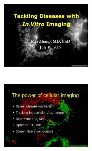
- 1. Tackling Diseases with In Vitro Imaging Mei Zhang, MD, PhD July 10, 2009 Mitochondria in live CV-1 cells The power of cellular imaging • Reveal disease mechanism • Tracking intracellular drug targets • Determine drug MoA • Optimize HTS hits • Screen library compounds Lysosomal contents in fused CT60 cells 1
- 2. Elucidation of cellular mechanisms for human disease 1. Niemann-Pick C disease (NPC) (late endosomal tubular trafficking defects) (@NIH) 2. Mucolipidosis IV (ML4) (protein trafficking defects and calcium accumulation in lysosomes) (@NIH) 3. Alzheimer’s disease (AD) (reduction of early endocytic recycling, accumulation of g-secretase substrates in ERC) (@MGH) 4. Cancer metastasis, angiogenesis, microenvironment (SDF1 and CXCR4 axel in metastasis) (@Synta) 5. Cancer cell stress (ROS and HSP70 in cancer) (@Synta) 6. Tumor vasculature (Microtubule and vascular structure) (@Synta) 7. Inflammation (TLR9 activation by CpG and cRel trafficking; CRACM1 and Stim1 trafficking) (@Synta) Lysosomes Lysosomes c-Rel c-Rel Niemann-Pick C disease (NPC) Mucolipidosis IV (ML4) (Trafficking defect) (Protein location and Ca++ defects) Endosomes Inflammation (TLR9, c-Rel trafficking) Reveal Disease Mechanism Alzheimer’s disease (AD) Pseudopodia (APP and g-secretase) ROS VE-Cadherin Viability Cancer metastasis, angiogenesis, microenvironment Tumor vasculature Cancer cell stress (CXCR4; HSP90) (VDA) (Elesclomol) 2
- 3. Localization of membrane and soluble proteins in intracellular organelles 1. Plasma membrane (CXCR4, S1P1, CRACM1) (@Synta) 2. Early endosomes (APP and APP-C99) (@MGH) 3. Late endosomes (NPC1, NPC2, MLN64, ML4, and activated CXCR4) (@NIH and Synta) 4. Lysosomes (NPC1, NPC2, MLN64, Mucolipin-1, and activated CXCR4) (@NIH and Synta) 5. Secretory granules (Insulin) (@Synta) 6. Lipid droplets (NPC defect, hexaminidase C short form) (@NIH) 7. ER (Mutant NPC1 and ML4) (@NIH) 8. Golgi apparatus (FcER) (@NIH) 9. Mitochondria (Ceramide, MLN64-cholesterol acceptor) (@NIH and Synta) 10. Microtubules (Alpha-tubulin, Spag6) (@NIH and Synta) 11. Actin filaments (16Q) (@NIH) 12. Nuclei (16Q) (@NIH) 13. Nucleoli (Proteasome) (@Synta) (Also: Hexosaminidase C) Lipid droplets (Sudan III) Early endosomes Mitochondria (APP-C99) (Mito-Tracker) Late endosomes (Also: APP) (NPC1) (Also: Ceramide) PM (CXCR4) (Also: NPC2, MLN64, Secretory granules ML4, and activated CXCR4) ER (ML4) (Also: CRACM1, S1P1) (Insulin) Tracking Nuclei (NFkB Cellular Targets Golgi (Also: Mutants NPC1, ML4) c-Rel) (Also: FcER) (Also: 16Q) Chromosome (pERK) Nucleoli Actin filaments (Proteasome-i) Microtubules (Also: 16Q) (a-tubulin) Centrosome (Tubulin-YFP) (Also: Spag6) 3
- 4. Study modes of action of new drugs in discovery and development 1. Apilimod (c-Rel translocation, ERK translocation, vacuolation) (@Synta) 2. Elesclomol (Proteasome inhibition, and induction of ROS and OH radical generation) (@Synta) 3. MTi (Existing or new replacement, fragmentation of MT and multinucleation) (@Synta) 4. CRAC inhibitors (Effect on cardiomyocytes; Stim-1 translocation by activation of ER calcium release) (@Synta) 5. Gamma-secretase inhibitors (Accumulation of substrates in ERC) (@MGH) 6. Natural compounds (Inducing insulin production) (@Synta) 7. Synergistic anti-cancer effects (Elesclomol with paclitaxel, ascorbate and other anticancer agents) (@Synta) ROS inducer (Elesclomol) IL12 inhibitor (Apilimod) MT inhibitors Determine MoA -secretase inhibitors CA4P CRAC HSP90i inhibitors 1+1>2 Anti-cancer synergy (Elesclomol+Taxol, or VitC) 4
- 5. Screening Focused Libraries and Optimizing HTS Hits • Apoptosis-rescuing, Screening Project (Leader, assay, screening, MoA) (@Synta) • New Microtubule Inhibitor, (VDA) Program, (MoA in Lead-Opt) (@Synta) • CRAC Inhibitor Backup Screening Program (Assay development, Hit-ID) (@Synta) • CXCR4 Inhibitor Screening Program (Leader, assay, MoA, chemistry, collaboration) (@Synta) • New ROS Inducer Screening Program (MoA in Lead-Opt) (@Synta) Apoptosis-rescuing Screening Project (Leader, assay, screening, MoA) New Microtubule Inhibitor (VDA) Program (MoA in Lead-Opt) Screening and Optimization CXCR4 Inhibitor CRAC Inhibitor Backup Screening Program Screening Program (Leader, assay, MoA, (Assay development, Hit-ID) chemistry, collaboration) New ROS Inducer Screening Program (MoA in Lead-Opt) 5
- 6. Topics • Revealing disease mechanisms for lysosomal storage disorders • Tracking cellular targets of g-secretase inhibitor for Alzheimer’s disease • Determining MoA of drug Elesclomol for metastatic melanoma • Screening and optimization for CXCR4 inhibitors Water immersions from Zeiss in LCBB NIDDK Topic 1 Discovery of novel mechanisms of NPC1 and ML4 diseases Co-culture of rat neurons and astrocytes 6
- 7. NPC1 disease Co-culture of rat neurons and astrocytes Niemann-Pick Disease, Type C (NPC) and the Late Endocytic Pathway Niemann-Pick Disease, type C • Age: 0-60 Recycling • Genes: NPC1 and NPC2 • Symptoms: dementia • Cellular abnormality: Accumulation of cholesterol in lysosomes • Diagnosis: – Mutations in NPC1 or NPC2 – Cholesterol esterification – Cholesterol in lysosomes (filipin staining) • Therapy: miglustat (on trials) Degradation 7
- 8. Confocal / digital scopes, cell warmers Zeiss 410 Lab-Tek covers Controllers CoolSnapHQ Lens warmer Sandwich and circulator Cellular phenotype of NPC: Cholesterol accumulation in lysosomes Normal NPC CT60 Cells Human Fibroblasts Patient Fibroblasts (NPC1 Null CHO) Cholesterol Filipin Staining Filipin 8
- 9. Cholesterol-laden lysosomes are multilamellar compartments EM structure of a lysosome Anti-LAMP / Filipin Freeze Fracture A human NPC fibroblast Freeze Fracture (Filipin) From Nancy and Joan NPC1 Protein and the SSD Millard, et al. JBC 2005 Davis, et al. JBC 2000 P691S NPC1 GFP 9
- 10. NPC1-GFP, but not the P691S mutant, is functional in clearing cholesterol accumulation. NPC1-GFP Anti-NPC1 Cholesterol SSD-NPC1-GFP (P691S) Anti-NPC1 Cholesterol CT60 mutant cells Cessation of mutant NPC1-GFP trafficking in NPC cells 10
- 11. NPC1-GFP regenerates LET trafficking in NPC cells NPC1-GFP adenovirus is fully functional. NPC1-GFP Cholesterol (Filipin) Without NPC1-GFP With NPC1-GFP Adenovirus CT60 cells 11
- 12. Adenovirus-NPC1-GFP infected rat cortical neuron (axon-like neurite) Adenovirus-NPC1-GFP infected rat astrocytes 12
- 13. A Networking Trouble? - A novel NPC cell defect Normal cell fusion NP-C cell fusion Can NPC cells exchange lysosomal content ? If not, what inhibits the dynamic networking? Rhodamine-dextran loads NPC1-GFP-containing late endosomes and large lysosomes NPC1-GFP RD SSD-NPC1-GFP RD 13
- 14. Procedure for examining lysosomal content exchange • Normal cells Incubate with fluorescent dextran for 24 hrs and wash for 2 hrs • NP-C cells • LDL Trypsinize and mix cells • LPDS LPDS: Plate mixed cells Lipoprotein-deficient serum (bovine) Fuse with Polyethylene Glycol (PEG) Fix at various time points (5 min to 48 hrs) Complete exchange of contents in fused WT CHO Normal cell fusion Living Cells, 2 hrs after fusion Red Channel Green Channel 14
- 15. NPC1 transports and clears cholesterol out of lysosomes; by doing this, NPC1 acts to: • Maintain the dynamic networking and communication among lysososmes; • Properly sort lysosomal glycolipids and direct lipid trafficking to the proper places; • Keep efficient long distance transportation along microtubules; • Maintain CNS function (PNAS February 17, 2009 vol. 106 no. 7 2377–2382 Benny Liua, … and John M. Dietschy.) LET and microtubules Disruption of microtubules prohibits content exchange of late endocytic compartments in fused cells Nocodazole treated WT CHO cells, 2 hrs after fusion 15
- 16. Content exchange among lysosomes is blocked in fused CT60 cells 2 h after fusion Cholesterol-depletion partially regenerate tubular trafficking LDL (cholesterol- enriched) LPDS (cholesterol- depleted) 16
- 17. Lysosomal content exchange is recovered in fused CT60 cells depleted of cholesterol 2 h after fusion Content exchange among lysosomes is regulated by endosomal cholesterol content LDL LPDS - 17
- 18. Glycolipids traffics in NPC1 compartment NPC1 Glycolipids mAb GM2 Lac-Cer GD3 CTH fibroblasts NPC1, NPC2, Endocytosis and Drug Discovery 18
- 19. Cholesterol depletion study on mice Survival Liver Neurode- function generations Controls (Saline, Allo., No improv. No improv. No improv. Ezetimibe) CYCLO at Improv. Improv. Improv. day 7. beta-cyclodextrin (‘CYCLO’) CYCLO at No improv. Improv. No improv. day 49. PNAS February 17, 2009 vol. 106 no. 7 2377–2382 Benny Liua, … and John M. Dietschy Cholesterol depletion improved NPC1 disease mouse model (npc1-/-) PNAS February 17, 2009 vol. 106 no. 7 2377–2382 Benny Liua, … and John M. Dietschy 19
- 20. NPC1 disease - a trafficking disorder! • NP-C disease has a defective late endosomal tubular trafficking function due to cholesterol accumulation. • Reducing cholesterol improves NPC1 in vitro as well as in vivo (Dr. John Dietschy, PNAS 2009 106:2377). cholesterol NPC1 transports and clears cholesterol out of lysosomes; by doing this, NPC1 acts to: • Maintain the dynamic networking and communication among lysososmes; • Properly sort lysosomal glycolipids and direct lipid trafficking to the proper places; • Keep efficient long distance transportation along microtubules; • Maintain CNS function (PNAS February 17, 2009 vol. 106 no. 7 2377–2382 Benny Liua, … and John M. Dietschy.) LET and microtubules 20
- 21. Mucolipidosis IV disease Mucolipin-1 is a channel protein cytoplasm HN2 COOH F408del membrane D362Y V446L 4(32aa del) lumen TRP cation channel domain 21
- 22. Mucolipin-1 Resides in the Late Endocytic Compartment Mucolipin-1 Rhodamine-dextran GFP-Mucolipin-1 Mucolipin-1-GFP GFP- Mucolipin1 Mucolipin1- GFP GFP CHO Cells Ion flow direction is depended upon mucolipin-1 topology on lysosomal membrane. Ion flow Or Ion flow Late endosome / Lysosome Late endosome / Lysosome 22
- 23. Topological Analysis on Mucolipin-1 Indicates Ion Flow from the Late Endocytic Compartment to the Cytoplasm Ion flow GFP-mucolipin-1 NPC2-RFP Late endosome / Lysosome Mucolipin-1-GFP NPC2-RFP NPC2-GFP as a tool… No leupeptin Leupeptin 12h NPC2-RFP Niemann-Pick Disease type C2 protein RFP is protease resistant! 23
- 24. Mucolipin-1-GFP is delivered to lysosomes via a biosynthetic pathway that bypasses the plasma membrane, which is different from LAMPII LAMP2-GFP Mucolipin-1-GFP Transfection 24 hr Transfection 72 hr Association of late endosomal mucolipin-1-GFP with the ER GFP-Mucolipin-1 24hr Rhodamine-dextran 24
- 25. F408del-GFP mutant is delivered to the late endocytic compartment in MLIV fibroblasts F408del-GFP V446L-GFP D362Y-GFP F408del Rho-dextran Calcium Orange Labeling: Calcium accumulation in the late endocytic compartment in MLIV fibroblasts Normal Niemann-Pick C KpnI cut uncut (CWN) (GM3123) KpnI cut 2527 2629 2527 2629 HeteroHomoNormal MLIV MLIV (GM2527) (GM2629) 25
- 26. Calcium Orange is trapped in the lumen of F408del-GFP defined late endosomes and lysosomes Expression of mucolipin-1 prevents calcium accumulation in the late endocytic compartment in MLIV fibroblasts GFP-Mucolipin-1 Calcium Orange F408del-GFP Calcium Orange Mucolipin-1-GFP Calcium Orange Note: Loaded dye first then transfect: Calcium Orange Mucolipin-1-GFP 26
- 27. Calcium starvation damages MLIV fibroblasts Ca2+ Ca2+ Ca2+ Ca2+ 200mg/L Free 200mg/L Free A B G H MLIV Normal fibroblasts1 human (GM2527) fibroblasts 1 C D I J Normal MLIV human fibroblasts2 fibroblasts (GM2629) 2 E F NPC1 fibroblasts Calcium depletion induces death of MLIV fibroblasts Ca2+ 200mg/L Ca2+ Free live dead live dead Normal human fibroblasts 1 Normal human fibroblasts 2 Live NPC1 fibroblasts Dead MLIV fibroblasts1 (GM2527) MLIV fibroblasts2 (GM2629) 27
- 28. Mucolipidosis IV has defective protein trafficking and endocytic calcium accumulation • ML4 disease has defective mucolipin protein trafficking and some mutants are stuck in the ER. • ML4 disease presents intracellular calcium homeostasis defect. • Modulating calcium homeostasis may improve ML4 patients. The Whole Story in One Big Movie 28
- 29. Topic 2 Drug modes of action study with in vitro imaging - Elesclomol is a special proteasome inhibitor Microtubules in CHO cells expressing tubulin-YFP Elesclomol Elesclomol was known as: • HSP70 inducer • Cytoskeleton disorganizer • Paclitaxel enhancer (in vivo) Taxol treated MCF7 cells expressing tubulin-YFP 29
- 30. Elesclomol induces accumulation of Tubulin-YFP at the centrosome in CHO cells DMSO Elesclomol CHO cells (12hr, 500nM) Elesclomol and known proteasome inhibitors cause similar phenotypes in CHO cells 5hr 24hr DMSO Elesclomol Elesclomol ALLN MG132 Lactacystin MG132 Lactacystin 30
- 31. IXMicro solution is now involved in centrosome ! HCS analysis for the centrosomal 1. effect Multiple known proteasome inhibitors 2. 80 compounds from MicroSource (‘Cancer Plate’) 31
- 32. Quantitative analysis of centrosomal proteasome inhibition by Elesclomol and other proteasome inhibitors “Plate 3, 24-26hr treatment” Z factor = 0.50-0.68 Centrosomal Average Intensity Elesclomol vs. derivatives and proteasome inhibitors 32
- 33. Quantitative analysis of centrosomal proteasome inhibition by 80 control compounds (Cancer Plate) “Plate 4, 24-26hr treatment” Centrosomal Average Intensity Elesclomol vs. 80 anti-cancer drugs 33
- 34. CHEMISTRY O H N ID MOLENAME 80 cpds for centrosome assay Chemistry 34 NH3 1501189 DEOXYURIDIN O O Pt NH3 HO O Chemistry 1 100537 SOLASODINE O Cl O HN Cl Chemistry 35 1502106 CARBOPLATIN O- O O O O N+ OH N O NH3 O OH OH OH Cl O CHLORAMPHEN O OH Pt O Chemistry 13 1500174 OL HO O TAMOXIFEN NH3 O Chemistry 24 1500557 CITRATE Cl Cl N Chemistry 2 200013 ROTENONE O O Chemistry 36 1502107 CISPLATIN HO P OH HO P OH S O O HN OH O OH OH H O N HCl HN HN N O N OH O OH O N CHLOROQUIN H2N N N HO O O OH Chemistry 14 1500179 DIPHOSPHAT AKLAVINE Chemistry 25 1500573 THIOGUANINE N+ N N- Chemistry 3 200022 HYDROCHLORIDE Cl Chemistry 37 1502109 ZIDOVUDINE [AZ Cl - H2O O N NH2 O O O O P N N NH Cl H2N N+ NH2 N O OH O CYCLOPHOSPH O Chemistry 15 1500213 MIDE HYDRAT ACRIFLAVINIUM O O OH Chemistry 26 1500618 HYDROCHLORIDE OH Chemistry 4 201138 DEGUELIN(-) O HN O N S O Chemistry 38 1502111 5-AZACYTIDIN O OH HO OH H N 2 N N O O O OH O OH H O O OH O O O OH O NH DIETHYLCARBA S H 2N HO Chemistry 16 1500242 AZINE CITRAT HO OH PENTAMIDINE OH NH Chemistry 27 1500641 ISETHIONATE O Chemistry 5 201664 CELASTROL O HCl O N Chemistry 39 1502112 CYCLOHEXIMID O O H Cl H N O O O O N- HO + O N O OH O O OH Chemistry 17 1500272 EMETINE MYCOPHENOLIC OH O NH2 Chemistry 28 1500674 ACID Chemistry 6 300038 JUGLONE H N O H2N N N Chemistry 40 1502113 AZASERINE OH H N N O S O O N O O - O NH H + F NH2 N N OH F O O O O HO O O Chemistry 18 1500305 FLUOROURAC Chemistry 29 1500679 AMINOPTERIN H2N OH p- SANGUINARINE FLUOROPHENY Chemistry 7 310035 SULFATE O OH O Chemistry 41 1502114 LANINE O N HN HN H O O O O N N + HN NH 1,2- O N O O O H2N O O N HCl DIMETHYLHYDR O O Cl - O NH2 NH N BERBERINE ZINE H O O Chemistry 19 1500344 HYDROXYURE O N N Chemistry 30 1500811 CHLORIDE Chemistry 42 1502116 HYDROCHLORID O O Cl Chemistry 8 330001 DACTINOMYCIN OH O OH O HO O O S N OH O O Cl HO METHYLMETHANE MECHLORETHA O PHENETHYL Chemistry 9 330003 SULFONATE Chemistry 20 1500375 NE Chemistry 43 1502209 CAFFEATE (CAP Chemistry 31 1500898 EMODIN O N N S N HCl O - N N+ H N N N N S O HN N N OH O NH 2H C l O N O N HN H N N OH N OH O N MERCAPTOPUR O Chemistry 21 1500387 E PUROMYCIN Chemistry 44 1502232 CAMPTOTHECI Chemistry 10 1500133 AZATHIOPRINE Chemistry 32 1501105 HYDROCHLORIDE HCl O N O O N O S O HCl O O O O O S NH HO O O O OH HO OH HO OH OH O Cl N HO O H OH QUINACRINE O OH Chemistry 11 1500152 BUSULFAN Chemistry 22 1500522 HYDROCHLORI Chemistry 45 1502244 AMYGDALIN Chemistry 33 1501136 NERIIFOLIN O OH O O O OH O OH O F O NH H NH Cl HO OH HO H HO OH O Cl O N O HO NH N OH HO O N O + CHLORAMPHENIC O O - N O O OL OH Chemistry 23 1500543 STREPTOZOS 5-FLUORO-5'- Chemistry 46 1502245 ELLAGIC ACID Chemistry 12 1500173 HEMISUCCINATE Chemistry 34 1501189 DEOXYURIDINE The proteasome effect might be a consequence of ROS induction… Multi- Tubulin-YFP ubiquitinated In Vitro Proteosensor Hsp70 accumulation Protein Proteasome accumulation in induction accumulation inhibitory in cells cells activity in cells Proteasome + + + + + inhibitors Elesclomol + + + + - 34
- 35. Topic 3 Tracking cellular targets of -secretase inhibitors for Alzheimer’s disease -secretase inhibition and APP accumulation in the ERC 35
- 36. Early Endosomal Recycling Pathway Recycling APP Processing and Alzheimer’s Disease 36
- 37. C99-GFP accumulates intracellularly in stable clone when -secretase is inhibited (24h treatment) DMSO DAPT C99-GFP, accumulates following treatment with PS/-secretase inhibitors DMSO DAPT L-685,458 APP-C99-GFP actin 37
- 38. Accumulation of APP-C99-GFP occurs first on the cell surface and then in the recycling compartment DAPT treatment 1 hr 3.5 hr 10.5 hr Bottom Morphological changes of APP-C99-GFP -containing compartments during Nocodazole or BFA treatment (Clone-27C) DAPT only DAPT + Nocodazole DAPT + BFA 38
- 39. AD mutants APP-CTF-GFP accumulate intracellularly when treated with DAPT EGg V717F V717I D M S O D A P T APP-C99-GFP accumulates both in endosomes and on the plasma membrane following treatment with the PS/-secretase inhibitor DAPT Top 2 3 4 5 Bottom 39
- 40. Accumulated C99-GFP is in the early endosomes when -secretase is inhibited C99-GFP Rho-Tfn C99-GFP Rho-dextran APP-C99-GFP accumulates in ERC + PM following treatment with DAPT and L-685,458 in cells stably transfected with APP-C99-GFP APP-C99-GFP Transferrin DMSO C99-GFP actin DAPT L-685, 458 40
- 41. APP-CTF-YFP accumulates in the recycling compartment following treatment with two different PS/-secretase inhibitors in cells transiently transfected with full length APP-YFP DMSO DAPT L-685,458 APP Plasma Membrane APP(full-length) BACE1 APP A Anti-GFP APP- CTF blotting APP-CTF * (free YFP) Secretase Complex (PS1, PEN2, actin Nicastrin, APH1) APP-YFP + Transferrin Development of gamma-secretase inhibitors Eli Lilly with LY450139 in Phase II Merck with MK0752 in Phase II Eisai with E2012 in Phase I Elan in preclinical 41
- 42. Summary GFP- and digital imaging-based live cell analysis is an ideal technology to identify defects of intracellular trafficking of proteins. GFP- and microscopy-based cell biology technologies can be applied to protease inhibitor assays (lysosomal protease and early endocytic g-secretase) in live cells. Conclusions Inhibition of gamma-secretase causes APP-CTF accumulates in the early endocytic pathway. This cellular phenotype (APP-C-GFP accumulation) is applicable to HCS for secretase inhibitor drug discovery. 42
- 43. Topic 4 Screening and optimization for CXCR4 inhibitors Dual color masking of Chem-1-CXCR4 cells for ‘happy feet’ formation CXCR4 and SDF1 mediate cancer metastasis CXCR4 SDF1a (CXCL12) Note: CXCL12 = SDF1 Luker and Luker (2005) Cancer Letters 238:30-41 43
- 44. Key assays in SDF1 signaling pathways 3 Assays: 1. Calcium Mobilization 2. Cell Migration 1 4 3. Receptor Binding 4. cAMP production 5. Endocytosis 2 5 The platform High content analysis Cell-based assays High throughput screen Medicinal chemistry Outsource collaboration Computational Chemistry Academic collaboration 44
- 45. Transflour assay Cyto-Nuc Translocation HCA, HCS Assays Transwell migration HCA - HCS Pseudopodia formation Endothelial tube formation The BioImaging Network: JPEG, BMP, Excel JPEG, BMP, Excel Regular Microscopy Data IXMicro HCS Data in in Local Drive Local Drive (Biol) JPEG, AVI (Biol) Videomicroscopy Data in Local Drive (Biol) BioImaging AcuityExpress Network Drive (Biol and IT) (Biol) JPEG, AVI, Excel JPEG, BMP, Excel IncuCyte Data In Local Drive (Biol) Tape Recording (IT) 45