2. +
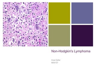
Lymphoma
Lymphomas are malignant neoplasms derived
from lymphoid tissues (lymphocytes,
histiocytes and their precursors/derivatives).
Typically they present as solid tumors.
3. +
Hodgkin vs. Non-Hodgkin
Histological: The distinction between Hodgkin’s Disease and
Non-Hodgkin’s Lymphoma is made upon histological
examination of the cancerous material. During examination if a
specific type of cells, the Reed-Sternberg cells are detected,
then the lymphoma is classified as Hodgkin’s Disease.
4. +
Hodgkin vs. Non-Hodgkin
Clinical: People with Hodgkin's Disease often have fevers,
night sweats, and weight loss. Other signs and symptoms
include fatigue, itching, anemia, and an enlarged spleen.
People who suffer from non-Hodgkin's Lymphoma can develop
fevers and night sweats as the disease progresses. More
common symptoms include abdominal pain, nausea, vomiting,
cough, and signs of central nervous system involvement
(seizures and altered mental status).
5. +
Cause
Accumulation of genetic lesions that affect:
proto-oncogenes
tumor suppressor genes
Chromosomal translocations
t(14;18) overexpression of bcl-2 an apoptotic inhibitor oncogene
follicular lymphoma
t(11;14) overexpression of bcl-1 mantle cell lymphoma
t(8;14) c-myc dysregulation – in Burkitt’s Lymphoma
t(2;5) expression of an aberrant fusion protein – anaplastic
large cell lymphomas
6. +
Cause
Infectious agents like
Epstein-Barr virus (EBV).
Human T-cell leukemia virus
H. Pylori
HHV8
HIV
Environmental factors such as chemicals.
Medical treatments such as radiation therapy and chemotherapy.
Genetic diseases, like Klinefelter's syndrome, Chédiak-Higashi
syndrome, ataxia telangiectasia syndrome.
Autoimmune diseases, like Sjögren’s syndrome, celiac sprue, rheumatoid
arthritis and systemic lupus erythematosus (SLE).
8. +
Origin
NHL represents a progressive clonal expansion of B cells or T
cells and/or NK cells arising from an accumulation of lesions
affecting proto-oncogenes or tumor suppressor genes, resulting
in cell immortalization.
Almost 85% B-cell origin;
15% T/NK cells;
Rarely from Macrophages.
9. +
Clinical Presentation
Depends upon type of tumor and the areas of involvement.
Some have mild presentation with lymphadenopathy waxing and
waning over years whereas others can have a very aggressive
presentation.
Aggressive Tumors: Commonly have an acute presentation with
rapid growth in size of tumor, systemic ‘B’ symptoms of fever, night
sweats, weight loss, etc.). Examples: diffuse large B cell
lymphoma, Burkitt lymphoma and adult T cell leukemia-lymphoma.
Indolent Tumors: Slow growing lymphadenopathy, hepatomegaly,
splenomegaly, or cytopenias. Examples: Follicular lymphoma,
chronic lymphocytic leukemia.
10. +
Oncological Emergencies
Patients may present with some emergent problem(s) that require
immediate intervention and therapy. These include:
Spinal cord compression
Pericardial tamponade
Hypercalcemia (eg, adult T cell leukemia-lymphoma)
Superior or inferior vena cava obstruction
Hyperleukocytosis (eg, B or T cell lymphoblastic leukemia/lymphoma)
Acute airway obstruction (eg, mediastinal lymphoma)
Lymphomatous meningitis and/or CNS mass lesions
Hyperuricemia and tumor lysis syndrome
Hyperviscosity syndrome (eg, lymphoplasmacytic lymphoma with
Waldenstrom macroglobulinemia)
11. +
Physical Examination
The physical examination needs to be directed to all potentially involved
lymphoid sites, these include:
Waldeyer's ring (tonsils, base of the tongue, nasopharynx)
Standard lymph node sites (cervical, supraclavicular, axillary, inguinal, femoral)
Liver and spleen
Abdominal nodal sites (mesenteric, retroperitoneal)
Less commonly involved nodal sites
Chest and Lungs.
Mediastinal Adenopathy
Abdomen and Pelvis.
Retroperitoneal, mesenteric and Pelvic nodes.
Extranodal Sites:
GI tract
Skin
Testicular
12. +
Laboratory
CBC:
Normal counts in early disease
Cytopenias due to bone marrow infiltration or autoimmune causes
Lymphocytosis with circulating malignant cells
Thrombocytosis (paraneoplastic syndrome or reactive)
Chemistries:
LDH (assoc with tumor burden – poor prognosis)
Beta-2 microglobulin (poor prognosis)
LFTs (hepatic involvement, hyper metabolism, chronic inflam)
Calcium (raised in T-ALL)
Viral serology:
HIV or HTLV-1 may be present depending on tumor histopathology
PET Scan:
post-treatment – differentiate between recurrence and residual disease
15. +
Biopsy
Biopsy is required for diagnosis and classification of NHL.
Modalities:
FNA
Definitive Tissue Biopsy
Studies on Excised Tissue
Histology
Immunophenotype
Genetic Studies
16. +
Biopsy
Histologic evaluation includes:
Assessment of the morphology;
Pattern of lymph node involvement.
Morphology: Morphological changes have prognostic impact.
Favorable Prognosis Unfavorable Prognosis
Nodular/follicular architecture Diffuse architecture
Small cell size Large cell size
Cleaved nucleus Un-cleaved nucleus
17. +
Biopsy
Pattern(s) of lymph node involvement:
Nodular/follicular pattern
Diffuse pattern
Change from a nodular to a diffuse pattern in adjacent nodes
Change from a lower to a higher grade of involvement within a
single node
23. +
Treatment
Indolent – I and II (contiguous)
Involved field radiation
10 year failure free survival: 50-60%
Indolent – II, III, IV
Asymptomatic – deferral therapy with careful observation (early
treatment does not improve prognosis)
Median progression in 4-6 years
Symptomatic: Purine nucleoside analogues (Fludarabine)
Oral alkylating agents (Cyclophosphamide)
Combination chemotherapy
24. +
Treatment
Aggressive – I and II (contiguous)
3 cycles of CHOP followed by involved field radiation.
Aggressive – II, III, IV
combination chemotherapy (CHOP is better than ProMACE-
CytaBOM, m-BACOD and MACOP-B; otherwise use a doxorubicin
based regime)
consider involved field radiation
Autologous/allogenic bone marrow or peripheral stem cell
25. +
Prognosis
International Prognostic Index (IPI)
A scoring system originally designed to assess prognosis in high-grade
lymphomas. Also useful for low and intermediate grade lymphomas.
Score of 1 for each:
Age - > 60 yrs.
LDH – elevated
Performance status – ECOG score 2-4
Ann Arbor State – III or IV
More than 1 extra nodal site
0-1: 75% chance of relapse free and overall survival at 5 years
2-3: 50%
4-5: 25%
26. +
Common Lymphomas
Diffuse Large Cell Lymphoma – 50% of NHL
Most of B cell origin
Dual age peak: twenties and sixties
Rapidly enlarging, symptomatic mass at a single site
Commonly extranodal: GI, skin, bone, brain – although liver,
spleen and marrow not often involved at presentation
Requires intensive therapy and CNS prophylaxis
27. +
Common Lymphomas
Follicular Lymphomas – 20-40% of adult NHL
Generally low grade
Usually age > 50 years
Characteristic t(14;18) in 90% of cases
Generalized, painless, lymphadenopathy. Spleen and marrow
often involved at time of diagnosis (75%).
Long natural history (6-8 years) – but not affected by treatment
Treatment options: watchful waiting, purine nucleoside analogues,
monoclonal antibodies to CD20
Becomes aggressive if progress to diffuse lymphoma – but still not
treatable
28. +
Common Lymphomas
Lymphoblastic Lymphoma
40% of all childhood lymphomas: mostly males under 20 years
high grade lymphoma; closely related to T-cell Acute Lymphocytic
Leukemia – and treated accordingly
present with a rapidly progressive mediastinal mass (50-70%)
early bone marrow spread, and onward to blood and meninges
Small Lymphocytic Lymphoma
4% of all NHL
older age group
generalized lymphadenopathy with enlarged liver and spleen
The only non-follicular low-grade lymphoma
prolonged survival – but not treatable
29. +
Common Lymphomas
Mantle Cell Lymphoma
B-Cell tumor believed to arise from the mantle of the follicle (as
opposed to the other lymphomas that arise from the middle)
Older males: disseminated disease
Aggressive and incurable
T(11; 14) - cyclin D1 driven monoclonal expansion
Burkitt’s Lymphoma
Endemic in parts of Africa – presents with maxillary/madibular mass
North America – presents with a progressive abdominal mass
Children and young adults (30% of childhood NHL)
Fastest growing human neoplasm
Intensive chemotherapy: 50% long term survival
20-30% risk of CNS involvment – provide intrathecal prophylaxis