SlideShare a Scribd company logo
1 of 75
Appropriate use of USG,CT and
MRI in liver imaging.
 Dr.K.Vinayachandran Nair MD,DM.
Encouraging us and asking right questions.
 Dr.Jagdeesh MRCPCH ,FRCR. Passion about
liver nodules. Also lending many slides.
 Few slides taken from google images.
I have to tell stories of
3 heroes… !
• Appropriate use of USG,CT and
MRI in liver imaging.
 Ultrasound of liver what we can diagnose and
what we can not.
 Primary liver lesions in cirrhotic liver.
 CT versus MRI in liver lesions.
 65 year old male with dyspepsia , bloating ,
not alcoholic.
Coarse texture ,
multiple tiny nodules.
Nodularity of liver contour.
Cirrhosis of liver.
 Surface nodularity with
high frequency probe.
 Caudate to right lobe
ratio more than 0.65
specific for cirrhosis.
 Portal vein diameter is
not sensitive in
detecting portal
hypertension.
 Color doppler is to
confirm direction of
flow , reversal
indicates portal
hypertension.
 Splenomegaly and
collaterals are
indicative of portal
hypertension.
 Increase of less than
20% in diameter of
portal vein with deep
inspiration indicates
portal hypertension.
 Very difficult criteria
…!
 Sensitivity and specificity varies from 60-
100% according to different studies.
 World journal gastroenterology..2010.
 Other criteria include
 Cork screw / enlarged hepatic artery.
 Loss of phasicity and pronounced cardiac
periodicity.
 Reduced portal vein velocity.
 Congestion index (ratio of area to flow velocity)
 Is controversial. May not be beneficial
 USG is not useful in detecting small (1-2 cm)
HCCs (sensitivity of 13 %).
 More sensitive in detecting larger ( more than 4
cm) HCCs(sensitivity of 75%)
 Can not differentiate between dysplastic nodule
and HCC.
AJR July 2002
Small HCC Large HCC
Diffuse type HCC , no mass detected
on USG. Liver transplantation
surgery showed diffuse HCC.
Slide taken from AJR
IT CAN DIFFERENTIATE TUMOR
FROM BLAND THROMBUS.
Portal vein
 Agents such as
perflubutane microbubble
cause contrast related
enhancement of focal liver
lesions.
 Enhancement pattern is
used to assess cellular
differentiation of HCCs.
 This can be repeated
realtime.
AJR
Enhancement and washout in a
poorly differentiated HCC
AJR
 A technique to assess
fibrosis of liver.
 Employs modified
ultrasound probe which
assesses velocity of a shear
wave created by a
vibratory source.
 Values of above 12.5kPa
are indicative of cirrhosis.
 50 year old male with
Progressive abdominal
distension.
 ? Mass in right upper
quadrant
Lack of visualization of
hepatic veins and
narrowing of caliber of IVC
indicating Budd Chairi
syndrome. A difficult
diagnosis on USG needs
IVCgram for confirmation.
 20 year old nursing student studying in
Karnataka has come to emergency with right
upper quadrant pain , not jaundiced --?
cholecystitis
 LFT showed elevated transaminases.
 Hepatitis A IgM marker positive.
 Final diagnosis – anicteric viral hepatitis.
Radiology assistant
www.radiologyassistant.nl
 Reason for showing this case is that there are
no specific sonographic findings for hepatitis.
 Reduced echotexture of liver , gall bladder
wall thickening , hepatomegaly , starry sky
appearance of liver due to periportal edema
are described findings.
 27 years old male working as executive in a
company comes with elevated liver
transaminases.
 Claims to be teetotaller.
Increased liver texture
indicating fatty liver.
 Liver echogenicity exceeds that
of right kidney and spleen.
And
 There is beam attenuation.
 Most often subjective..!!
 Grade 1– increased texture with good
visualization of diaphragm and intrahepatic
vessels.
 Grade 2- mild impairment of visualization.
 Grade 3– severe impairment of visualization.
 Here there is fatty infiltration , hepatomegaly
and elevated liver enzymes. (AST and ALT).
 Histologically, there is inflammation and
fibrosis.
 Radiologically it is not possible to separate
NASH from ordinary fatty liver.
 Fatty liver involves about 15% of population.
 NASH involves about 2-5% of population.
Focal fatty infiltration can
simulate mass.
Focal fatty sparing can
also mimic mass.
 25 year old healthy male for employment
health checkup.
 When USG shows hemangioma typical
features in a nononcology patient this
modality alone is considered sufficient.
 Not sensitive in detecting small HCCs.
 Can not differentiate between HCC and
dysplastic nodule.
 Entire liver can not be assessed , few USG
blind areas in liver.
 Moderate sensitivity in detecting cirrhosis.
 Hemangioma in a nononcology patient , cyst
and to certain extent abscesses can be
confidently diagnosed with USG, for other
focal lesions one needs to assess further.
Varying signals; varying sizes, varying enhancement
Siphon out the dysplastic nodules & HCC!!
How!
Lesions MRI features:-post contrast behaviour
Intermediate/vague features/cross over features
Varying signals; varying sizes, varying enhancement
Siphon out the dysplastic nodules & HCC!!
How!
Lesions MRI features:-post contrast behaviour
Intermediate/vague features/cross over features
Varying signals; varying sizes, varying enhancement
Siphon out the dysplastic nodules & HCC!!
How!
Lesions MRI features:-post contrast behaviour
Intermediate/vague features/cross over features
 T2,
 T2FS
 T1
 T1 in and opposed phase
 Dynamic post contrastT1FS(fat
saturation) with subtracted
images
 Regenerative Nodules ; Siderotic nodules
 Dysplastic Nodules
 HCC
 Size
- diameter of less than 2 cm are more likely to be benign than
malignant
 Vascularity
- shift from predominantly venous perfusion to predominantly
arterial perfusion
- dedifferentiated nodules - markedly enhanced on
early arterial phase
 Hepatocellular Function
- hepatocellular contrast agents
 most common cirrhosis-associated hepatocellular nodules
 T2 : Isointense to liver; hypointense ( iron)
 T1: Variable
 Postcontrast:
- No arterial enhancement
- isointense enhancement to the liver
- washout isointense to liver
 Low grade and high grade
 T2 : hypointense or isointense to liver,
 T1: Variable
 Postcontrast:
- Patchy ill defined nodular arterial
enhancement
- isointense enhancement to the liver
- hypointense to liver in the later phases, no
definite tumor capsule
 T2 : Hyperintense ( 94%) – isointense to spleen
 T1: Variable
 Postcontrast:
- Intense arterial enhancement
- isointense on the portal venous phase
- wash out on the later phase with persistent
tumor capsule
 T1- and T2-weighted MR images, the mosaic pattern appears
as areas of variable signal intensities
 lesions enhance in a heterogeneous fashion during the
arterial and later phases
Diffuse type HCC a
permeative tumor often with portal vein
thrombus.
LESION T2 T1 ART DEL
Reg N Low-iso Int - High No enhancement No wash out
Dys N Low-iso Int - High Heterogenenous
moderate
enhancement
Irregular wash
out with no
capsule
HCC Int - High Low - Int Intense
enhancement
Wash out with
enhancing
capsule
 CT versus MRI mosaic pattern- 88%
T1- and T2-weighted MR
images, the mosaic pattern
appears as areas of variable
signal intensities
lesions enhance in a
heterogeneous fashion during
the arterial and later phases
 When doing survey for metastasis , CT is
better as it can assess extra hepatic lesions
better than MRI.

 In patient with chronic liver disease , for
characterization of regenerative ,dysplastic
nodules and HCC , MRI is better than CT.
 There are several arterial phase enhancing
lesions.
 TAHD – transient hepatic attenuation
difference.
 Confluent hepatic fibrosis.
 Contrast MR has sensitivity of near 81% and
specificity of near 85%.
 CT has sensitivity of near 73% and sensitivity
of near 93%.
 Imaging not sensitive for lesions less than 20
mm and diffuse lesions.
 USG is the first line modality in liver imaging.
Used to rule out liver disease and diagnose
with confidence common lesions like cysts
and hemangiomas.
 Multiphasic CT is the standard reference
study in liver disease. Standard in assessing
and follow up of oncology patients. When
high resolution MRI is not available can be
used to image cirrhosis.
 MRI is modality of choice in assessing
cirrhosis liver . It is increasingly preferred in
follow up of patients and in children where
radiation is an issue.
 Tissue specific contrast agents are being
available and are helpful.
 Biopsy or follow up is advised in doubtful
lesions or histopathological confirmation is
necessary for management.
 Decisions should be patient specific.
 Diffusion weighted imaging
 Mixed extracellular and hepatocellular contrast agents
-gadoxetic acid disodium
 MR spectroscopy
 Liver perfusion
 Diffusion imaging liver.
Liver imaging snapshots role of CT USG MRI in liver imaging.

More Related Content

What's hot

Imaging of portal hypertension
Imaging of portal hypertensionImaging of portal hypertension
Imaging of portal hypertensionDev Lakhera
 
Presentation1.pptx, radiological imaging of cholangiocarcinoma.
Presentation1.pptx, radiological imaging of cholangiocarcinoma.Presentation1.pptx, radiological imaging of cholangiocarcinoma.
Presentation1.pptx, radiological imaging of cholangiocarcinoma.Abdellah Nazeer
 
Radiology in portal hypertension
Radiology in portal hypertensionRadiology in portal hypertension
Radiology in portal hypertensionSunil Kumar
 
Diagnostic Imaging of Cholangiocarcinoma
Diagnostic Imaging of CholangiocarcinomaDiagnostic Imaging of Cholangiocarcinoma
Diagnostic Imaging of CholangiocarcinomaMohamed M.A. Zaitoun
 
imaging of benign hepatic masses
imaging of benign hepatic massesimaging of benign hepatic masses
imaging of benign hepatic massesNavni Garg
 
radiological imaging of pancreatic malignancy - solid neoplasms radiological ...
radiological imaging of pancreatic malignancy - solid neoplasms radiological ...radiological imaging of pancreatic malignancy - solid neoplasms radiological ...
radiological imaging of pancreatic malignancy - solid neoplasms radiological ...RagubharathiRavi
 
Radiologic mimics of cirrhosis
Radiologic mimics of cirrhosisRadiologic mimics of cirrhosis
Radiologic mimics of cirrhosisPankaj Kaira
 
Imaging in small bowel tumors Dr. Muhammad Bin Zulfiqar
Imaging in small bowel tumors Dr. Muhammad Bin ZulfiqarImaging in small bowel tumors Dr. Muhammad Bin Zulfiqar
Imaging in small bowel tumors Dr. Muhammad Bin ZulfiqarDr. Muhammad Bin Zulfiqar
 
Utrasound Gall-bladder & biliary tract
Utrasound Gall-bladder & biliary tractUtrasound Gall-bladder & biliary tract
Utrasound Gall-bladder & biliary tractDr. Mohit Goel
 
Liver lesions
Liver lesionsLiver lesions
Liver lesionsairwave12
 
Radiology Spots PPT- 2 by Dr Chandni Wadhwani
Radiology Spots PPT- 2 by Dr Chandni WadhwaniRadiology Spots PPT- 2 by Dr Chandni Wadhwani
Radiology Spots PPT- 2 by Dr Chandni WadhwaniChandni Wadhwani
 
patterns of enhancement in hepatocellular carcinoma
patterns of enhancement in hepatocellular carcinomapatterns of enhancement in hepatocellular carcinoma
patterns of enhancement in hepatocellular carcinomaHaseeb Manzoor
 
Hepatocellular carcinoma—role of interventional radiologist Dr. Muhammad Bin ...
Hepatocellular carcinoma—role of interventional radiologist Dr. Muhammad Bin ...Hepatocellular carcinoma—role of interventional radiologist Dr. Muhammad Bin ...
Hepatocellular carcinoma—role of interventional radiologist Dr. Muhammad Bin ...Dr. Muhammad Bin Zulfiqar
 
Venous Doppler Lower limb Dr Mukesh Tilgam
Venous Doppler Lower limb Dr Mukesh TilgamVenous Doppler Lower limb Dr Mukesh Tilgam
Venous Doppler Lower limb Dr Mukesh TilgamDrmukesh Tilgam
 
Portal hypertension radiological diagnosis and interventions
Portal hypertension radiological diagnosis and interventionsPortal hypertension radiological diagnosis and interventions
Portal hypertension radiological diagnosis and interventionsSourav Talukder
 
Renal masses imaging
Renal masses imagingRenal masses imaging
Renal masses imagingSatish Naga
 

What's hot (20)

Imaging of portal hypertension
Imaging of portal hypertensionImaging of portal hypertension
Imaging of portal hypertension
 
Presentation1.pptx, radiological imaging of cholangiocarcinoma.
Presentation1.pptx, radiological imaging of cholangiocarcinoma.Presentation1.pptx, radiological imaging of cholangiocarcinoma.
Presentation1.pptx, radiological imaging of cholangiocarcinoma.
 
Radiology in portal hypertension
Radiology in portal hypertensionRadiology in portal hypertension
Radiology in portal hypertension
 
Diagnostic Imaging of the Liver
Diagnostic Imaging of the LiverDiagnostic Imaging of the Liver
Diagnostic Imaging of the Liver
 
Diagnostic Imaging of Cholangiocarcinoma
Diagnostic Imaging of CholangiocarcinomaDiagnostic Imaging of Cholangiocarcinoma
Diagnostic Imaging of Cholangiocarcinoma
 
imaging of benign hepatic masses
imaging of benign hepatic massesimaging of benign hepatic masses
imaging of benign hepatic masses
 
radiological imaging of pancreatic malignancy - solid neoplasms radiological ...
radiological imaging of pancreatic malignancy - solid neoplasms radiological ...radiological imaging of pancreatic malignancy - solid neoplasms radiological ...
radiological imaging of pancreatic malignancy - solid neoplasms radiological ...
 
Spots with keys (2)
Spots with keys (2)Spots with keys (2)
Spots with keys (2)
 
Radiologic mimics of cirrhosis
Radiologic mimics of cirrhosisRadiologic mimics of cirrhosis
Radiologic mimics of cirrhosis
 
Imaging in small bowel tumors Dr. Muhammad Bin Zulfiqar
Imaging in small bowel tumors Dr. Muhammad Bin ZulfiqarImaging in small bowel tumors Dr. Muhammad Bin Zulfiqar
Imaging in small bowel tumors Dr. Muhammad Bin Zulfiqar
 
Utrasound Gall-bladder & biliary tract
Utrasound Gall-bladder & biliary tractUtrasound Gall-bladder & biliary tract
Utrasound Gall-bladder & biliary tract
 
Liver lesions
Liver lesionsLiver lesions
Liver lesions
 
Radiology spotters
Radiology spottersRadiology spotters
Radiology spotters
 
Radiology Spots PPT- 2 by Dr Chandni Wadhwani
Radiology Spots PPT- 2 by Dr Chandni WadhwaniRadiology Spots PPT- 2 by Dr Chandni Wadhwani
Radiology Spots PPT- 2 by Dr Chandni Wadhwani
 
patterns of enhancement in hepatocellular carcinoma
patterns of enhancement in hepatocellular carcinomapatterns of enhancement in hepatocellular carcinoma
patterns of enhancement in hepatocellular carcinoma
 
Benign focal lesions in liver
Benign focal lesions in liverBenign focal lesions in liver
Benign focal lesions in liver
 
Hepatocellular carcinoma—role of interventional radiologist Dr. Muhammad Bin ...
Hepatocellular carcinoma—role of interventional radiologist Dr. Muhammad Bin ...Hepatocellular carcinoma—role of interventional radiologist Dr. Muhammad Bin ...
Hepatocellular carcinoma—role of interventional radiologist Dr. Muhammad Bin ...
 
Venous Doppler Lower limb Dr Mukesh Tilgam
Venous Doppler Lower limb Dr Mukesh TilgamVenous Doppler Lower limb Dr Mukesh Tilgam
Venous Doppler Lower limb Dr Mukesh Tilgam
 
Portal hypertension radiological diagnosis and interventions
Portal hypertension radiological diagnosis and interventionsPortal hypertension radiological diagnosis and interventions
Portal hypertension radiological diagnosis and interventions
 
Renal masses imaging
Renal masses imagingRenal masses imaging
Renal masses imaging
 

Viewers also liked

Advanced imaging modalities of the liver
Advanced imaging modalities of the liverAdvanced imaging modalities of the liver
Advanced imaging modalities of the liverEnass Khattab
 
CT and MRI Imaging of Hepatic metastases
CT and MRI Imaging of Hepatic metastasesCT and MRI Imaging of Hepatic metastases
CT and MRI Imaging of Hepatic metastasesDr Priyanka Vishwakarma
 
Liver Neoplasms
Liver   NeoplasmsLiver   Neoplasms
Liver NeoplasmsDeep Deep
 
Presentation1.pptx, radiological anatomy of the abdomen and pelvis.
Presentation1.pptx, radiological anatomy of the abdomen and pelvis.Presentation1.pptx, radiological anatomy of the abdomen and pelvis.
Presentation1.pptx, radiological anatomy of the abdomen and pelvis.Abdellah Nazeer
 
Abdominal imaging hcc t loke
Abdominal imaging hcc t lokeAbdominal imaging hcc t loke
Abdominal imaging hcc t lokeJFIM
 
Cronograma Aulas Li-RADs 2016/1
Cronograma Aulas Li-RADs 2016/1Cronograma Aulas Li-RADs 2016/1
Cronograma Aulas Li-RADs 2016/1Lucas Samuel
 
Liver radiology MRI
Liver radiology MRILiver radiology MRI
Liver radiology MRIMahesh Kumar
 
Liver,bile duct,pancreas and spleen
Liver,bile duct,pancreas and spleenLiver,bile duct,pancreas and spleen
Liver,bile duct,pancreas and spleenghalan
 
Focal hepatic lesions
Focal hepatic lesionsFocal hepatic lesions
Focal hepatic lesionsYasser Asiri
 
Liver tumors - A basic guide to diagnose and treat liver tumors
Liver tumors - A basic guide to diagnose and treat liver tumorsLiver tumors - A basic guide to diagnose and treat liver tumors
Liver tumors - A basic guide to diagnose and treat liver tumorsBhavin Vasavada
 
Ct Imaging of Abdomen Dr. Muhammad Bin Zulfiqar
Ct Imaging of Abdomen Dr. Muhammad Bin ZulfiqarCt Imaging of Abdomen Dr. Muhammad Bin Zulfiqar
Ct Imaging of Abdomen Dr. Muhammad Bin ZulfiqarDr. Muhammad Bin Zulfiqar
 

Viewers also liked (20)

MR imaging of liver
MR imaging of liverMR imaging of liver
MR imaging of liver
 
Advanced imaging modalities of the liver
Advanced imaging modalities of the liverAdvanced imaging modalities of the liver
Advanced imaging modalities of the liver
 
CT and MRI Imaging of Hepatic metastases
CT and MRI Imaging of Hepatic metastasesCT and MRI Imaging of Hepatic metastases
CT and MRI Imaging of Hepatic metastases
 
Focal liver lesion
Focal liver lesionFocal liver lesion
Focal liver lesion
 
Abdomin Liver CT
Abdomin Liver CT Abdomin Liver CT
Abdomin Liver CT
 
Ultrasound liver
Ultrasound liverUltrasound liver
Ultrasound liver
 
Liver Neoplasms
Liver   NeoplasmsLiver   Neoplasms
Liver Neoplasms
 
Presentation1.pptx, radiological anatomy of the abdomen and pelvis.
Presentation1.pptx, radiological anatomy of the abdomen and pelvis.Presentation1.pptx, radiological anatomy of the abdomen and pelvis.
Presentation1.pptx, radiological anatomy of the abdomen and pelvis.
 
CT ABDOMEN ANATOMY
 CT ABDOMEN ANATOMY CT ABDOMEN ANATOMY
CT ABDOMEN ANATOMY
 
Liver ultrasound
Liver ultrasoundLiver ultrasound
Liver ultrasound
 
Abdominal imaging hcc t loke
Abdominal imaging hcc t lokeAbdominal imaging hcc t loke
Abdominal imaging hcc t loke
 
Cronograma Aulas Li-RADs 2016/1
Cronograma Aulas Li-RADs 2016/1Cronograma Aulas Li-RADs 2016/1
Cronograma Aulas Li-RADs 2016/1
 
Liver radiology MRI
Liver radiology MRILiver radiology MRI
Liver radiology MRI
 
Liver,bile duct,pancreas and spleen
Liver,bile duct,pancreas and spleenLiver,bile duct,pancreas and spleen
Liver,bile duct,pancreas and spleen
 
Dr maryam
Dr maryamDr maryam
Dr maryam
 
Phase pdf
Phase pdfPhase pdf
Phase pdf
 
Focal hepatic lesions
Focal hepatic lesionsFocal hepatic lesions
Focal hepatic lesions
 
Liver tumors - A basic guide to diagnose and treat liver tumors
Liver tumors - A basic guide to diagnose and treat liver tumorsLiver tumors - A basic guide to diagnose and treat liver tumors
Liver tumors - A basic guide to diagnose and treat liver tumors
 
CT Abdomen and Pelvis
CT Abdomen and PelvisCT Abdomen and Pelvis
CT Abdomen and Pelvis
 
Ct Imaging of Abdomen Dr. Muhammad Bin Zulfiqar
Ct Imaging of Abdomen Dr. Muhammad Bin ZulfiqarCt Imaging of Abdomen Dr. Muhammad Bin Zulfiqar
Ct Imaging of Abdomen Dr. Muhammad Bin Zulfiqar
 

Similar to Liver imaging snapshots role of CT USG MRI in liver imaging.

Veeru ca pancreas
Veeru ca pancreasVeeru ca pancreas
Veeru ca pancreasVeeru Reddy
 
Imaging in Benign hepatic lesions
Imaging in Benign hepatic lesionsImaging in Benign hepatic lesions
Imaging in Benign hepatic lesionsGirendra Shankar
 
Case of pancreatic adenocarcinoma and other pancreatic tumors
Case of pancreatic adenocarcinoma and other pancreatic tumorsCase of pancreatic adenocarcinoma and other pancreatic tumors
Case of pancreatic adenocarcinoma and other pancreatic tumorsAlex Allu
 
Pancreatic neoplasms
Pancreatic neoplasmsPancreatic neoplasms
Pancreatic neoplasmsAjai Sasidhar
 
radiologicmimicsofcirrhosis-161222165901.pdf
radiologicmimicsofcirrhosis-161222165901.pdfradiologicmimicsofcirrhosis-161222165901.pdf
radiologicmimicsofcirrhosis-161222165901.pdfDrYaqoobBahar
 
INVESTIGATIONS OF A PATIENT WITH OBSTRUCTIVE JAUNDICE -LETS GO
INVESTIGATIONS OF A PATIENT WITH OBSTRUCTIVE JAUNDICE -LETS GOINVESTIGATIONS OF A PATIENT WITH OBSTRUCTIVE JAUNDICE -LETS GO
INVESTIGATIONS OF A PATIENT WITH OBSTRUCTIVE JAUNDICE -LETS GODr.Manojit Sarkar
 
Grish hcc presentation
Grish hcc presentationGrish hcc presentation
Grish hcc presentationsadiqsikora
 
Imaging of non endocrine tumour of pancreas
Imaging of non endocrine tumour of pancreasImaging of non endocrine tumour of pancreas
Imaging of non endocrine tumour of pancreasDev Lakhera
 
Renal cell carcinoma
Renal cell carcinomaRenal cell carcinoma
Renal cell carcinomagbanstha
 
Solid lesions of the Pancreas
Solid lesions of the PancreasSolid lesions of the Pancreas
Solid lesions of the PancreasAtit Ghoda
 
Imaging of adrenal masses abdulj final
Imaging of adrenal masses abdulj finalImaging of adrenal masses abdulj final
Imaging of adrenal masses abdulj finalabduljelil nejmu
 
LIVER SEGMENTAL ANATOMY AND IMAGINGIN IN LIVER CIRRHOSIS.pptx
LIVER SEGMENTAL ANATOMY AND IMAGINGIN IN LIVER CIRRHOSIS.pptxLIVER SEGMENTAL ANATOMY AND IMAGINGIN IN LIVER CIRRHOSIS.pptx
LIVER SEGMENTAL ANATOMY AND IMAGINGIN IN LIVER CIRRHOSIS.pptxDr vaibhavi patel
 
GIT Kurdistan Board GEH Journal club Lower PVT 2014.
GIT Kurdistan Board GEH Journal club Lower PVT 2014.GIT Kurdistan Board GEH Journal club Lower PVT 2014.
GIT Kurdistan Board GEH Journal club Lower PVT 2014.Shaikhani.
 
cystickidneydiseases-161210202710 (1).pdf
cystickidneydiseases-161210202710 (1).pdfcystickidneydiseases-161210202710 (1).pdf
cystickidneydiseases-161210202710 (1).pdfHARYANVITRAVLLER
 
Cystic kidney diseases
Cystic kidney diseasesCystic kidney diseases
Cystic kidney diseasessahar Hamdy
 

Similar to Liver imaging snapshots role of CT USG MRI in liver imaging. (20)

Veeru ca pancreas
Veeru ca pancreasVeeru ca pancreas
Veeru ca pancreas
 
Liver ppt..pptx- Liver Computed Tomography
Liver ppt..pptx- Liver Computed TomographyLiver ppt..pptx- Liver Computed Tomography
Liver ppt..pptx- Liver Computed Tomography
 
Renal malignancies
Renal  malignanciesRenal  malignancies
Renal malignancies
 
Imaging in Benign hepatic lesions
Imaging in Benign hepatic lesionsImaging in Benign hepatic lesions
Imaging in Benign hepatic lesions
 
Case of pancreatic adenocarcinoma and other pancreatic tumors
Case of pancreatic adenocarcinoma and other pancreatic tumorsCase of pancreatic adenocarcinoma and other pancreatic tumors
Case of pancreatic adenocarcinoma and other pancreatic tumors
 
Pancreatic neoplasms
Pancreatic neoplasmsPancreatic neoplasms
Pancreatic neoplasms
 
radiologicmimicsofcirrhosis-161222165901.pdf
radiologicmimicsofcirrhosis-161222165901.pdfradiologicmimicsofcirrhosis-161222165901.pdf
radiologicmimicsofcirrhosis-161222165901.pdf
 
INVESTIGATIONS OF A PATIENT WITH OBSTRUCTIVE JAUNDICE -LETS GO
INVESTIGATIONS OF A PATIENT WITH OBSTRUCTIVE JAUNDICE -LETS GOINVESTIGATIONS OF A PATIENT WITH OBSTRUCTIVE JAUNDICE -LETS GO
INVESTIGATIONS OF A PATIENT WITH OBSTRUCTIVE JAUNDICE -LETS GO
 
Grish hcc presentation
Grish hcc presentationGrish hcc presentation
Grish hcc presentation
 
Imaging of non endocrine tumour of pancreas
Imaging of non endocrine tumour of pancreasImaging of non endocrine tumour of pancreas
Imaging of non endocrine tumour of pancreas
 
Renal cell carcinoma
Renal cell carcinomaRenal cell carcinoma
Renal cell carcinoma
 
Solid lesions of the Pancreas
Solid lesions of the PancreasSolid lesions of the Pancreas
Solid lesions of the Pancreas
 
Bile Duct Tumor
Bile Duct TumorBile Duct Tumor
Bile Duct Tumor
 
Imaging of adrenal masses abdulj final
Imaging of adrenal masses abdulj finalImaging of adrenal masses abdulj final
Imaging of adrenal masses abdulj final
 
LIVER SEGMENTAL ANATOMY AND IMAGINGIN IN LIVER CIRRHOSIS.pptx
LIVER SEGMENTAL ANATOMY AND IMAGINGIN IN LIVER CIRRHOSIS.pptxLIVER SEGMENTAL ANATOMY AND IMAGINGIN IN LIVER CIRRHOSIS.pptx
LIVER SEGMENTAL ANATOMY AND IMAGINGIN IN LIVER CIRRHOSIS.pptx
 
GIT Kurdistan Board GEH Journal club Lower PVT 2014.
GIT Kurdistan Board GEH Journal club Lower PVT 2014.GIT Kurdistan Board GEH Journal club Lower PVT 2014.
GIT Kurdistan Board GEH Journal club Lower PVT 2014.
 
MRI in Cirrhotic Liver
MRI in Cirrhotic LiverMRI in Cirrhotic Liver
MRI in Cirrhotic Liver
 
sandra liver.pptx
sandra liver.pptxsandra liver.pptx
sandra liver.pptx
 
cystickidneydiseases-161210202710 (1).pdf
cystickidneydiseases-161210202710 (1).pdfcystickidneydiseases-161210202710 (1).pdf
cystickidneydiseases-161210202710 (1).pdf
 
Cystic kidney diseases
Cystic kidney diseasesCystic kidney diseases
Cystic kidney diseases
 

More from Krishna Kiran Karanth (8)

Practical tips in sonography
Practical tips in sonographyPractical tips in sonography
Practical tips in sonography
 
Workshop pediatic neuroimaging
Workshop pediatic neuroimagingWorkshop pediatic neuroimaging
Workshop pediatic neuroimaging
 
Pediatric neuroimaging
Pediatric neuroimagingPediatric neuroimaging
Pediatric neuroimaging
 
MRI IN FOOT PAIN
MRI IN FOOT PAINMRI IN FOOT PAIN
MRI IN FOOT PAIN
 
Cartilage imaging simplified
Cartilage imaging simplifiedCartilage imaging simplified
Cartilage imaging simplified
 
MRI OF SHOULDER INJURY
MRI OF SHOULDER INJURYMRI OF SHOULDER INJURY
MRI OF SHOULDER INJURY
 
MRI in neck imaging
MRI in neck imaging MRI in neck imaging
MRI in neck imaging
 
BASICS OF MRI
BASICS OF MRIBASICS OF MRI
BASICS OF MRI
 

Recently uploaded

Chandrapur Call girls 8617370543 Provides all area service COD available
Chandrapur Call girls 8617370543 Provides all area service COD availableChandrapur Call girls 8617370543 Provides all area service COD available
Chandrapur Call girls 8617370543 Provides all area service COD availableDipal Arora
 
Call Girls Varanasi Just Call 9907093804 Top Class Call Girl Service Available
Call Girls Varanasi Just Call 9907093804 Top Class Call Girl Service AvailableCall Girls Varanasi Just Call 9907093804 Top Class Call Girl Service Available
Call Girls Varanasi Just Call 9907093804 Top Class Call Girl Service AvailableDipal Arora
 
All Time Service Available Call Girls Marine Drive 📳 9820252231 For 18+ VIP C...
All Time Service Available Call Girls Marine Drive 📳 9820252231 For 18+ VIP C...All Time Service Available Call Girls Marine Drive 📳 9820252231 For 18+ VIP C...
All Time Service Available Call Girls Marine Drive 📳 9820252231 For 18+ VIP C...Arohi Goyal
 
Manyata Tech Park ( Call Girls ) Bangalore ✔ 6297143586 ✔ Hot Model With Sexy...
Manyata Tech Park ( Call Girls ) Bangalore ✔ 6297143586 ✔ Hot Model With Sexy...Manyata Tech Park ( Call Girls ) Bangalore ✔ 6297143586 ✔ Hot Model With Sexy...
Manyata Tech Park ( Call Girls ) Bangalore ✔ 6297143586 ✔ Hot Model With Sexy...vidya singh
 
Top Quality Call Girl Service Kalyanpur 6378878445 Available Call Girls Any Time
Top Quality Call Girl Service Kalyanpur 6378878445 Available Call Girls Any TimeTop Quality Call Girl Service Kalyanpur 6378878445 Available Call Girls Any Time
Top Quality Call Girl Service Kalyanpur 6378878445 Available Call Girls Any TimeCall Girls Delhi
 
Premium Call Girls Cottonpet Whatsapp 7001035870 Independent Escort Service
Premium Call Girls Cottonpet Whatsapp 7001035870 Independent Escort ServicePremium Call Girls Cottonpet Whatsapp 7001035870 Independent Escort Service
Premium Call Girls Cottonpet Whatsapp 7001035870 Independent Escort Servicevidya singh
 
VIP Hyderabad Call Girls Bahadurpally 7877925207 ₹5000 To 25K With AC Room 💚😋
VIP Hyderabad Call Girls Bahadurpally 7877925207 ₹5000 To 25K With AC Room 💚😋VIP Hyderabad Call Girls Bahadurpally 7877925207 ₹5000 To 25K With AC Room 💚😋
VIP Hyderabad Call Girls Bahadurpally 7877925207 ₹5000 To 25K With AC Room 💚😋TANUJA PANDEY
 
Book Paid Powai Call Girls Mumbai 𖠋 9930245274 𖠋Low Budget Full Independent H...
Book Paid Powai Call Girls Mumbai 𖠋 9930245274 𖠋Low Budget Full Independent H...Book Paid Powai Call Girls Mumbai 𖠋 9930245274 𖠋Low Budget Full Independent H...
Book Paid Powai Call Girls Mumbai 𖠋 9930245274 𖠋Low Budget Full Independent H...Call Girls in Nagpur High Profile
 
Night 7k to 12k Navi Mumbai Call Girl Photo 👉 BOOK NOW 9833363713 👈 ♀️ night ...
Night 7k to 12k Navi Mumbai Call Girl Photo 👉 BOOK NOW 9833363713 👈 ♀️ night ...Night 7k to 12k Navi Mumbai Call Girl Photo 👉 BOOK NOW 9833363713 👈 ♀️ night ...
Night 7k to 12k Navi Mumbai Call Girl Photo 👉 BOOK NOW 9833363713 👈 ♀️ night ...aartirawatdelhi
 
Bangalore Call Girl Whatsapp Number 100% Complete Your Sexual Needs
Bangalore Call Girl Whatsapp Number 100% Complete Your Sexual NeedsBangalore Call Girl Whatsapp Number 100% Complete Your Sexual Needs
Bangalore Call Girl Whatsapp Number 100% Complete Your Sexual NeedsGfnyt
 
Call Girls Ooty Just Call 9907093804 Top Class Call Girl Service Available
Call Girls Ooty Just Call 9907093804 Top Class Call Girl Service AvailableCall Girls Ooty Just Call 9907093804 Top Class Call Girl Service Available
Call Girls Ooty Just Call 9907093804 Top Class Call Girl Service AvailableDipal Arora
 
Call Girls Gwalior Just Call 8617370543 Top Class Call Girl Service Available
Call Girls Gwalior Just Call 8617370543 Top Class Call Girl Service AvailableCall Girls Gwalior Just Call 8617370543 Top Class Call Girl Service Available
Call Girls Gwalior Just Call 8617370543 Top Class Call Girl Service AvailableDipal Arora
 
Call Girls Horamavu WhatsApp Number 7001035870 Meeting With Bangalore Escorts
Call Girls Horamavu WhatsApp Number 7001035870 Meeting With Bangalore EscortsCall Girls Horamavu WhatsApp Number 7001035870 Meeting With Bangalore Escorts
Call Girls Horamavu WhatsApp Number 7001035870 Meeting With Bangalore Escortsvidya singh
 
Lucknow Call girls - 8800925952 - 24x7 service with hotel room
Lucknow Call girls - 8800925952 - 24x7 service with hotel roomLucknow Call girls - 8800925952 - 24x7 service with hotel room
Lucknow Call girls - 8800925952 - 24x7 service with hotel roomdiscovermytutordmt
 
Call Girls Cuttack Just Call 9907093804 Top Class Call Girl Service Available
Call Girls Cuttack Just Call 9907093804 Top Class Call Girl Service AvailableCall Girls Cuttack Just Call 9907093804 Top Class Call Girl Service Available
Call Girls Cuttack Just Call 9907093804 Top Class Call Girl Service AvailableDipal Arora
 
Call Girls Kochi Just Call 9907093804 Top Class Call Girl Service Available
Call Girls Kochi Just Call 9907093804 Top Class Call Girl Service AvailableCall Girls Kochi Just Call 9907093804 Top Class Call Girl Service Available
Call Girls Kochi Just Call 9907093804 Top Class Call Girl Service AvailableDipal Arora
 
Call Girls Siliguri Just Call 9907093804 Top Class Call Girl Service Available
Call Girls Siliguri Just Call 9907093804 Top Class Call Girl Service AvailableCall Girls Siliguri Just Call 9907093804 Top Class Call Girl Service Available
Call Girls Siliguri Just Call 9907093804 Top Class Call Girl Service AvailableDipal Arora
 
Call Girls Faridabad Just Call 9907093804 Top Class Call Girl Service Available
Call Girls Faridabad Just Call 9907093804 Top Class Call Girl Service AvailableCall Girls Faridabad Just Call 9907093804 Top Class Call Girl Service Available
Call Girls Faridabad Just Call 9907093804 Top Class Call Girl Service AvailableDipal Arora
 
Call Girls Ludhiana Just Call 9907093804 Top Class Call Girl Service Available
Call Girls Ludhiana Just Call 9907093804 Top Class Call Girl Service AvailableCall Girls Ludhiana Just Call 9907093804 Top Class Call Girl Service Available
Call Girls Ludhiana Just Call 9907093804 Top Class Call Girl Service AvailableDipal Arora
 
(Rocky) Jaipur Call Girl - 09521753030 Escorts Service 50% Off with Cash ON D...
(Rocky) Jaipur Call Girl - 09521753030 Escorts Service 50% Off with Cash ON D...(Rocky) Jaipur Call Girl - 09521753030 Escorts Service 50% Off with Cash ON D...
(Rocky) Jaipur Call Girl - 09521753030 Escorts Service 50% Off with Cash ON D...indiancallgirl4rent
 

Recently uploaded (20)

Chandrapur Call girls 8617370543 Provides all area service COD available
Chandrapur Call girls 8617370543 Provides all area service COD availableChandrapur Call girls 8617370543 Provides all area service COD available
Chandrapur Call girls 8617370543 Provides all area service COD available
 
Call Girls Varanasi Just Call 9907093804 Top Class Call Girl Service Available
Call Girls Varanasi Just Call 9907093804 Top Class Call Girl Service AvailableCall Girls Varanasi Just Call 9907093804 Top Class Call Girl Service Available
Call Girls Varanasi Just Call 9907093804 Top Class Call Girl Service Available
 
All Time Service Available Call Girls Marine Drive 📳 9820252231 For 18+ VIP C...
All Time Service Available Call Girls Marine Drive 📳 9820252231 For 18+ VIP C...All Time Service Available Call Girls Marine Drive 📳 9820252231 For 18+ VIP C...
All Time Service Available Call Girls Marine Drive 📳 9820252231 For 18+ VIP C...
 
Manyata Tech Park ( Call Girls ) Bangalore ✔ 6297143586 ✔ Hot Model With Sexy...
Manyata Tech Park ( Call Girls ) Bangalore ✔ 6297143586 ✔ Hot Model With Sexy...Manyata Tech Park ( Call Girls ) Bangalore ✔ 6297143586 ✔ Hot Model With Sexy...
Manyata Tech Park ( Call Girls ) Bangalore ✔ 6297143586 ✔ Hot Model With Sexy...
 
Top Quality Call Girl Service Kalyanpur 6378878445 Available Call Girls Any Time
Top Quality Call Girl Service Kalyanpur 6378878445 Available Call Girls Any TimeTop Quality Call Girl Service Kalyanpur 6378878445 Available Call Girls Any Time
Top Quality Call Girl Service Kalyanpur 6378878445 Available Call Girls Any Time
 
Premium Call Girls Cottonpet Whatsapp 7001035870 Independent Escort Service
Premium Call Girls Cottonpet Whatsapp 7001035870 Independent Escort ServicePremium Call Girls Cottonpet Whatsapp 7001035870 Independent Escort Service
Premium Call Girls Cottonpet Whatsapp 7001035870 Independent Escort Service
 
VIP Hyderabad Call Girls Bahadurpally 7877925207 ₹5000 To 25K With AC Room 💚😋
VIP Hyderabad Call Girls Bahadurpally 7877925207 ₹5000 To 25K With AC Room 💚😋VIP Hyderabad Call Girls Bahadurpally 7877925207 ₹5000 To 25K With AC Room 💚😋
VIP Hyderabad Call Girls Bahadurpally 7877925207 ₹5000 To 25K With AC Room 💚😋
 
Book Paid Powai Call Girls Mumbai 𖠋 9930245274 𖠋Low Budget Full Independent H...
Book Paid Powai Call Girls Mumbai 𖠋 9930245274 𖠋Low Budget Full Independent H...Book Paid Powai Call Girls Mumbai 𖠋 9930245274 𖠋Low Budget Full Independent H...
Book Paid Powai Call Girls Mumbai 𖠋 9930245274 𖠋Low Budget Full Independent H...
 
Night 7k to 12k Navi Mumbai Call Girl Photo 👉 BOOK NOW 9833363713 👈 ♀️ night ...
Night 7k to 12k Navi Mumbai Call Girl Photo 👉 BOOK NOW 9833363713 👈 ♀️ night ...Night 7k to 12k Navi Mumbai Call Girl Photo 👉 BOOK NOW 9833363713 👈 ♀️ night ...
Night 7k to 12k Navi Mumbai Call Girl Photo 👉 BOOK NOW 9833363713 👈 ♀️ night ...
 
Bangalore Call Girl Whatsapp Number 100% Complete Your Sexual Needs
Bangalore Call Girl Whatsapp Number 100% Complete Your Sexual NeedsBangalore Call Girl Whatsapp Number 100% Complete Your Sexual Needs
Bangalore Call Girl Whatsapp Number 100% Complete Your Sexual Needs
 
Call Girls Ooty Just Call 9907093804 Top Class Call Girl Service Available
Call Girls Ooty Just Call 9907093804 Top Class Call Girl Service AvailableCall Girls Ooty Just Call 9907093804 Top Class Call Girl Service Available
Call Girls Ooty Just Call 9907093804 Top Class Call Girl Service Available
 
Call Girls Gwalior Just Call 8617370543 Top Class Call Girl Service Available
Call Girls Gwalior Just Call 8617370543 Top Class Call Girl Service AvailableCall Girls Gwalior Just Call 8617370543 Top Class Call Girl Service Available
Call Girls Gwalior Just Call 8617370543 Top Class Call Girl Service Available
 
Call Girls Horamavu WhatsApp Number 7001035870 Meeting With Bangalore Escorts
Call Girls Horamavu WhatsApp Number 7001035870 Meeting With Bangalore EscortsCall Girls Horamavu WhatsApp Number 7001035870 Meeting With Bangalore Escorts
Call Girls Horamavu WhatsApp Number 7001035870 Meeting With Bangalore Escorts
 
Lucknow Call girls - 8800925952 - 24x7 service with hotel room
Lucknow Call girls - 8800925952 - 24x7 service with hotel roomLucknow Call girls - 8800925952 - 24x7 service with hotel room
Lucknow Call girls - 8800925952 - 24x7 service with hotel room
 
Call Girls Cuttack Just Call 9907093804 Top Class Call Girl Service Available
Call Girls Cuttack Just Call 9907093804 Top Class Call Girl Service AvailableCall Girls Cuttack Just Call 9907093804 Top Class Call Girl Service Available
Call Girls Cuttack Just Call 9907093804 Top Class Call Girl Service Available
 
Call Girls Kochi Just Call 9907093804 Top Class Call Girl Service Available
Call Girls Kochi Just Call 9907093804 Top Class Call Girl Service AvailableCall Girls Kochi Just Call 9907093804 Top Class Call Girl Service Available
Call Girls Kochi Just Call 9907093804 Top Class Call Girl Service Available
 
Call Girls Siliguri Just Call 9907093804 Top Class Call Girl Service Available
Call Girls Siliguri Just Call 9907093804 Top Class Call Girl Service AvailableCall Girls Siliguri Just Call 9907093804 Top Class Call Girl Service Available
Call Girls Siliguri Just Call 9907093804 Top Class Call Girl Service Available
 
Call Girls Faridabad Just Call 9907093804 Top Class Call Girl Service Available
Call Girls Faridabad Just Call 9907093804 Top Class Call Girl Service AvailableCall Girls Faridabad Just Call 9907093804 Top Class Call Girl Service Available
Call Girls Faridabad Just Call 9907093804 Top Class Call Girl Service Available
 
Call Girls Ludhiana Just Call 9907093804 Top Class Call Girl Service Available
Call Girls Ludhiana Just Call 9907093804 Top Class Call Girl Service AvailableCall Girls Ludhiana Just Call 9907093804 Top Class Call Girl Service Available
Call Girls Ludhiana Just Call 9907093804 Top Class Call Girl Service Available
 
(Rocky) Jaipur Call Girl - 09521753030 Escorts Service 50% Off with Cash ON D...
(Rocky) Jaipur Call Girl - 09521753030 Escorts Service 50% Off with Cash ON D...(Rocky) Jaipur Call Girl - 09521753030 Escorts Service 50% Off with Cash ON D...
(Rocky) Jaipur Call Girl - 09521753030 Escorts Service 50% Off with Cash ON D...
 

Liver imaging snapshots role of CT USG MRI in liver imaging.

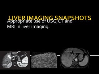
  • 1. Appropriate use of USG,CT and MRI in liver imaging.
  • 2.  Dr.K.Vinayachandran Nair MD,DM. Encouraging us and asking right questions.  Dr.Jagdeesh MRCPCH ,FRCR. Passion about liver nodules. Also lending many slides.  Few slides taken from google images.
  • 3. I have to tell stories of 3 heroes… ! • Appropriate use of USG,CT and MRI in liver imaging.
  • 4.  Ultrasound of liver what we can diagnose and what we can not.  Primary liver lesions in cirrhotic liver.  CT versus MRI in liver lesions.
  • 5.
  • 6.  65 year old male with dyspepsia , bloating , not alcoholic.
  • 7.
  • 8. Coarse texture , multiple tiny nodules. Nodularity of liver contour. Cirrhosis of liver.
  • 9.  Surface nodularity with high frequency probe.
  • 10.  Caudate to right lobe ratio more than 0.65 specific for cirrhosis.
  • 11.  Portal vein diameter is not sensitive in detecting portal hypertension.  Color doppler is to confirm direction of flow , reversal indicates portal hypertension.
  • 12.  Splenomegaly and collaterals are indicative of portal hypertension.
  • 13.  Increase of less than 20% in diameter of portal vein with deep inspiration indicates portal hypertension.  Very difficult criteria …!
  • 14.  Sensitivity and specificity varies from 60- 100% according to different studies.  World journal gastroenterology..2010.
  • 15.  Other criteria include  Cork screw / enlarged hepatic artery.  Loss of phasicity and pronounced cardiac periodicity.  Reduced portal vein velocity.  Congestion index (ratio of area to flow velocity)
  • 16.  Is controversial. May not be beneficial  USG is not useful in detecting small (1-2 cm) HCCs (sensitivity of 13 %).  More sensitive in detecting larger ( more than 4 cm) HCCs(sensitivity of 75%)  Can not differentiate between dysplastic nodule and HCC. AJR July 2002
  • 18. Diffuse type HCC , no mass detected on USG. Liver transplantation surgery showed diffuse HCC. Slide taken from AJR
  • 19. IT CAN DIFFERENTIATE TUMOR FROM BLAND THROMBUS. Portal vein
  • 20.  Agents such as perflubutane microbubble cause contrast related enhancement of focal liver lesions.  Enhancement pattern is used to assess cellular differentiation of HCCs.  This can be repeated realtime. AJR
  • 21. Enhancement and washout in a poorly differentiated HCC AJR
  • 22.  A technique to assess fibrosis of liver.  Employs modified ultrasound probe which assesses velocity of a shear wave created by a vibratory source.  Values of above 12.5kPa are indicative of cirrhosis.
  • 23.  50 year old male with Progressive abdominal distension.  ? Mass in right upper quadrant
  • 24. Lack of visualization of hepatic veins and narrowing of caliber of IVC indicating Budd Chairi syndrome. A difficult diagnosis on USG needs IVCgram for confirmation.
  • 25.  20 year old nursing student studying in Karnataka has come to emergency with right upper quadrant pain , not jaundiced --? cholecystitis
  • 26.
  • 27.  LFT showed elevated transaminases.  Hepatitis A IgM marker positive.  Final diagnosis – anicteric viral hepatitis.
  • 29.  Reason for showing this case is that there are no specific sonographic findings for hepatitis.  Reduced echotexture of liver , gall bladder wall thickening , hepatomegaly , starry sky appearance of liver due to periportal edema are described findings.
  • 30.  27 years old male working as executive in a company comes with elevated liver transaminases.  Claims to be teetotaller.
  • 31.
  • 33.  Liver echogenicity exceeds that of right kidney and spleen. And  There is beam attenuation.
  • 34.  Most often subjective..!!  Grade 1– increased texture with good visualization of diaphragm and intrahepatic vessels.  Grade 2- mild impairment of visualization.  Grade 3– severe impairment of visualization.
  • 35.  Here there is fatty infiltration , hepatomegaly and elevated liver enzymes. (AST and ALT).  Histologically, there is inflammation and fibrosis.  Radiologically it is not possible to separate NASH from ordinary fatty liver.  Fatty liver involves about 15% of population.  NASH involves about 2-5% of population.
  • 36. Focal fatty infiltration can simulate mass. Focal fatty sparing can also mimic mass.
  • 37.  25 year old healthy male for employment health checkup.
  • 38.
  • 39.  When USG shows hemangioma typical features in a nononcology patient this modality alone is considered sufficient.
  • 40.
  • 41.
  • 42.  Not sensitive in detecting small HCCs.  Can not differentiate between HCC and dysplastic nodule.  Entire liver can not be assessed , few USG blind areas in liver.  Moderate sensitivity in detecting cirrhosis.
  • 43.  Hemangioma in a nononcology patient , cyst and to certain extent abscesses can be confidently diagnosed with USG, for other focal lesions one needs to assess further.
  • 44.
  • 45. Varying signals; varying sizes, varying enhancement Siphon out the dysplastic nodules & HCC!! How! Lesions MRI features:-post contrast behaviour Intermediate/vague features/cross over features Varying signals; varying sizes, varying enhancement Siphon out the dysplastic nodules & HCC!! How! Lesions MRI features:-post contrast behaviour Intermediate/vague features/cross over features Varying signals; varying sizes, varying enhancement Siphon out the dysplastic nodules & HCC!! How! Lesions MRI features:-post contrast behaviour Intermediate/vague features/cross over features
  • 47.  T1 in and opposed phase
  • 48.  Dynamic post contrastT1FS(fat saturation) with subtracted images
  • 49.
  • 50.  Regenerative Nodules ; Siderotic nodules  Dysplastic Nodules  HCC
  • 51.  Size - diameter of less than 2 cm are more likely to be benign than malignant  Vascularity - shift from predominantly venous perfusion to predominantly arterial perfusion - dedifferentiated nodules - markedly enhanced on early arterial phase  Hepatocellular Function - hepatocellular contrast agents
  • 52.  most common cirrhosis-associated hepatocellular nodules  T2 : Isointense to liver; hypointense ( iron)  T1: Variable  Postcontrast: - No arterial enhancement - isointense enhancement to the liver - washout isointense to liver
  • 53.
  • 54.  Low grade and high grade  T2 : hypointense or isointense to liver,  T1: Variable  Postcontrast: - Patchy ill defined nodular arterial enhancement - isointense enhancement to the liver - hypointense to liver in the later phases, no definite tumor capsule
  • 55.
  • 56.  T2 : Hyperintense ( 94%) – isointense to spleen  T1: Variable  Postcontrast: - Intense arterial enhancement - isointense on the portal venous phase - wash out on the later phase with persistent tumor capsule
  • 57.
  • 58.
  • 59.  T1- and T2-weighted MR images, the mosaic pattern appears as areas of variable signal intensities  lesions enhance in a heterogeneous fashion during the arterial and later phases
  • 60.
  • 61. Diffuse type HCC a permeative tumor often with portal vein thrombus.
  • 62. LESION T2 T1 ART DEL Reg N Low-iso Int - High No enhancement No wash out Dys N Low-iso Int - High Heterogenenous moderate enhancement Irregular wash out with no capsule HCC Int - High Low - Int Intense enhancement Wash out with enhancing capsule
  • 63.  CT versus MRI mosaic pattern- 88% T1- and T2-weighted MR images, the mosaic pattern appears as areas of variable signal intensities lesions enhance in a heterogeneous fashion during the arterial and later phases
  • 64.  When doing survey for metastasis , CT is better as it can assess extra hepatic lesions better than MRI. 
  • 65.  In patient with chronic liver disease , for characterization of regenerative ,dysplastic nodules and HCC , MRI is better than CT.
  • 66.  There are several arterial phase enhancing lesions.  TAHD – transient hepatic attenuation difference.  Confluent hepatic fibrosis.
  • 67.  Contrast MR has sensitivity of near 81% and specificity of near 85%.  CT has sensitivity of near 73% and sensitivity of near 93%.  Imaging not sensitive for lesions less than 20 mm and diffuse lesions.
  • 68.
  • 69.  USG is the first line modality in liver imaging. Used to rule out liver disease and diagnose with confidence common lesions like cysts and hemangiomas.
  • 70.  Multiphasic CT is the standard reference study in liver disease. Standard in assessing and follow up of oncology patients. When high resolution MRI is not available can be used to image cirrhosis.
  • 71.  MRI is modality of choice in assessing cirrhosis liver . It is increasingly preferred in follow up of patients and in children where radiation is an issue.  Tissue specific contrast agents are being available and are helpful.
  • 72.  Biopsy or follow up is advised in doubtful lesions or histopathological confirmation is necessary for management.  Decisions should be patient specific.
  • 73.  Diffusion weighted imaging  Mixed extracellular and hepatocellular contrast agents -gadoxetic acid disodium  MR spectroscopy  Liver perfusion

Editor's Notes

  1. Good evening! Thank you for having me here to talk to you on Primary .....
  2. Size As lesions grow, the likelihood of high-grade dysplasia or frank malignancy increases. As a general rule, lesions with a diameter of less than 2 cm are more likely to be benign than malignant and, if malignant, are usually well differentiated (16,17). By comparison, lesions with a diameter of more than 2 cm are more likely to be malignant than benign and tend to be characterized by moderate to poor differentiation. Vascularity Regenerative nodules and low-grade dysplastic nodules are predominantly portally perfused and, after gadolinium administration, show enhancement similar to that of the surrounding liver (18,19). As dedifferentiation progresses within these nodules, angiogenic pathways are activated that induce new vessel formation, which manifests as an increased density of unpaired arteries and capillary units (16,18,20). This development leads to an increasing shift from predominantly venous perfusion to predominantly arterial perfusion as low-grade dysplastic nodules and hepatocellular carcinomas become high-grade lesions (18). The increasingly dedifferentiated nodules appear more markedly enhanced on early arterial phase images obtained after the intravenous injection of a contrast agent, with more pronounced washout on venous phase images and equilibrium phase images (21,22). The major shift in angiogenesis typically occurs during the transition from low-grade to high-grade dysplasia (18). Hepatocellular Function Regenerative nodules generally have normal hepatocellular function and therefore demonstrate avid uptake of hepatocellular contrast agents. As dedifferentiation proceeds, the number of expressed organic ion transporters decreases, with a resultant progressive reduction in the uptake of hepatocellular agents (22–25). Kupffer Cell Density The density of Kupffer cells within regenerative lesions is similar to that in the surrounding nonneoplastic hepatic parenchyma. The cell density is visible at contrast-enhanced imaging because Kupffer cells avidly accumulate particulate agents through phagocytic mechanisms. With dedifferentiation, the Kupffer cell density changes; however, such changes are unpredictable during the early stages of carcinogenesis. According to empirically derived values reported in the literature, dysplastic nodules and well-differentiated hepatocellular carcinomas have variable Kupffer cell densities, ranging from diminished to elevated levels (26–28). Moderately and poorly differentiated hepatocellular carcinomas tend to have a diminished Kupffer cell density (26,27).
  3. form in response to necrosis, altered circulation, or other stimuli ( most have a diameter of less than 2 cm, Steatotic regenerative nodules tend to occur in multiples (Fig 2). A single fatty nodule is suggestive of a dysplastic or malignant process (Fig 3).
  4. Lesions with dysplastic features that do not satisfy the histologic criteria for malignancy or invasion are described as either(a) dysplastic foci (<1 mm in diameter) or (b) dysplastic nodules (≥1 mm in diameter). Dysplastic nodules usually occur in the setting of cirrhosis and may be classified as low or high grade, according to the degree of dysplasia.
  5. Fig. 37.2. Dysplastic nodules, cirrhotic liver, MRI findings. A Coronal SSTSE image (SSTSE): Multiple low signal intensity nodules are present with a cirrhotic liver with splenomegaly and ascites (*). B Axial fat-suppressed TSE image (TSE fatsat): All nodules show low signal intensity. C Axial arterial phase image (ART): The largest nodule shows increased enhancement (arrow); other lesions show variable enhancement. D Axial delayed phase image (DEL): The largest nodule (arrow) does not show any enhancing tumor capsule. E Axial opposed-phase image (T1 opposed-phase): Most hepatic nodules are bright. F Axial in-phase image (T1 in-phase): Several nodules lose their signal due to iron accumulation, i.e. siderotic nodules (arrows); note also the dark Gamna-Gandy bodies in the spleen. G Detailed view of the arterial phase (ART): The largest nodule clearly shows enhancement (arrow). H Detailed view of the delayed phase (DEL): The largest nodule does not show a tumor capsule (arrow) Fig. 37.3. Dysplastic nodules, histopathology, drawings. A Photomicrograph shows a large nodule surrounded by fibrous septa. H&E stain, ×20. B A detailed photomicrograph shows increased cellularity with variable size of the nuclei indicating at least dysplastic changes. H&E stain, ×100. C Situation I shows the presence of several dysplastic (DN) and regenerative (RN) nodules in a cirrhotic liver. D Situation II shows the presence of a focus of HCC with the largest DN (arrow) 37 Cirrhosis III – Dysplastic Nodules 81
  6. Only 52% have these classic features
  7. Fig. 46.2. HCC, cirrhotic liver, large, mosaic pattern, typical MRI findings. A Axial SSTSE image (SSTSE): HCC is predominantly hyperintense to the liver. The tumor capsule is hypointense and not visible. B Axial in-phase image (T1 inphase): HCC is predominantly hyperintense to the cirrhotic liver with a darker tumor capsule. C Axial arterial phase image (ART): HCC shows intense enhancement in some areas, indicating themosaic pattern. D Axial delayed phase image (DEL): HCC shows washout with enhanced thick tumor capsule. E A detailed view of the SSTSE image (SSTSE): HCC shows areas with high and low signal indicating the mosaic pattern. F Axial opposedphase T1-w GRE image (T1 opposed-phase): HCC as well as the cirrhotic liver show no signs of fatty infiltration. G Axial portal phase image (POR): HCC shows washout with enhanced thick tumor capsule. H A detailed view of the axial delayed phase 2D T1-w GRE image (DEL): HCC is surrounded by an enhanced thick tumor capsule (arrow)
  8. Not sensitive for lesions less than 20 mm and carcinomatosis.