Coronary Artery Bypass Graft Under Cardiopulmonary Bypass
•Download as PPT, PDF•
39 likes•4,291 views

coronary artery bypass graft under cardiopulmonary bypass

Recommended
More Related Content
What's hot
What's hot (20)
Viewers also liked
Viewers also liked (20)
Similar to Coronary Artery Bypass Graft Under Cardiopulmonary Bypass
Similar to Coronary Artery Bypass Graft Under Cardiopulmonary Bypass (20)
More from Dharmraj Singh
Recently uploaded
Recently uploaded (20)
Coronary Artery Bypass Graft Under Cardiopulmonary Bypass
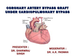
- 1. CCOORROONNAARRYY AARRTTEERRYY BBYYPPAASSSS GGRRAAFFTT UUNNDDEERR CCAARRDDIIOOPPUULLMMOONNAARRYY BBYYPPAASSSS MMOODDEERRAATTOORR :: DDRR.. AA..KK.. PPAASSWWAANN PPRREESSEENNTTEERR :: DDRR.. DDHHAARRMMRRAAJJ SSIINNGGHH
- 2. • Main cause of CAD is atherosclerotic narrowing of major branches of coronary arteries resulting in luminal obstruction and myocardial ischemia. • Risk factors for increased morbidity and mortality are - poor LV function - h/o CHF or EF<30% - h/o DM, HTN etc. - obesity - redo procedures - emergency procedures - advanced age
- 3. ardiac ischemia occurs when coronary blood flow does not increase to an extent to meet increased myocardial oxygen demand V subendocardial blood flow is intermittent and occurs only during diastole oronary blood flow is directly proportional to coronary perfusion pressure CPP = Aor tic DP - LVEDP
- 4. oronary blood flow is inversely proportional to coronary vascular resistance and thus coronary stenosis increases resistance and decreases blood flow esistance to coronary blood flow depends on- - length of stenosis -degree of stenosis -presence or absence of collaterals -co-existing diseases like DM or HTN
- 5. • Patients may present with typical anginal pain, atypical pain or discomfort, silent or asymptomatic attack with evidence of ECG changes only. • 50% decrease in diameter causes symptoms on exertion and 75% decrease in diameter causes angina at rest
- 6. • Myocardial oxygen demand is increased by Increase in- heart rate - contractility -chamber pressure(afterload) - chamber size(preload) • Ways to increase myocardial oxygen supply are maintaining DBP, Hb conc, oxygen saturation, decreasing oxygen demand by decreasing HR and ventricular wall tension
- 8. CABG is a surgical procedure in which one or more blocked coronary vessels are bypassed by a blood vessel graft to restore normal blood flow to myocardium and most of these grafts come from patient’s own body. ost common conduits used are aphenous vein for bypassing right coronary artery and circumflex coronary artery nternal mammary artery for bypassing left anterior descending coronary artery f more conduits are needed upper extremity vessels can
- 11. According to AHA, indications of CABG are- • Disease of the left main coronary artery • Multi vessel disease • Non-discrete or diffuse disease not amenable to t/t with PCI
- 12. CABG CAN BE PERFORMED EITHER ONPUMP OR OFFPUMP(BEATING HEART CABG)
- 13. PREOPERATIVE EVALUATION- • Histor y and physical examination to evaluate LV dysfunction and LV/RV failure, respiratory disease, prior cardiac surgery • Chest radiograph to detect resp. disease, CHF, abnormal cardiothoracic ratio etc. • Resting ECG to detect rhythm disturbances, conduction defects, decision of intra-op lead selection
- 14. xercise ECG showing significant ST segment changes in early stages, sustained changes, abnormal changes in HR or BP, development of angina or arrythmia indicate severe CAD CHO shows segmental wall motion abnormality tress ECHO with exercise or dobutamine and contrast ECHO detect abnormal areas of perfusion
- 15. yocardial per fusion scans using thallium-201 or Tc 99m locate and quantitate ischemic areas ngiography defines location and degree of occlusion and coronary artery spasm ontrast ventriculography shows areas of hypokinesia, akinesia and dyskinesia F= EDV-ESV/EDV [N-50-75%] 25-50%- symptoms on exercise
- 16. INTRA-OP MONITORING- BP- Dominant hand radial art preffered . CG- ST segment changes or new T wave changes are diagnostic of ischemia imultaneous observation of an inferior lead [II, III, aVF ] and anterior lead [V4,V5] detects approximately 90% of events. osterior heart ischemia is difficult to detect
- 17. •CVP – Internal juglar vein • PA CATHETER- Appearance of new V wave in pulmonary artery pressure waveform indicates development of MVR due to ischemic papillary muscle dysfunction Imp in post-op period where TEE can not be used Intra-op monitoring may require frequent balloon inflations
- 18. TEE – an assess regions supplied by all three major coronary arteries egional wall motion abnormality can precede ECG and PA wave form changes ntra op stress TEE with low dose dobutamine can demonstrate myocardial contractile reserve and helps revascularize myocardium that will be benefited from increased blood supply
- 19. ARDIO-PULMONARY-BYPASS-It provides both artificial ventilation and perfusion by diverting blood away from vascular system and performing the function of both lungs and heart temporarily The goal of this CPB is to provide a bloodless and motionless surgical field
- 20. PREMEDICATION arcotic or Anxiolytic agent or both to mitigate pain and anxiety. upplemental intra-venous drugs- commonly midazolam and fentanyl- are often necessary during radial artery cannulation before induction of anesthesia. atients with low cardiac output secondary to CHF sedation should be performed judiciously to avoid myocardial depression and resultant hypotension.
- 21. NDUCTION- oal is to avoid undue hypotension and to attenuate hemodynamic response to laryngoscopy and intubation ypotension may be due to hypovolemic state and reduction in sympathetic tone in response to inducing agents particularly in patients with poor LV function. Fall in BP >20% of baseline needs use of inotropes.
- 22. ypertension may be due to pre-induction anxiety and sympathetic stimulation ll anesthetic agents except ketamine cause decreased blood pressure by decreasing sympathetic tone , systemic vascular resistance , inducing bradycardia or directly depressing myocardial function.
- 23. elected agent should be given in small incremental doses and titrated first against loss of consciousness then to an acceptable fall in BP. uscle relaxation and controlled ventilation ensures adequate oxygenation and prevents hypercapnia.
- 24. IGH DOSE NARCOTICS- entanyl 50-100 mcg/kg or sufentanil 15-25mcg/kg roduces prolonged post-op respiratory depression, high incidence of awareness, rigidity, fail to control hypertensive response to stimulation
- 25. OTAL INTRAVENOUS ANESTHESIA- nfusion of propofol,0.5-1.5 mg/kg f/b 25-100 mcg/kg/min and remifentanil 1 mcg/kg bolus f/b 0.25-1 mcg/kg infusion. otal dose of fentanyl should be 5-7 mcg/kg se of short acting agents results in early extubation and lesser hospital stay rugs are costlier and remifentanil should be
- 26. MIXED INTRAVENOUS/INHALATION ANESTHESIA- • Propofol 0.5-1.5 mg/kg or thiopentone 2-3 mg/kg and midazolam 0.05 mg/kg • Opioids are given intermittently and total dose of fentanyl and remifentanil should not exceed 15 and 5 mcg/kg respectively.
- 27. • Iso, sevo or desflurane are used for maintenance • It results in easy control of depth of anesthesia and hemodynamic stability and early extubation OTHERS- • In frail patients, combination of ketamine and midazolam provides hemodynamic stability, good amnesia, analgesia and minimal respiratory depression.
- 28. RE CARDIO-PULMONARY BYPASS PERIOD- heck bilateral breath sounds djust fresh gas flow heck pressure points rotect eyes heck all monitors and tubings after final position dminister antibiotics
- 29. kin incision can cause sympathetic stimulation, so adequate depth of anesthesia is necessary ternal incision and splitting accompanies high level of sympathetic stimulation ternal splitting can cause awareness and recall, so amnesic agents like benzodiazepines or propofol is to be used
- 30. achycardia and raised BP can be treated by nitroglycerine boluses or by esmolol igh doses of fentanyl can reduce response to pain ungs are to be deflated during sternal splitting to avoid damage
- 31. ternal spread can cause kinking or malpositioning of PA cath. issection of post ganglionic sympathetic fibres from aorta to cannulate it can cause intense stimulation
- 32. HEMODYNAMIC CHANGES ASSOCIATED IN THIS PERIOD HYPOTENSION-may be due to Hypovolemia Decreased venous return due to increased airway pressure , tension pneumothorax , handling of heart and great vessels Impaired myocardial contractility Ischemia Dysarrythmia Measurement error due to kinked catheter, wrist positioning error etc.
- 33. T/T of hypotension- • Rule out technical and mechanical factors • Check for dysarrythmia • Use of inotropes • Fluid loading • Decrease inhalational agents
- 34. HYPERTENSION-may be due to Light anesthesia Hypercapnia Hypoxia Hypervolemia T/T of hypertension- • Increasing anesthetic depth • Vasodilator agents like nitroglycerine, nitroprusside • Using b-blockers
- 35. SINUS BRADYCARDIA- may be due to Vagotonic effects of narcotics Use of b-blockers Hypoxia Ischemia • T/t of bradycardia is indicated if there is fall in BP or HR<40 even with no fall in BP • Atropine 0.4-0.6 mg i.v is indicated
- 36. SINUS TACHYCARDIA- may be due to Light anesthesia Hypovolemia, anemia Inotropic drugs , pancuronium , isoflurane Hypoxia Hypercapnia Ischemia Management of tachycardia includes • Checking ventilation abnormalities • Increasing depth of anesthesia • Volume loading • Using b-blockers
- 37. DYSRYTHMIAS may be due to- Mechanical stimulation of heart Preexisting dysrythmia Electrolyte imbalance Increased catecholamines Ischemia These can be treated by treating underlying causes, using lidocaine,b-blockers and by synchronized cardioversion.
- 38. RIMING of circuit is to be done by balanced salt solution(1200-1800ml for adults) ther components like albumin or hetastarch, mannitol, heparin and bicarbonate are added t decreases hematocrit to 22-25% n patients who are severely anemic or pediatric patients blood is used as prime
- 39. ENEFITS OF HEMODILUTION- ecreased viscosity improves microcirculation and compensates for increased viscosity due to hypothermia RISKS- ecreased SVR decreases BP ilution of drugs and coagulation factors
- 40. HEPARINIZATION- • Heparin 300-400u/kg is administered through a central vein targeting ACT level min of 480s • ACT is the time from adding whole blood to a tube containing a contact phase activator (celite or kaolin) up to the time when first clot appears. • Repeat ACT is measured after 5 mins and if it is less, 100u/kg is to be administered again
- 41. • Whole blood heparin conc. of about 3-4u/ml is sufficient for CPB. • Heparin resistance is seen in cases of AT-III deficiency which can be treated with infusion of 2-3 units of FFP , AT-III concentrates , recombinant AT-III etc.
- 42. ANNULATION- or tic cannula is inser ted first to allow rapid volume infusion in cases of hemorrhage during venous cannulation BP is lowered to avoid risk of dissection and PEEP applied to avoid air entrainment by increasing intracardiac pressure
- 43. omplications during aortic cannulation can be ortic dissection leeding mbolisation of atheromatous plaque ysrythmia ypotension
- 44. REBYPASS CHECKLIST- nticoagulation (min ACT of 480sec) is needed osition of cannulae is to be checked by checking waveforms rine noted and urobag is to be emptied quality of carotid pulse is to be checked upplemental doses of anesthetic agents are to be administered to compensate for dilution
- 45. NITIAL BYPASS CHECKLIST- ace is to be checked for colour , edema , conjunctival chemosis A pressure should be less than 15 mm Hg rterial blood pressure should be mean 30-40 mm Hg VP should be<5 mm hg ardiac contractility and distensibility is to be checked
- 46. MAINTENANCE OF BYPASS- • ACT repeated every 30-60 mins, if less supplemental heparin is added • Blood gas values to be evaluated every 30-60 mins • PaO2 maintained between 100-300 mm Hg & PaCO2 between 35-40 mm Hg. • Blood glucose and hematocrit is measured every 30- 60 min
- 47. ufficient anesthetic depth is maintained to prevent awareness, spontaneous movement, hypertensive and tachycardic responses epth maintained by adding anesthetic agents and muscle relaxants directly into the circuit and adding volatile agents by connecting vapouriser to oxygenator
- 48. • INTRA OPERATIVE AWARENESS may be due to underdosing , dilution or absorption of drugs and increased requirement during rewarming . • It can be prevented monitoring BIS and supplementing drug. • Ventilation should cease when total bypass begins.
- 49. • Pump flow rate is to be maintained at 50-70 ml/kg/min or 2.2-3.1 l/min/square mt • Urine output should be at least 0.5ml/kg/hr • Core temp. is to be monitored at nasopharynx or tympanic membrane( jugular bulb temp is gold standard) • De-airing of heart is to be done before weaning from CPB by increasing venous pressure by inflating lungs
- 50. MYOCARDIAL PROTECTION- • To provide a motionless field for surgery, heart is stopped in diastole by administering a potassium rich cardioplegia soln. • It interrupts myocardial electromechanical activity, reduces oxygen consumption by 90% and cold cardioplegia soln. reduces it by 97%.
- 51. • For most complete cardioplegia , both antegrde (through aortic root) and retrograde(through coronary sinus) approach is used • Arrest can be reversed by reperfusing heart by warm normokalemic blood(hot shot)
- 52. PREPARATION FOR WEANING- (Elements of Romanoff and Royster’s) Pneumonic is CVP • COLD- patient’s temp. should be 36-37 degrees, hyperthermia is deleterious • CONDUCTION- HR of 80-100 bpm is optimal, bradycardia may need epicardial pacing wire for AV pacing or inotropes, tachycardia needs t/t of cause, AV block may need AV pacing and supraventricular tachycardia needs pharmacotherapy and cardioversion
- 53. • CONTRACTILITY is estimated by TEE and CO by PA catheter • CELLS-Hb should be at least 7-8g% • COAGULATION- long bypass period and extreme hypothermia increase risk, PT,PTT,PC should be normal • VENTILATION OF LUNGS- must be established after PA blood flow is restored
- 54. ISUALISATION of heart and TEE for regional and global contractility OLUME EXPANSION-if necessary ACER AND PRESSOR AGENTS should be readily available OTASSIUM must be corrected as hypokalemia can cause dysrythmias and hyperkalemia can cause conduction blocks
- 55. WEANING FROM BYPASS- • Before termination, patient should be rewarmed, heart is de-aired, regular cardiac electrical activity confirmed or supported by pacemaker, lab values confirmed and corrected • Ventilation of lungs is established, venous drainage is slowly reduced and cardiac filling volume is gradually increased • Vasopressors or inotropic support may be needed
- 56. • When patient becomes hemodynamically stable, protamine is administered to reverse anticoagulation • 1-1.3mg of protamine per 100 units of heparin is administered slowly over 10-15 mins • ACT should be brought to baseline values • When pre-loading is optimal and contractility is adequate, aortic inflow line is clamped to separate from bypass
- 57. • Elevated BP should be avoided to prevent stress on suture lines • If CO is not optimal, preload can be increased in 100ml increments as rewarming is associated with vasodilation • Increase in hemodynamic instability and use of inotropes may need reinstitution of CPB
- 58. EVENTS IN POST BYPASS PERIOD- 1.Cardiovascular decompensation- Ischemia and infarction may be due to • Thrombosis or particulate or air emboli in graft • Kinking or spasm of graft • Incomplete revascularization due to distal disease • Inoperable vessels
- 59. LV dysfunction is amenable to combination of inotropes and vasodilators to increase CO RV dysfunction may be due to inadequate protection, ischemia, infarction, pulmonary air emboli, preexisting pulmonary HTN RV failure needs inotropic support as well as pulmonary vasodilation nitroglycerin, nitroprusside, prostaglandin E1 (PGE1), B-type natriuretic peptide (e.g., nesiritide), sildenafil, or inhaled agents such as nitric oxide and prostacyclin (prostaglandin I2 [PGI2, epoprostenol]).
- 60. YPOTENSION may be due to low SVR, severe anemia, low viscosity, acid-base disturbances and is treated with vasoconstrictors YSRYTHMIAS - AF is most common and converted to sinus rhythm by synchronized cardioversion, amiodarone etc. VF or flutter needs defibrillation and drugs like amiodarone and lidocaine Bradycardia and heart block need AV sequential
- 61. 2.Bleeding and coagulopathy- • Inadequate surgical hemostasis is most common cause • Platelet dysfunction due to hemodilution, hypothermia, contact activation, adhesion and sequestration
- 62. • Activation of coagulation cascade by contact factors • Fibrinolysis by release of t-PA from damaged endothelium • Consumption of factors • Treated by FFP and platelet concentrates
- 63. 3.pulmonar y complications- • Atelectasis causing decreased oxygenation, lungs are to be reinflated by hand before machine ventilation • Hemothorax, pneumothorax may need chest tube insertion
- 64. • Cardiogenic pulmonary edema due to fluid overload in patients with preexisting HF • Noncardiogenic pulmonary edema due to inflammatory response, multiple emboli, increased permeability, transfusion reaction
- 65. .Metabolic disturbances- ypokalemia due to diuretics, mannitol, hyperglycemia treated with insulin :- treated with KCl @ 10-20 meq/hr yperkalemia due to cardioplegia, blood products, impaired renal function: - treated with hyperventilation, calcium, diuretics, glucose and insulin infusion
- 66. ypocalcemia due to citrate in blood products, hemodilution, alkalosis:- treated with 10% calcium chloride 5-10mg/kg ypomagnesemia due to hemodilution:- treated with 2-4 g of magnesium yperglycemia is deleterious and is due to stress of surgery and inflammatory response, glucose level > 200mg/dl:- should be treated with insulin
- 67. 6.Ef fect on CNS- • MC complication is transient neuropsychiatric dysfunction, strokes are uncommon • Causes are micro and macro emboli, global hypoperfusion, cerebral hyperthermia, cerebral edema, inflammation, BBB disruption • Intra-op awareness should be avoided
- 68. Temperature regulation- • Hypothermia causes increase in SVR, shivering increasing oxygen consumption and coagulopathy • So normothermia should be achieved at end of bypass • Rewarming should be gradual • Hyperthermia should be avoided as it delays neuronal metabolic recovery, increases excitotoxic neurotransmitter release, oxygen free radical production, intracellular acidosis, increased BBB permeability.
- 69. enal ef fects- luid loss, myocardial depression and vasodilation by anesthetic agents, long term use of ACEIs, inflammatory response, loss of pulsatile flow decrease renal perfusion luid replacement, vasoconstrictors, frusemide 10-20 mg or mannitol 0.5-1mg/kg can be used
- 70. RANSPORT TO ICU- ortable monitoring equipment, infusion pumps, full oxygen cylinder with a self-inflating bag for ventilation should be ready pon arrival to icu patient is attached to ventilator, breath sounds checked, orderly transfer of monitors and infusions should follow
- 71. OFF PUMP CABG ANDIDATES- pts with anterior lesions,single /double vessel ds. ts with high risk of stroke, renal failure ,pulmonary dysfunction, severe valvular ds. urgeon employed nstitute
- 72. Major differences from onpump ollowing sternotomy , goal of heparin anticoagulation achived is – > 2 times of baseline ACT or > 300 sec or same as onpump(>400 secs). Only focal area of heart is stablized via epicardial stablizers. Distal anastmosisis done then aorta is partially clamped to perform proximal anastmosis .(BP kept <100 mmHg). emodynamic disturbances & arrhythmias more frequent and need to be adderssed.
- 73. Transport from OT omplications during transpor t- nadvertent extubation ull off of monitors oss of i.v lines njury to body parts isconnection of pacemaker wires
- 74. ARE IN ICU- ost patients require mechanical ventilation for 2-12 hrs, sedation and analgesia should be continued ypertension unresponsive to sedation and analgesics should be aggressively treated with vasodilators xtubation is considered when patient becomes conscious, muscle paralysis has worn off, blood gas values are acceptable, surgical hemostasis is adequate and the patient is hemodynamically stable
- 75. ABG procedures relieve chest pain and angina, enable patients to resume a healthy life style, lower the risk of further heart attack and its consequences hey do not prevent coronary disease from recurring, hence medications along with appropriate lifestyle changes are strongly recommended to reduce the risk of recurrence.
- 76. UTCOMES OF CONVENTIONAL CABG-P OSITIVE- elief of angina in 90% 0% angina free after 5 years 5% survival after 1 year ow chance of restenosis EGATIVE-
Editor's Notes
- a