Beginners Guide to TikTok for Search - Rachel Pearson - We are Tilt __ Bright...
Arturotalledo1908
1. Auger Electron Spectroscopy (AES) for
investigation of materials and hard coatings
Arturo Talledo and Carsten Benndorf
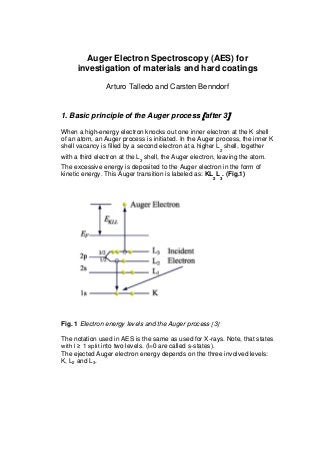
1. Basic principle of the Auger process after 3
When a high-energy electron knocks out one inner electron at the K shell
of an atom, an Auger process is initiated. In the Auger process, the inner K
shell vacancy is filled by a second electron at a higher L
2
shell, together
with a third electron at the L
3
shell, the Auger electron, leaving the atom.
The excessive energy is deposited to the Auger electron in the form of
kinetic energy. This Auger transition is labeled as: KL
2
L
3
. (Fig.1)
Fig. 1 Electron energy levels and the Auger process 3
The notation used in AES is the same as used for X-rays. Note, that states
with l ≥ 1 split into two levels. (l=0 are called s-states).
The ejected Auger electron energy depends on the three involved levels:
K, L2 and L3.
2. However, the binding energy of an electron in the presence of
a core hole is greater than that of the same level in a neutral atom. In
approximation, the following equation is used to estimate the kinetic
energy of Auger electrons. (z: atomic number of an atom)
EABC = EA(z) -1/2 EB(z) + EB(z+1) - ½ EC(z) + EC(z+1)
2. How can we detect Auger electrons?
To measure Auger electrons we need
a. a vacuum system providing UHV (ultra high vacuum below 10 -8
mbar).
b. an electron gun (2.5 to 5 keV ) or an X-ray source to knock out
inner shell electrons of our sample.
c. an analyzer, capable to measure the energy and the intensity of the
ejected Auger electrons. In our laboratory we use for the detection
of Auger electrons a cylindrical mirror analyzer (CMA) with an
integrated electron gun.
3. The principal design of a CMA (cylindrical mirror
analyzer)
The CMA consists of two coaxially aligned cylinders, an outer (CO) and an
inner cylinder (CI). Dimensions about OC diameter 8 cm, CI diameter 4
cm, length 8 cm. Integrated in the inner cylinder is the electron gun. The
CMA is surrounded by a magnetic shielding (-metal) to reduce the earth
magnetic field. The sample to being investigated is close to the front of the
CMA (about 1.5 cm). The emitted electrons (secondary electrons including
the Auger electrons) enter through slits of the inner cylinder CI into the
analyzer. Depending on the energy of the electrons and the voltage
between outer and inner cylinder some electrons can reach the outgoing
slits and be detected with the channeltron detector.
The cylindrical mirror analyzer fulfils two focussing conditions:
3. 1. The measured electron energy is a linear function of the applied
voltage between outer (negative) and inner (positive or ground)
cylinder.
2. For a specific geometry (which is used for commercial CMA’s),
electrons with the same energy but slightly different entrance
angles (deviation from 420
) are also focussed to the exit slit.
The following fig. 2. is reproduced from Ranke 2.
Fig.2 Schematic drawing of a CMA and the AES detection system 2
3. How are the AES spectra recorded?
With the excitation of Auger electrons by electron impact not only Auger
electrons are produced but rather also “ true” secondary electrons. These
true secondary electrons provide a large background to the spectrum, so
that the Auger electrons are only contributing with small signals. The
background could be eliminated when we do not record the N(E) (Intensity
versus energy) spectrum but the differentiated spectrum
4. dN(E)/dE. For this purpose, the voltage applied to the outer cylinder is
modulated with a small AC voltage ( in the range of 3 Vpp and about 1.5
kHz). The signal, which comes from the channeltron is fed to a lock-in
amplifier (which is able to exactly amplify at the same frequency as the
modulation). The DC output from the lock-in gives the dN(E)/dE spectrum.
4. What is the role of the Channeltron amplifier?
The signal which reaches the exit slit of the CMA is very weak and needs
to be amplified. This could be done with a SEV (Secondary electron
amplifier) or a channeltron. Electrons with sufficient energy (50 eV) hitting
a surface are producing secondary electrons which could be emitted from
the surface. They are accelerated inside the channeltron and hitting once
more the surface producing more electrons. The multiplication of one
electron hitting the entrance of the channeltron could be enhanced by a
factor of 107
in a time scale of few ns. This would be sufficient even to
count single electrons.
5. Measuring and recording the Auger Spectra with a PC
(personal computer)
We use a DAQ (Data Acquisition) system from the company LabJack
(U12) and the software from DAQFactory to acquire the data from our
AES. This allows us to scan the AES spectra with a resolution of 12 bits
and also record the signals from the CMA with the same resolution. An
example of a recently recorded AES spectrum in our laboratory from a Ni
sample is given below (fig.3).
We notice beside the signals from Ni, impurities from oxygen, carbon and
sulphur. These are either contaminations from the surroundings (oxygen
and carbon) or due to segregation at higher temperature (600 0
C) from
very small concentrations inside the bulk Ni. The S segregation to the
surface is due to the lowering of the surface energy by the adsorbate. This
segregation effect plays also a significant role for the mechanical strength
of metals and alloys.
5. Fig.3 Auger electron spectrum measured at the UNI from a Ni(110)
single crystal
The Auger spectroscopy allows determining the elemental composition of
a sample and using the atomic sensitivity factors (ASF) of the elements of
the specific AES transition also their fraction.
The following figure 4 summarizes the core level binding energies and
Auger transitions (L3M45M45) of Ni 4 with a measured energy of 847 eV
(fig. 3):
6. Fig. 4 Energy levels for Ni and the L3 M45 M45 Auger process 4
6. Photo from the Auger system used in the UNI
Fig. 4
Photo from the UHV system
used for measuring Auger
electron spectra. The stainless
steel bell jar vacuum system is
pumped with an ionization
pump, pumping speed 300
l/min and a Ti sublimation
pump. Without noise from
roughing or turbomolecular
pumps the vacuum can be
kept in the 10 -10
mbar range
for weeks.
Behind the vacuum vessel we
Fig 4 Photo from our AES system at the UNI
7. observe the equipment used to detect the AES. From top: Lock-in
amplifier, Oscilloscope, electron gun power supply, scanning and
modulation power supply and (in blue colour) power supply for LEED (low
energy electron diffraction).
7. Pierre Auger, who first detected this kind of
electrum emission
The Auger effect is named for its discoverer,
Pierre Auger, who observed radiationless
relaxation of excited ions in a cloud chamber,
during the 1920s.
8.References
From the Nobel prize winner 2007 in Chemistry, Gerhart Ertl:
1. G. Ertl and J. Küppers, “Low Energy Electrons and Surface
Chemistry”, VCH Verlagsgesellschaft mbH, 1985 ISBN 3-527-
26056-0
2. Wolfgang Ranke, Fritz-Haber-Institut Berlin in: http://www.fhi-
berlin.mpg.de/acnew/department/pages/teaching/pages/teaching__winterse
mester__2004_2005/ranke_aes_modulation_techniques_291004.pdf
3. http://saturno.fmc.uam.es/web/superficies/problemas/auger.pdf
4. http://www.xpsfitting.com/2012/08/auger-peaks-and-auger-parameter.html