1. Abstract: The lack of biological complexity and limited life span
of conventional human hepatocyte culture models are major
limitations in toxicological research. Recent advances in three-
dimensional (3D) monoculture and co-culture systems show
promise, but may suffer from the lack of reproducibility and
spatial control. In this study, we generated 3D human liver
tissue mimetics composed of parenchymal and non-parenchymal
cell populations using a proprietary automated 3D fabrication
platform. The resulting liver tissues were well organized,
containing junctional protein E-Cadherin between hepatocytes,
CD31+ endothelial cell networks and desmin+ quiescent stellates.
The hepatocytes also demonstrated the ability to store both lipids
and glycogen. The tissues actively secreted albumin, cholesterol,
fibrinogen, and transferrin into the medium for six weeks. The
3D liver tissues expressed key Phase I enzymes CYP3A4, 2D6,
2B6, 1A2, and 2C9 over 28 days. Basal activity of CYP3A4, as
evidenced by the conversion of midazolam to 4-
hydroxymidazolam, was detected throughout the 28 day culture
period. In addition, exposure to rifampicin led to an increase in
both CYP3A4 mRNA and activity, with 4-fold induction still evident
after 28 days in culture. The 3D liver tissues exhibited a
clinically relevant injury response, evidenced by decreased
overall viability, to published hepatotoxic agents such as
Diclofenac and Troglitazone. These results demonstrate the
potential utility of human 3D bioprinted liver tissues in drug
discovery and development.
Safe Harbor Statement
Any statements contained in this report and presentations that do not describe historical facts may constitute forward-looking statements as that term is defined in the Private Securities Litigation Reform Act of 1995. Any forward-looking statements contained herein are based on
current expectations, but are subject to a number of risks and uncertainties. The factors that could cause actual future results to differ materially from current expectations include, but are not limited to, risks and uncertainties relating to the Company’s ability to develop, market and
sell products based on its technology; the expected benefits and efficacy of the Company’s products and technology; the timing of commercial launch and the market acceptance and potential for the Company’s products, and the risks related to the Company’s business, research,
product development, regulatory approval, marketing and distribution plans and strategies. These and other factors are identified and described in more detail in the Company’s filings with the SEC, including its prospectus supplement filed with the SEC on November 27, 2013, its
report on Form 10-Q filed February 6, 2014 and its transition report on Form 10-KT filed with the SEC on May 24, 2013 and our other filings with the Securities and Exchange Commission. You should not place undue reliance on these forward-looking statements, which speak
only as of the date of this Current Report. These cautionary statements should be considered with any written or oral forward-looking statements that we may issue in the future. Except as required by applicable law, including the securities laws of the United States, we do not
intend to update any of the forward-looking statements to conform these statements to reflect actual results, later events or circumstances or to reflect the occurrence of unanticipated events.
SOURCE Organovo Holdings, Inc.
Rhiannon N. Hardwick, Deborah G. Nguyen, Justin Robbins, Candace Grundy, Vivian Gorgen, Preeti Bangalore, Dean Perusse, Olivia Creasey, Shelby King, Susan Lin, Chirag
Khatiwala, Craig Halberstadt, and Sharon C. Presnell
Organovo, Inc., 6275 Nancy Ridge Drive Suite 110, San Diego, CA 92121
Functional Characterization of Three-Dimensional (3D) Human Liver Tissues
Generated by an Automated Bioprinting Platform
www.organovo.com
Abstract #9211
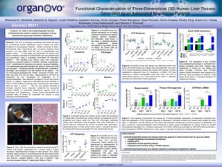
Figure 1: (Top, Left) Representative image of single exVive3D™
human liver tissue, measuring 2.5 x 2.5mm, with a 0.5mm
thickness. Representative images of H&E section (Top, Right)
showing distinct zones of non-parenchymal (N) and parenchymal
(P) cells, CD31+ microvascular structures in close association
with Desmin+ stellates (Bottom, Left), and E-Cadherin+ tight
junctions between Albumin+ hepatocytes (Bottom, Right).
Purpose: To create a more physiologically relevant,
multicellular liver model to enable investigation of drug-
induced liver injury and xenobiotic metabolism.
Figure 7: The capacity of exVive3D liver tissues for CYP3A4-mediated metabolism of midazolam increased over
time, and displayed a 2-3X induction response to Rifampicin. ExVive3D human liver tissues were treated for three
days +/- Rifampicin (10 µM) starting on the culture day indicated, followed by a 24-hour exposure to Midazolam (10
µM). Levels of 1-hydroxymidazolam were measured in either tissue homogenates or culture media by GC/MS
(SciAnalytical Startegies, Inc.). Relative expression of CYP3A4 mRNA was quantified by qRTPCR following induction,
and normalized to GAPDH. Data shown is the mean +/- standard deviation for independent tissues.
CONCLUSIONS:
• exVive3D bioprinted human liver tissues retain key aspects of native human liver for up to six weeks:
compartmentalized multicellular architecture
viability (ATP)
expression of liver-specific proteins
expression and function of key CYP450 enzymes
• exVive3D bioprinted human liver tissues respond to prototypical hepatotoxic agents.
Figure 6: The expression of key CYP450
drug metabolizing enzymes in exVive3D liver
tissues increases over time. Tissues were
maintained in culture for two weeks and
basal CYP2B6, CYP2C9, and CYP3A4
relative mRNA expression was measured by
qRTPCR, and normalized to GAPDH. Data
shown is the mean +/- standard deviation for
independent tissues.
Figure 2: exVive3D human liver
tissues maintained for six weeks
continue to express liver-specific
proteins: albumin, transferrin,
and fibrinogen. Data shown is
the mean +/- standard deviation
for independent tissues
normalized to ng protein per mL
of media, per million cells (at
time of fabrication) per day.
Figure 3: exVive3D human liver tissues remain viable and functional
for weeks after manufacturing. Tissue viability, as measured by ATP
production per tissue using Cell Titer Glo (Promega), stabilizes
rapidly after fabrication and increases over time. Production of
human albumin, assessed by ELISA, increases after fabrication,
stabilizing by Day 14. Data shown is the mean +/- standard deviation
for independent tissues generated from two hepatocyte donors.
Figure 4: exVive3D human liver
tissues can differentiate between
related toxic and non-toxic
compounds. Tissue viability was
determined by measurement of
ATP (Cell Titer Glo) following 7
days exposure to either vehicle,
Troglitazone (100 µM), or
Pioglitazone (100 µM). Data
shown is the mean +/- standard
deviation for independent tissues.
Figure 5: exVive3D human liver tissues can effectively model liver
injury. exVive3D human liver tissues were manufactured from each
of (3) human hepatocyte donors and treated with vehicle, a known
hepatotoxic compound (Toxic X), Diclofenac, or DMSO. ATP was
measured in tissue homogenates (Cell Titer Glo), and LDH in
supernatants (Abcam, Inc.). Data shown is the mean +/- standard
deviation for independent tissue replicates.