Critical Review (drug toxicity testing using organ on chip)
eb2015_Kidney_FINAL
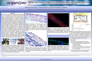
1. Figure 5. Renal proximal tubule epithelial cells in 3D renal
tissues exhibit features of polarization. Tissues were
stained with antibodies against E-cadherin (green). E-
cadherin is observed at the lateral membranes between
adjacent RPTEC cells, corresponding to localization at tight
junctions.
Figure 4. Extensive endothelial cell networks are
observed in 3D renal proximal tubule tissues. Tissues
were stained with antibodies against CD31 (green,
endothelial cells) and TE7 (red, fibroblasts). Networks with
putative lumens lined with endothelial cells are marked (*).
Figure 6. γ-glutamyl-transferase (GGT) activity over time
in 3D renal tissues or tissues composed of interstitial cells
alone. GGT is expressed on the apical surface of RPTEC
cells and is involved in glutathione homeostasis and
xenobiotic metabolism. GGT activity increases over time
in culture for 3D renal tissues as assessed by a
colorimetric assay. Schematic of pathway from IM Frey et
al., Physiological Genomics 2007 28(3): 301-310.
Safe Harbor Statement
Any statements contained in this report and presentations that do not describe historical facts may constitute forward-looking statements as that term is defined in the Private Securities Litigation Reform Act of 1995. Any forward-looking statements contained herein are based on current expectations, but are subject to a number of risks and uncertainties. The factors that could cause actual future results to differ materially from current expectations include, but are not limited to, risks and uncertainties relating to the Company’s ability to develop, market and sell products based on its technology; the expected benefits and efficacy of the Company’s
products and technology; the timing of commercial launch and the market acceptance and potential for the Company’s products, and the risks related to the Company’s business, research, product development, regulatory approval, marketing and distribution plans and strategies. These and other factors are identified and described in more detail in the Company’s filings with the SEC, including its prospectus supplement filed with the SEC on November 27, 2013, its report on Form 10-Q filed February 6, 2014 and its transition report on Form 10-KT filed with the SEC on May 24, 2013 and our other filings with the Securities and Exchange
Commission. You should not place undue reliance on these forward-looking statements, which speak only as of the date of this Current Report. These cautionary statements should be considered with any written or oral forward-looking statements that we may issue in the future. Except as required by applicable law, including the securities laws of the United States, we do not intend to update any of the forward-looking statements to conform these statements to reflect actual results, later events or circumstances or to reflect the occurrence of unanticipated events.
SOURCE Organovo Holdings, Inc.
CONCLUSIONS
• Morphologic and functional aspects of the human
proximal tubule are recapitulated in the 3D renal tissues
• Direct interface between tubular epithelium and
renal interstitial cells
• Polarized renal epithelium
• Microvascular structures within the interstitial tissue
• Stable / increasing tubular function (GGT activity)
over a 2 week period
• This model may be useful in the preclinical assessment
of human nephrotoxicity and drug-induced fibrosis
Abstract #9212 Shelby M. King, Olivia A. Creasey, Sharon C. Presnell, and Deborah G. Nguyen
Organovo, Inc., 6275 Nancy Ridge Drive Suite 110, San Diego, CA 92121
Design and characterization of a multicellular, three-dimensional (3D) tissue
model of the human kidney proximal tubule
www.organovo.com
Figure 2. Hematoxylin and eosin staining of 3D renal
proximal tubule tissues. The interstitial layer is
approximately 100 um thick and is composed of renal
fibroblasts and endothelial cells. Adjacent to the
interstitium is a monolayer of renal proximal tubule
epithelial cells (RPTEC). Putative brush borders are
observed on the apical surface of some epithelial cells
(arrows).
Figure 3. Masson’s trichrome stain of 3D renal proximal
tubule tissues. Abundant collagen deposition is evidenced
by blue staining in the interstitial layer. Aligned fibrils
(arrows) can be observed supporting extensive endothelial
cell networks.
Figure 1. The Novogen MMX™ Bioprinter (A) deposits multiple
types of cell aggregates in spatially-defined patterns. Following
bioprinting, the cells secrete extracellular matrix that maintains
relevant tissue microarchitecture in the absence of exogenous
scaffold material. B, Schematic of 3D renal tissues comprising
an interstitial layer of renal fibroblasts and endothelial cells
supporting a polarized epithelial monolayer in standard transwell
format. C, 3D Renal tissues 6 days after printing.
ABSTRACT
Renal toxicity is a major cause of drug attrition at the clinical trial
stage, and the primary site of this toxicity is within the proximal
tubule. Conventional renal cell culture models lack the
complexity of native tissue and thus have a limited capacity for
predicting tissue-level responses. In addition, the predictive
potential of pre-clinical animal trials is limited due to species-
specific differences between human and animal renal functions,
including differential sensitivity to insults. We designed and
fabricated a human three-dimensional (3D) tissue model of the
tubulointerstitial interface in which human renal interstitial tissue
is supporting proximal tubule epithelial cells to facilitate their
optimal morphology and function. Histological characterization
demonstrated that the interstitial layer is viable and well
organized, containing well-developed CD31+ endothelial cell
networks. Method optimization resulted in the formation of a
polarized layer of renal epithelium on top of the interstitial layer,
and formation of a basement membrane between the layers.
Gene expression analysis showed that the renal tissues
expressed key enzymes involved in metabolism and protein
processing (CYPs, renin-angiotensin system), suggesting that
physiologic function is retained. These bioprinted human
tissues may provide an opportunity to accurately study how
compounds affect the renal proximal tubule as well as modeling
pathogenic processes that involve tubular transport, cell-cell
interactions, and the development of tubulointerstitial fibrosis.
*
*
*
**