Critical Role of PET-Scan in Unravelling the Dual Pathology- Review of Litera...
Leydig Cell Tumor A Case Report
1. CASE REPORT
NJR I VOL 2 I ISSUE 1 I Jan-June, 2012
Leydig Cell Tumor: A Case Report
Amatya N 1
, Zhu M 2
, Sapkota P K 1
1
Radiology Unit, Helping Hands Community Hospital, Kathmandu, Nepal, 2
Department of
Diagnostic Ultrasound, West China Hospital of Sichuan University, Chengdu, China
Abstract
Leydig cell tumor is a relatively uncommon tumor clinically presenting with gynecomastia,
increased sex hormone levels (i.e. testosterone and estradiol) and other correlated symptoms.
However, preoperative ultrasonic diagnosis is difficult especially when the clinical
manifestations are unremarkable. We report a case of Leydig cell tumor of testis that
presented with atypical features.
Keywords: Leydig cell tumor, Neoplasm, Testosterone, Ultrasonography
Introduction
Leydig cell tumor (LCT) is a rare entity
among testicular neoplasm. It can occur at
any age, typically between 30 and 60 years of
age. Due to variable appearance, it can be
challenging to distinguish between germ cell
tumor and LCT. Sonographic imaging is still
a problem in diagnosing this entity and hence
easily can be misdiagnosed.1
We report a case
of LCT sonographically presenting with
atypical features of “thin tubular structures”
and color Doppler USG revealing a marked
hypervascularity pattern highlight the
ultrasonography-histological correlation.
Case Report
A 46 year old man presented with scrotal
swelling for more than one year. Genital
examination found that the right scrotum was
swollen with mild tenderness. The laboratory
examination revealed normal
____________________________________
Correspondence to: Dr. Nabin Amatya, MD
Radiology Unit, Helping Hands Community
Hospital Kathmandu, Nepal
Email:naabbin@hotmail.com
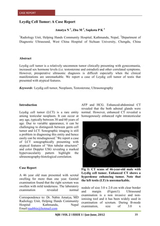
AFP and HCG. Enhanced-abdominal CT
revealed that the both adrenal glands were
normal. However, enhanced CT revealed a
homogenously enhanced right intratesticular
Fig 1: CT scans of 46-year-old male with
Leydig cell tumor. Enhanced CT shows a
hyperdense enhancing tumor. Note that
the left testis (LT) is unremarkable.
nodule of size 3.0 x 2.0 cm with clear border
and margin (Figure1). Ultrasound
examination is a non invasive and non-
ionising tool and it has been widely used in
examination of scrotum. During B-mode
examination, size of 3.0 x
39
2. Amatya et al. Leydig Cell Tumor: A Case Report
NJR I VOL 2 I ISSUE 1 I Jan-June, 2012
Fig 2a: Sonogram showing a
homogeneously hypoechoic tumor (white
arrow).
2.1 cm solid nodule was found in the right
testis. The nodule was well-defined and
heterogeneous hypoechoic with thin tubular
structure (Figure 2a). Color Doppler
demonstrated hypervascularity inside the
nodule (Figure 2b). Pathological examination
revealed an intratumoral abundance of blood
vessels and prominent vascularity
surrounding the lesion which corresponded to
the ultrasonographic imaging feature. Tumor
cells were seen to grow in sheets and
infiltrated growth around the seminiferous
tubule (Figure3).
Discussion
The common tumor among non–germ cell
neoplasms of the testis is Leydig cell tumor,
accounting for 1%-3% of testicular tumors in
adults and 4% in prepubertal children. About
90% of LCTs are benign.2-4
Clinically,
testosterone, estradiol and other sex hormone
levels may rise and the patients present with
precocious puberty, gynecomastia,
impotence, and virilization. In our case,
laboratory examination was unremarkable for
testosterone level.
Undoubtedly, ultrasound plays an important
role in the diagnosis of scrotal disease. In the
literature, various patterns of echogenicity
have been described for Leydig cell tumor.
These include hypoechoic nodule, isoechoic
Fig 2b: Doppler image showing multi-
vascularity of the tumor.
Fig 3: Histological section (x200,
hematoxylin-eosin staining) showing
multiple vessels (V) and the seminiferous
tubule (asterisk) in the lesion (T).
nodule or hyperechoic appearances ,
infrequently presenting with calcification.5-8
In our case, the nodule was small and
heterogeneously hypoechoic. In addition, in
our case we observed the “thin tubular
structure” which is an atypical feature.
Seminoma is the most common germ cell
tumor and its sonographic appearance is
typically a homogeneous well-defined
hypoechoic lesion. LCT is indistinguishable
from germ cell tumors sonographically,
although a high level of testosterone and
estradiol, gynecomastia and an intratesticular
tumor seen in ultrasound may be suggestive
40
3. Amatya et al. Leydig Cell Tumor: A Case Report
NJR I VOL 2 I ISSUE 1 I Jan-June, 2012
of LCT. Doppler study showed profuse blood
flow signal inside the mass, similar to that of
the testicular malignancy. In the literature,
the following types of vascular patterns of
LCT have been reported: (a) Hypo-vascular
appearance. (b) A large feeding vessel
leading into the mass. (c) The prominent
peripheral and circumferential blood flow,
contrasted with the lack of internal
vascularity. (d) Marked hypervascularity.5,7,9
In our case, marked hypervascularity was
seen.
Conclusion
The clinical features LCTs are characteristic
and should alert the clinician to the likelihood
of an underlying LCT, which should be
evaluated by laboratory and ultrasonographic
examination.
Acknowledgements
This study was supported by Dr Li Fei and
Associate Prof Liu Rong Bo from the
Department of Radiology of the West China
Hospital, Chengdu, China.
References
1. Woodward PJ, Sohaey R, O’Donoghue
MJ, et al. Tumors and tumor like lesions
of the testis: radiologic-pathologic
correlation. Radiographics 2002;
22:189–216.
2. Kim I, Young RH, Scully RE. Leydig
cell tumors of the testis: a
clinicopathological analysis of 40 cases
and review of the literature. Am J Surg
Pathol 1985;9:177–192.
3. Henderson CG, Ahmed AA, Sesterhenn
I, et al. Enucleation for prepubertal
Leydig cell tumor. J Urol 2006;176:703-
5.
4. Sugimoto K, Matsumoto S, Nose K, et
al. A malignant Leydig cell tumor of the
testis. Int Urol Nephrol 2006;38:291-2.
5. Maizlin ZV, Belenky A, Kunichezky M,
et al: Leydig cell tumors of the testis:
gray scale and color Doppler
sonographic appearance. J Ultrasound
Med, 2004; 23:959-64.
6. Corrie D, Norbeck JC, Thompson IM, et
al.Ultrasound detection of bilateral
Leydig cell tumors in palpable normal
testes. J Urol 1987;137:747 –8.
7. Ricci ZJ, Stein MW, Koenigsberg M, et
al. Unusual sonographic appearance of a
Leydig cell tumor of the testis. Pediatr
Radiol 2004;34:177–8.
8. Laks MP, Lustrin E, Molho L, et al.
Ultrasound and CT of a calcified Leydig
cell tumor. J Comput Assist Tomogr
1992;16:836-7.
9. Horstman WG, Melson GL, Middleton
WD, et al. Testicular tumors: findings
with color Doppler US. Radiology 1992;
185:733 –7.
41