Thymoquinone ameliorates oxidative damage and histopathological changes of de...
Caffeine Blocks HIV-1 Tat-Induced Amyloid Beta Production and Tau Phosphorylation
1. BRIEF REPORT
Caffeine Blocks HIV-1 Tat-Induced Amyloid Beta Production
and Tau Phosphorylation
Mahmoud L. Soliman1
& Jonathan D. Geiger1
& Xuesong Chen1
Received: 3 June 2016 /Accepted: 2 September 2016
# Springer Science+Business Media New York 2016
Abstract The increased life expectancy of people living with
HIV-1 who are taking effective anti-retroviral therapeutics is
now accompanied by increased Alzheimer’s disease (AD)-
like neurocognitive problems and neuropathological features
such as increased levels of amyloid beta (Aβ) and phosphor-
ylated tau proteins. Others and we have shown that HIV-1 Tat
promotes the development of AD-like pathology. Indeed,
HIV-1 Tat once endocytosed into neurons can alter morpho-
logical features and functions of endolysosomes as well as
increase Aβ generation. Caffeine has been shown to have
protective actions against AD and based on our recent findings
that caffeine can inhibit endocytosis in neurons and can pre-
vent neuronal Aβ generation, we tested the hypothesis that
caffeine blocks HIV-1 Tat-induced Aβ generation and tau
phosphorylation. In SH-SY5Y cells over-expressing wild-
type amyloid beta precursor protein (AβPP), we demonstrated
that HIV-1 Tat significantly increased secreted levels and in-
tracellular levels of Aβ as well as cellular protein levels of
phosphorylated tau. Caffeine significantly decreased levels of
secreted and cellular levels of Aβ, and significantly blocked
HIV-1 Tat-induced increases in secreted and cellular levels of
Aβ. Caffeine also blocked HIV-1 Tat-induced increases in
cellular levels of phosphorylated tau. Furthermore, caffeine
blocked HIV-1 Tat-induced endolysosome dysfunction as in-
dicated by decreased protein levels of vacuolar-ATPase and
increased protein levels of cathepsin D. These results further
implicate endolysosome dysfunction in the pathogenesis of
AD and HAND, and by virtue of its ability to prevent and/or
block neuropathological features associated with AD and
HAND caffeine might find use as an effective adjunctive ther-
apeutic agent.
Keywords Caffeine . HIV-1 Tat . Amyloid beta . Tau
phosphorylation . Endolysosomes . BACE-1
Introduction
Human immunodeficiency virus-1 (HIV-1) continues to be a
serious global health concern with more than 40 million peo-
ple worldwide living with HIV-1 infection. Although effective
antiretroviral therapies (ART) have increased the life span of
people living with HIV-1 infection, HIV-1 infected individuals
are now experiencing a family of HIV-1 associated
neurocognitive disorders (HAND), the prevalence of which
in the USA is greater than 50 % (Ellis et al. 2010; Heaton
et al. 2010). Increasingly, the incidence of Alzheimer’s disease
(AD)-like clinical symptomatology and neuropathological
features such as increased levels of amyloid beta (Aβ) protein
(Esiri et al. 1998; Nebuloni et al. 2001; Gelman and Schuenke
2004; Green et al. 2005; Achim et al. 2009; Clifford et al.
2009; Pulliam 2009; Xu and Ikezu 2009) and phosphorylated
tau protein (Brew et al. 2005; Anthony et al. 2006; Patrick
et al. 2011) are being noted in HIV-1 infected patients the vast
majority of whom are taking ART.
HIV-1 transactivator of transcription (Tat) protein is a non-
structural transcriptional regulator essential for the replication
of HIV-1. HIV-1 Tat can be transported across the blood-brain
barrier from the systemic circulation (Kim et al. 2003; Banks
et al. 2005), can be secreted by infected macrophages and
microglia, and has been detected in brain of patients with
HIV-1 associated dementia (Westendorp et al. 1995; Ellis
* Jonathan D. Geiger
jonathan.geiger@med.und.edu
1
Department of Biomedical Sciences, School of Medicine and Health
Sciences, University of North Dakota, 504 Hamline St., Grand
Forks, ND 58203, USA
J Neuroimmune Pharmacol
DOI 10.1007/s11481-016-9707-4
2. et al. 2000; Nath 2002). High concentrations of HIV-1 Tat
levels (>4000 pg/ml) were observed in CSF of HIV infected
individuals regardless of viral load (Johnson et al. 2013). HIV-
1 has been shown to increase neuronal Aβ generation
(Rempel and Pulliam 2005; Giunta et al. 2009; Aksenov
et al. 2010) and tau phosphorylation (Giunta et al. 2009;
Fields et al. 2015). HIV-1 Tat enters neurons rapidly by
receptor-mediated endocytosis with the assistance of low-
density lipoprotein receptor-related protein (LRP-1) (Liu
et al. 2000; Vendeville et al. 2004; King et al. 2006;
Deshmane et al. 2011). Once endocytosed, HIV-1 Tat accu-
mulates in endolysosomes (Vendeville et al. 2004) – acidic
organelles where amyloidogenic processing of amyloid beta
precursor protein (AβPP) to Aβ occurs in neurons (Rajendran
and Annaert 2012; Morel et al. 2013). We have shown that
HIV-1 Tat contributes directly to the development of
endolysosome dysfunction in neurons (Hui et al. 2012), a
common pathological feature present in AD (Cataldo et al.
2000; Tate and Mathews 2006; Boland et al. 2008) and in
HAND (Gelman et al. 2005; Spector and Zhou 2008; Zhou
and Spector 2008). Furthermore, we have shown that
endolysosome dysfunction plays an important role in HIV-1
Tat-induced Aβ generation in neurons (Hui et al. 2012; Chen
et al. 2013).
Caffeine, the most commonly ingested psychoactive drug
in the world, has been shown to be protective against AD
pathogenesis (Cao et al. 2009; Arendash and Cao 2010;
Eskelinen and Kivipelto 2010; Wostyn et al. 2011; Cao et al.
2012; Carman et al. 2014; Flaten et al. 2014).
Epidemiologically, caffeine ingestion has a reciprocal rela-
tionship with the prevalence and severity of AD (Ritchie
et al. 2007; Eskelinen et al. 2009; Santos et al. 2010a,
2010b; Gelber et al. 2011). In animal models, caffeine has
been shown to prevent AD-like features as well as reverse
the features once formed (Arendash et al. 2006, 2009;
Espinosa et al. 2013; Han et al. 2013; Laurent et al. 2014).
Although different mechanisms underlying the protective ac-
tions of caffeine have been implicated, we reported recently
that caffeine blocks LDL endocytosis and that this inhibition
plays an important role in caffeine’s protective effects against
LDL-induced neuronal generation of Aβ (Li et al. 2015). Our
findings are consistent with the notion that amyloidogenic
processing of AβPP occurs predominantly within
endolysosomes after AβPP is internalized (Rajendran and
Annaert 2012; Morel et al. 2013).
Because HIV-1 Tat enters neurons via receptor mediated
endocytosis (Liu et al. 2000) and HIV-1 Tat accumulation in
endolysosomes affects the morphology and function of these
organelles including Aβ generation and tau phosphorylation
(Kenessey et al. 1997; Oyama et al. 1998; Hamano et al. 2008;
Wang et al. 2009; Hui et al. 2012; Chen et al. 2013; Chesser
et al. 2013), and because our recent finding that caffeine
blocks LDL endocytosis and LDL-induced neuronal
generation of Aβ (Li et al. 2015), here we tested the hypoth-
esis that caffeine blocks HIV-1 Tat-induced endolysosome
dysfunction and AD-like pathology including Aβ generation
and tau phosphorylation.
Material and Methods
Cultures of Human Neuroblastoma Cells Human neuro-
blastoma cells (SH-SY5Y) expressing wild-type AβPP were
kindly supplied by Dr. Norman Haughey (Johns Hopkins,
Baltimore, MD). Cells were cultured in Eagle’s minimum es-
sential medium (MEM) supplemented with 10 % fetal calf
serum, penicillin/streptomycin, nonessential amino acids,
and sodium pyruvate (1 mM) at 37 °C in 5 % CO2/95 % air.
For the experiments, 4 × 10⁶
cells were seeded on 60 mm2
dishes and cultured for 48 h. Cells were treated with HIV-1
Tat1–72 or as a control a mutant form of HIV-1 Tat (TatΔ31–61)
for 2 days, in the absence or presence of caffeine. Highly
purified recombinant Tat1–72 and TatΔ31–61 were prepared as
previously described (Ma and Nath 1997) and were kindly
provided to us by Dr. Avindra Nath (NINDS).
Quantification of Aβ Levels Secreted and intracellular Aβ
levels were measured using human Aβ1–40 and Aβ1–42
ELISA kits as per the manufacturer’s protocol (Invitrogen,
Carlsbad, CA). For secreted Aβ measurements, media from
cultured cells was collected, diluted 1:4 with standard diluent
buffer, and each sample was analyzed in duplicate. Total cel-
lular protein levels were determined by a DC protein assay
(Bio-Rad). Aβ levels were normalized to total protein content
in each sample. For intracellular Aβ measurement, cells were
trypsinized and collected by centrifugation at 5000 × g and the
cell pellet was homogenized thoroughly with 8-times mass of
ice-cold 5 M guanidine-HCl/50 mM Tris–HCl. The samples
were diluted with ice-cold reaction buffer (Dulbecco’s
phosphate-buffered saline with 5 % BSA and 0.03 %
Tween-20 supplemented with 1 × protease inhibitor cocktail)
and centrifuged at 16,000 × g for 20 min at 4 °C. The super-
natant was collected, diluted at 1:1 with standard diluent buff-
er, and quantified by colorimetric sandwich ELISA kits.
Intracellular Aβ levels were normalized to total protein con-
tent in the samples.
Immunoblotting Cells were lysed with RIPA buffer (Pierce)
plus 10 mM NaF, 1 mM Na3VO4 and Protease Inhibitor
Cocktail (Sigma). After centrifugation (14,000 × g for
10 min at 4 °C), supernatants were collected and protein con-
centrations were determined with a DC protein assay (Bio-
Rad). Proteins (10 μg) were separated by SDS-PAGE (12 %
gel) and following transfer to polyvinylidene difluoride mem-
branes (Millipore), membranes were incubated overnight at
4 °C with antibodies including anti-tau-5 (Abcam), anti-
3. phospho tau (AT8, Thermo Scientific), anti-cathepsin D
(Abcam), anti-LAMP-1 (Sigma), and anti-vacuolar-ATPase
(Santa Cruz). GAPDH (Abcam) was used as a loading control.
The immunoblots were developed with enhanced chemilumi-
nescence, and bands were visualized and analyzed by
LabWorks 4.5 software on a UVP Bioimaging System
(Upland). Quantification of results was performed by densi-
tometry and the results were analyzed as total integrated den-
sitometric volume values (arbitrary units).
Immunoelution of Tat HIV-1 Tat1–72 were immunoadsorbed
by adding 1:100 (v:v) dilutions of mouse monoclonal anti-
body raised against the N-terminal portion of the Tat protein
bound to protein G-coated agarose beads. Following incuba-
tion for 90 min at room temperature, samples were centrifuged
at 14,000 × g for 10 min and supernatants were used as
controls.
Statistical Analysis All data were expressed as means and
SEM values. Statistical significance for multiple comparisons
was determined by one-way ANOVA plus a Tukey post hoc
test. p < 0.05 was considered to be statistically significant.
Results
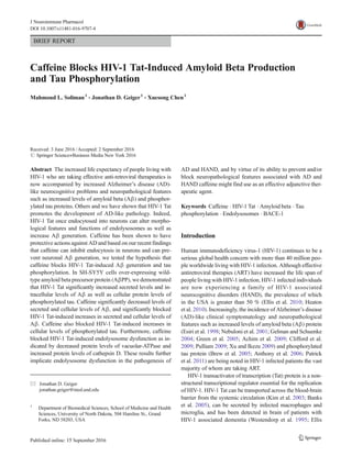
Caffeine Blocked HIV-1 Tat-Induced Increases in Aβ
Generation and Tau Phosphorylation Consistent with pre-
vious findings by others and us that HIV-1 Tat increases neu-
ronal Aβ generation (Rempel and Pulliam 2005; Giunta et al.
2009; Aksenov et al. 2010; Chen et al. 2013) and tau phosphor-
ylation (Giunta et al. 2009; Fields et al. 2015), we demonstrated
in SH-SY5Y human neuroblastoma cells over-expressing wild-
type AβPP that HIV-1 Tat treatment significantly increased
levels of secreted Aβ1–40 and Aβ1–42 (Fig. 1a). The HIV-1
Tat concentration used here is consistent with the Tat levels
(>4000 pg/ml) measured in CSF of infected individuals regard-
less of viral load (Johnson et al. 2013). To test for HIV-1 Tat
specificity, a mutant form of HIV-1 Tat (TatΔ31–61) that is not
directly neurotoxic (Buscemi et al. 2007) and immunoeluted
Tat1–72 were used as controls and neither significantly affected
levels of Aβ (Fig. 1a). In addition, HIV-1 Tat1–72, but not
TatΔ31–61 or immunoeluted Tat1–72 significantly increased pro-
tein levels of phosphorylated tau (Fig. 1b).
In preliminary studies using caffeine at concentrations of
20, 200 and 2000 μM we found 200 μM to be the most
effective concentration that consistently and significantly de-
creased Aβ levels as well as HIV-1 Tat-induced increases in
levels of Aβ (data not shown). In the absence of HIV-1 Tat
treatment, caffeine (200 μM) significantly decreased secreted
levels of Aβ1–40 (Fig. 1c) and intracellular levels of Aβ1–42
(Fig. 1d). Caffeine (200 μM) blocked HIV-1 Tat-induced in-
creases in levels of secreted Aβ1–40 and Aβ1–42 (Fig. 1c) and
levels of intracellular Aβ1–40 and Aβ1–42 (Fig. 1d).
Furthermore, while caffeine (200 μM) alone did not affect
protein levels of phosphorylated tau, but it did block HIV-1
Tat-induced increases in tau phosphorylation (Fig. 1e).
Caffeine Blocked HIV-1 Tat-Induced Endolysosome
Dysfunction Consistent with our previous findings that
HIV-1 Tat de-acidified endolysosomes (Hui et al. 2012;
Chen et al. 2013), we found here that HIV-1 Tat (100 nM
for 2 days) significantly decreased protein levels of
vacuolar-ATPase; a major mechanism by which
endolysosomes maintain their acidic environment (Fig. 2a).
Caffeine (200 μM) significantly blocked HIV-1 Tat-induced
decreases in protein levels of vacuolar-ATPase (Fig. 2a), but
did not by itself affect protein levels of vacuolar-ATPase. In
addition, caffeine (200 μM) alone did not affect protein levels
of endolysosome enzyme cathepsin D, but did significantly
block HIV-1 Tat-induced increases in protein levels of cathep-
sin D (Fig. 2b). There appeared to be a trend that caffeine
blocks Tat-induced increase in protein levels of lysosome as-
sociated membrane protein LAMP-1 (Fig. 2c), although dif-
ferences did not reach significant level.
Discussion
HIV-1 virus does not infect neurons, and HIV-associated neu-
rodegenerative pathology is not proportional to viral load (van
de Bovenkamp et al. 2002). Thus, HIV-1 viral proteins and
other factors have been implicated in the neurological compli-
cations associated with HIV-1 infections. Among HIV-1 viral
proteins, HIV-1 Tat is present in brains of HIV-1 infected
individuals and its levels stay elevated in CSF even when
HIV-1 viral levels are immeasurable (Johnson et al. 2013).
Others and we have shown consistently that HIV-1 Tat is
neurotoxic and it continues to be linked to the pathogenesis
of HAND (Nuovo et al. 1994; Nath et al. 1996; Merino et al.
2011). Increasingly, HIV-1 infection and ART treatment has
been shown to contribute to the development of AD-like pa-
thology including increases in Aβ levels (Rempel and Pulliam
2005; Giunta et al. 2009; Aksenov et al. 2010; Chen et al.
2013; Kim et al. 2013; Fields et al. 2015). Of mechanistic
significance, we have shown that HIV-1 Tat-induced
endolysosome dysfunction following receptor-mediated en-
docytosis underlies HIV-1 Tat-induced increases in neuronal
generation of Aβ (Hui et al. 2012; Chen et al. 2013). Here, we
demonstrated that caffeine, a protective agent against AD,
blocks HIV-1 Tat-induced increases in Aβ generation
and tau phosphorylation as well as decreases in protein
levels of vacuolar-ATPase. Our findings suggest that caf-
feine exerts it protective effects against the development
of AD-like pathology, in part, by blocking HIV-1 Tat-
induced endolysosome dysfunction.
4. Endolysosomes are acidic organelles that contain various
pH-dependent lytic enzymes and vacuolar H+
-ATPase helps
maintain an acidic environment necessary for maintenance of
protein turnover and cellular homeostasis (Appelqvist et al.
2013). Indeed, endolysosomes have been implicated in the
pathogenesis of sporadic AD (Tate and Mathews 2006;
Boland et al. 2008) and HAND (Gelman et al. 2005; Spector
and Zhou 2008; Zhou and Spector 2008; Cysique et al. 2015).
Neurons, as long-lived post-mitotic cells, are especially vul-
nerable to perturbations of endolysosome pH and by maintain-
ing an acidic environment endolysosomes are able to control
the integrity of quality of critical proteins (Nixon and Cataldo
1995; Bonaldo and Sandri 2013). Endolysosome dysfunction
has been implicated in the development of the two pathological
hallmarks of AD; Aβ accumulation and neurofibrillary tangle
formation. Following endocytosis of AβPP, a ubiquitously
expressed type-I transmembrane protein with largely
uncharacterized physiological functions, amyloidogenic pro-
cessing of AβPP occurs (Rajendran and Annaert 2012; Morel
et al. 2013; Jiang et al. 2014) because this is where the
amyloidogenic enzymes BACE-1 and γ-secretase are almost
exclusively located. The acidic environment of endolysosomes
is favorable for amyloidogenic metabolism of AβPP (Nixon
2005; Rajendran et al. 2008; Shimizu et al. 2008; Sannerud
et al. 2011) and once formed Aβ can either accumulate in
endolysosomes or following exocytotic release it can
Fig. 1 Caffeine blocked HIV-1
Tat-induced increases in Aβ
generation and tau
phosphorylation. a HIV-1 Tat
(100 nM) treatment for 2 days
significantly increased secreted
levels of Aβ1–40 and Aβ1–42
(n = 4). Neither mutant Tat
(mutant TatΔ31–61 at 100 nM for
2 days) nor immunoeluted Tat
affect levels of secreted Aβ1–40
and Aβ1–42. b HIV-1 Tat (100 nM
for 2 days), but not mutant Tat or
immunoeluted Tat, increased tau
phosphorylation. c Caffeine
treatment (200 μM) not only
blocked HIV-1 Tat (100 nM for
2 days) induced increases in
levels of secreted Aβ1–40 and
Aβ1–42 (n = 4), but also blocked
HIV-1 Tat-induced increases in
intracellular accumulation of
Aβ1–40 and Aβ1–42 (n = 4). In
addition, caffeine (200 μM)
treatment alone significantly
decreased basal levels of secreted
Aβ1–40 as well as intracellular
accumulation of Aβ1–42. e
Caffeine treatment (200 μM)
alone did not affect tau
phosphorylation, but blocked
HIV-1 Tat (100 nM for 2 days)
induced increases in tau
phosphorylation (n = 4). One-way
ANOVA with a Tukey post-hoc
test was for statistical analysis
5. accumulate extracellularly. Thus, Aβ accumulation can be en-
hanced by factors that promote AβPP internalization (Grbovic
et al. 2003), that enhance protein levels and/or activities of
BACE-1 and/or γ-secretase, that prevent AβPP recycling back
to the cell surface (Ma et al. 2009), and that impair Aβ degra-
dation by lysosome-resident cathepsins (Miners et al. 2011);
(Torres et al. 2012). Tau is a microtubule-associated protein,
and when hyperphosphorylated it aggregates and contributes to
the formation of neurofibrillary tangles. Tau aggregates can be
degraded by cathepsin D in autophagosomes-lysosomes
(Hamano et al. 2008; Chesser et al. 2013). Thus, endolysosome
dysfunction can contribute to tau aggregation and neurofibril-
lary tangle formation (Jo et al. 2014); (Bi and Liao 2007), and
transcriptional activation of lysosome biogenesis can clear ag-
gregated tau (Polito et al. 2014).
HIV-1 Tat enters neurons via receptor-mediated endocyto-
sis mainly with the assistance of LRP-1 (Liu et al. 2000;
Deshmane et al. 2011). Once endocytosed, HIV-1 Tat can
accumulate into and exit from endolysosomes (Vendeville
et al. 2004). Further, we have shown that HIV-1 Tat can di-
rectly affect morphological and functional features of neuro-
nal endolysosomes including endolysosome de-acidification
and endolysosome enlargement (Hui et al. 2012), indicating
that HIV-1 Tat has a direct effect on endolysosome structure
and function. Here, we demonstrated, in AβPP over-
expressing SH-SY5Y cells, that HIV-1 Tat decreased protein
levels of vacuolar-ATPase, the proton pump that is essential
for maintaining the acidic environment of endolysosomes.
Thus, endolysosome de-acidification by HIV-1 Tat might be
caused by decreased vacuolar-ATPase. The HIV-1 Tat-in-
duced decreases in vacuolar-ATPase and endolysosome de-
acidification might then cause compensatory increases in ca-
thepsin D and LAMP-1 (Mangieri et al. 2014), as we observed
in these studies. Although SH-SY5Y cells over-expressing
human AβPP are human Aβ processing neuronal cell cultures
that are commonly used in AD-related studies, this non-
physiological over-expression of AβPP may not fully recon-
stitute pathogenic cascades of HAND and may lead to supple-
mentary pathogenic effects in addition to the accumulation of
Aβ. However, we did observe similar Tat-induced
endolysosome dysfunction and AD-like pathology in primary
cultured neurons (Hui et al. 2012; Chen et al. 2013).
Consistent with our previous findings in primary cultured
neurons showing that HIV-1 Tat altered structural and function-
al features of endolysosomes and increased endolysosome ac-
cumulation of Aβ (Chen et al. 2013), we demonstrated here
using SH-SY5Y cells over-expressing AβPP that HIV-1 Tat
increased levels of secreted Aβ as well as increased accumula-
tion of intracellular Aβ. LRP-1, a promiscuous receptor that
mediates HIV-1 Tat endocytosis, has been shown by others and
us to interact physically and functionally with AβPP (Waldron
et al. 2006, 2008; Klug et al. 2011) to increase HIV-1 Tat and
AβPP internalization (Chen et al. 2013). Once internalized into
acidic endosomes, AβPP can be cleaved by β- and γ-secretase
to form Aβ. The increased Aβ production may also have been
due to HIV-1 Tat-induced increases in protein levels of AβPP
and BACE-1 and/or alternatively to HIV-1 Tat-induced
endolysosome dysfunction; both are highly likely explanations
because endolysosomes play a critical role in Aβ generation
(Rajendran and Annaert 2012; Morel et al. 2013; Jiang et al.
2014). HIV-1 Tat-induced endolysosome dysfunction may also
contribute to increases in tau phosphorylation. Because tau and
Fig. 2 Caffeine blocked HIV-1 Tat-induced endolysosome dysfunction.
a Caffeine treatment (200 μM) blocked HIV-1 Tat-induced decreases in
protein levels of vacuolar-ATPase (n = 4;). Caffeine treatment alone did
not affect protein levels of vacuolar-ATPase. b Caffeine treatment
(200 μM) blocked HIV-1 Tat treatment (100 nM for 2 days) induced
increases in protein levels of endolysosome enzyme cathepsin D
(n = 4;). Caffeine treatment alone did not affect protein levels of either
cathepsin D. c Caffeine treatment (200 μM) attenuated HIV-1 Tat
treatment (100 nM for 2 days) induced increases in protein levels of
lysosome associated membrane protein LAMP-1 (n = 4). One-way
ANOVA with a Tukey post-hoc test was for statistical analysis
6. phosphorylated tau can be degraded in autophagosomes and
lysosomes (Chesser et al. 2013; Jo et al. 2014), endolysosome
dysfunction and impaired lysosome degradation following
HIV-1 Tat endocytosis could lead to accumulations of phos-
phorylated tau and the development of neurofibrillary tangles.
Our findings suggest that HIV-1 Tat induced endolysosome
dysfunction may play an important role in HIV-1 Tat-induced
AD-like pathology. As such, attenuating endolysosome func-
tion may present an effective strategy against HIV-1 Tat-in-
duced increases in Aβ generation and tau phosphorylation.
Based on our recent findings that caffeine inhibits LDL endo-
cytosis and LDL-induced Aβ generation in neurons (Li et al.
2015), we examined the effects of caffeine on HIV-1 Tat-in-
duced increases in Aβ generation and tau phosphorylation.
We demonstrated that caffeine blocked HIV-1 Tat induced
endolysosome dysfunction as well as AD-like pathological
features including increases in levels of secreted Aβ, intracel-
lular accumulation of Aβ, and increased levels of phosphory-
lated tau. Our findings suggest that caffeine could prevent
HIV-1 Tat-induced AD-like pathology in part by preventing
HIV-1 Tat-induced endolysosome dysfunction. It is estimated
that 1 cup of coffee consumption (40–100 mg of caffeine) can
result in plasma concentrations of free caffeine of about 1–
10 μM (Fredholm et al. 1999). However, because caffeine is
highly protein bound, total plasma caffeine concentrations
would be much higher (Blanchard 1982). Caffeine concentra-
tions used in this study (200 μM) are in the upper range of
plasma concentrations of caffeine (Fredholm et al. 1999).
Indeed, we confirmed in a previous study that 200 μM of
caffeine does not have neurotoxic effects (Li et al. 2015).
In short, our findings add to the ever-increasing reports that
caffeine provides robust protective actions against AD. Besides
its protective role against the development of AD, caffeine has
been shown to exert protective effect against HIV-1 pathogen-
esis (Daniel et al. 2005; Nunnari et al. 2005). Thus, caffeine
might be used in the treatment of AD–like symptoms and neu-
ropathology associated with HIV-1 infection.
Acknowledgments This work was supported by the following grants
received from the National Institutes of Health; P30GM103329,
R01MH100972 and R01MH105329.
Compliance with Ethical Standards
Disclosure Statement The authors have no current or potential con-
flicts of interest to report.
References
Achim CL, Adame A, Dumaop W, Everall IP, Masliah E (2009) Increased
accumulation of intraneuronal amyloid beta in HIV-infected pa-
tients. J NeuroImmune Pharmacol 4:190–199
Aksenov MY, Aksenova MV, Mactutus CF, Booze RM (2010) HIV-1
protein-mediated amyloidogenesis in rat hippocampal cell cultures.
Neurosci Lett 475:174–178
Anthony IC, Ramage SN, Carnie FW, Simmonds P, Bell JE (2006)
Accelerated tau deposition in the brains of individuals infected with
human immunodeficiency virus-1 before and after the advent of
highly active anti-retroviral therapy. Acta Neuropathol 111:529–538
Appelqvist H, Waster P, Kagedal K, Ollinger K (2013) The lysosome:
from waste bag to potential therapeutic target. J Mol Cell Biol 5:
214–226
Arendash GW, Cao C (2010) Caffeine and coffee as therapeutics against
Alzheimer's disease. J Alzheimers Dis 20(Suppl 1):S117–S126
Arendash GW, Schleif W, Rezai-Zadeh K, Jackson EK, Zacharia LC,
Cracchiolo JR, Shippy D, Tan J (2006) Caffeine protects
Alzheimer's mice against cognitive impairment and reduces brain
beta-amyloid production. Neuroscience 142:941–952
Arendash GW, Mori T, Cao C, Mamcarz M, Runfeldt M, Dickson A,
Rezai-Zadeh K, Tane J, Citron BA, Lin X, Echeverria V, Potter H
(2009) Caffeine reverses cognitive impairment and decreases brain
amyloid-beta levels in aged Alzheimer's disease mice. J Alzheimers
Dis 17:661–680
Banks WA, Robinson SM, Nath A (2005) Permeability of the blood-brain
barrier to HIV-1 tat. Exp Neurol 193:218–227
Bi X, Liao G (2007) Autophagic-lysosomal dysfunction and neurodegen-
eration in Niemann-pick type C mice: lipid starvation or indiges-
tion? Autophagy 3:646–648
Blanchard J (1982) Protein binding of caffeine in young and elderly
males. J Pharm Sci 71:1415–1418
Boland B, Kumar A, Lee S, Platt FM, Wegiel J, WH Y, Nixon RA (2008)
Autophagy induction and autophagosome clearance in neurons: re-
lationship to autophagic pathology in Alzheimer's disease. J
Neurosci 28:6926–6937
Bonaldo P, Sandri M (2013) Cellular and molecular mechanisms of mus-
cle atrophy. Dis Model Mech 6:25–39
Brew BJ, Pemberton L, Blennow K, Wallin A, Hagberg L (2005) CSF
amyloid beta42 and tau levels correlate with AIDS dementia com-
plex. Neurology 65:1490–1492
Buscemi L, Ramonet D, Geiger JD (2007) Human immunodeficiency
virus type-1 protein tat induces tumor necrosis factor-alpha-
mediated neurotoxicity. Neurobiol Dis 26:661–670
Cao C, Cirrito JR, Lin X, Wang L, Verges DK, Dickson A, Mamcarz M,
Zhang C, Mori T, Arendash GW, Holtzman DM, Potter H (2009)
Caffeine suppresses amyloid-beta levels in plasma and brain of
Alzheimer's disease transgenic mice. J Alzheimers Dis 17:681–697
Cao C, Loewenstein DA, Lin X, Zhang C, Wang L, Duara R, Wu Y,
Giannini A, Bai G, Cai J, Greig M, Schofield E, Ashok R, Small
B, Potter H, Arendash GW (2012) High blood caffeine levels in
MCI linked to lack of progression to dementia. J Alzheimers Dis
30:559–572
Carman AJ, Dacks PA, Lane RF, Shineman DW, Fillit HM (2014)
Current evidence for the use of coffee and caffeine to prevent age-
related cognitive decline and Alzheimer's disease. J Nutr Health
Aging 18:383–392
Cataldo AM, Peterhoff CM, Troncoso JC, Gomez-Isla T, Hyman BT,
Nixon RA (2000) Endocytic pathway abnormalities precede amy-
loid beta deposition in sporadic Alzheimer's disease and down syn-
drome: differential effects of APOE genotype and presenilin muta-
tions. Am J Pathol 157:277–286
Chen X, Hui L, Geiger NH, Haughey NJ, Geiger JD (2013)
Endolysosome involvement in HIV-1 transactivator protein-
induced neuronal amyloid beta production. Neurobiol Aging 34:
2370–2378
Chesser AS, Pritchard SM, Johnson GV (2013) Tau clearance mecha-
nisms and their possible role in the pathogenesis of Alzheimer dis-
ease. Front Neurol 4:122
7. Clifford DB, Fagan AM, Holtzman DM, Morris JC, Teshome M, Shah
AR, Kauwe JS (2009) CSF biomarkers of Alzheimer disease in
HIV-associated neurologic disease. Neurology 73:1982–1987
Cysique LA, Hewitt T, Croitoru-Lamoury J, Taddei K, Martins RN,
Chew CS, Davies NN, Price P, BJ B (2015) APOE epsilon4 mod-
erates abnormal CSF-abeta-42 levels, while neurocognitive impair-
ment is associated with abnormal CSF tau levels in HIV+ individ-
uals - a cross-sectional observational study. BMC Neurol 15:51
Daniel R, Marusich E, Argyris E, Zhao RY, Skalka AM, Pomerantz RJ
(2005) Caffeine inhibits human immunodeficiency virus type 1
transduction of nondividing cells. J Virol 79:2058–2065
Deshmane SL, Mukerjee R, Fan S, Sawaya BE (2011) High-performance
capillary electrophoresis for determining HIV-1 tat protein in neu-
rons. PLoS One 6:e16148
Ellis RJ, Gamst AC, Capparelli E, Spector SA, Hsia K, Wolfson T,
Abramson I, Grant I, McCutchan JA (2000) Cerebrospinal fluid
HIV RNA originates from both local CNS and systemic sources.
Neurology 54:927–936
Ellis RJ, Rosario D, Clifford DB, McArthur JC, Simpson D, Alexander T,
Gelman BB, Vaida F, Collier A, Marra CM, Ances B, Atkinson JH,
Dworkin RH, Morgello S, Grant I (2010) Continued high preva-
lence and adverse clinical impact of human immunodeficiency
virus-associated sensory neuropathy in the era of combination anti-
retroviral therapy: the CHARTER study. Arch Neurol 67:552–558
Esiri MM, Biddolph SC, Morris CS (1998) Prevalence of Alzheimer
plaques in AIDS. J Neurol Neurosurg Psychiatry 65:29–33
Eskelinen MH, Kivipelto M (2010) Caffeine as a protective factor in
dementia and Alzheimer's disease. J Alzheimers Dis 20(Suppl 1):
S167–S174
Eskelinen MH, Ngandu T, Tuomilehto J, Soininen H, Kivipelto M (2009)
Midlife coffee and tea drinking and the risk of late-life dementia: a
population-based CAIDE study. J Alzheimers Dis 16:85–91
Espinosa J, Rocha A, Nunes F, Costa MS, Schein V, Kazlauckas V,
Kalinine E, Souza DO, Cunha RA, Porciuncula LO (2013)
Caffeine consumption prevents memory impairment, neuronal dam-
age, and adenosine A2 A receptors upregulation in the hippocampus
of a rat model of sporadic dementia. J Alzheimers Dis 34:509–518
Fields JA, Dumaop W, Crews L, Adame A, Spencer B, Metcalf J, He J,
Rockenstein E, Masliah E (2015) Mechanisms of HIV-1 tat neuro-
toxicity via CDK5 translocation and hyper-activation: role in HIV-
associated neurocognitive disorders. Curr HIV Res 13:43–54
Flaten V, Laurent C, Coelho JE, Sandau U, Batalha VL, Burnouf S,
Hamdane M, Humez S, Boison D, Lopes LV, Buee L, Blum D
(2014) From epidemiology to pathophysiology: what about caffeine
in Alzheimer's disease? Biochem Soc Trans 42:587–592
Fredholm BB, Battig K, Holmen J, Nehlig A, Zvartau EE (1999) Actions
of caffeine in the brain with special reference to factors that contrib-
ute to its widespread use. Pharmacol Rev 51:83–133
Gelber RP, Petrovitch H, Masaki KH, Ross GW, White LR (2011) Coffee
intake in midlife and risk of dementia and its neuropathologic cor-
relates. J Alzheimers Dis 23:607–615
Gelman BB, Schuenke K (2004) Brain aging in acquired immunodefi-
ciency syndrome: increased ubiquitin-protein conjugate is correlated
with decreased synaptic protein but not amyloid plaque accumula-
tion. J Neurovirol 10:98–108
Gelman BB, Soukup VM, Holzer CE 3rd, Fabian RH, Schuenke KW,
Keherly MJ, Richey FJ, Lahart CJ (2005) Associated dementia. J
Acquir Immune Defic Syndr 39:422–425
Giunta B, Hou H, Zhu Y, Rrapo E, Tian J, Takashi M, Commins D, Singer
E, He J, Fernandez F, Tan J (2009) HIV-1 tat contributes to
Alzheimer's disease-like pathology in PSAPP mice. Int J Clin Exp
Pathol 2:433–443
Grbovic OM, Mathews PM, Jiang Y, Schmidt SD, Dinakar R, Summers-
Terio NB, Ceresa BP, Nixon RA, Cataldo AM (2003) Rab5-
stimulated up-regulation of the endocytic pathway increases intra-
cellular beta-cleaved amyloid precursor protein carboxyl-terminal
fragment levels and abeta production. J Biol Chem 278:31261–
31268
Green DA, Masliah E, Vinters HV, Beizai P, Moore DJ, Achim CL (2005)
Brain deposition of beta-amyloid is a common pathologic feature in
HIV positive patients. AIDS 19:407–411
Hamano T, Gendron TF, Causevic E, Yen SH, Lin WL, Isidoro C, Deture
M, Ko LW (2008) Autophagic-lysosomal perturbation enhances tau
aggregation in transfectants with induced wild-type tau expression.
Eur J Neurosci 27:1119–1130
Han K, Jia N, Li J, Yang L, Min LQ (2013) Chronic caffeine treatment
reverses memory impairment and the expression of brain BNDF and
TrkB in the PS1/APP double transgenic mouse model of
Alzheimer's disease. Mol Med Rep 8:737–740
Heaton RK et al. (2010) HIV-associated neurocognitive disorders persist
in the era of potent antiretroviral therapy: CHARTER study.
Neurology 75:2087–2096
Hui L, Chen X, Haughey NJ, Geiger JD (2012) Role of endolysosomes in
HIV-1 tat-induced neurotoxicity. ASN Neuro 4:243–252
Jiang S, Li Y, Zhang X, Bu G, Xu H, Zhang YW (2014) Trafficking
regulation of proteins in Alzheimer's disease. Mol Neurodegener
9:6
Jo C, Gundemir S, Pritchard S, Jin YN, Rahman I, Johnson GV (2014)
Nrf2 reduces levels of phosphorylated tau protein by inducing au-
tophagy adaptor protein NDP52. Nat Commun 5:3496
Johnson TP, Patel K, Johnson KR, Maric D, Calabresi PA, Hasbun R,
Nath A (2013) Induction of IL-17 and nonclassical T-cell activation
by HIV-tat protein. Proc Natl Acad Sci U S A 110:13588–13593
Kenessey A, Nacharaju P, Ko LW, Yen SH (1997) Degradation of tau by
lysosomal enzyme cathepsin D: implication for Alzheimer neurofi-
brillary degeneration. J Neurochem 69:2026–2038
Kim TA, Avraham HK, Koh YH, Jiang S, Park IW, Avraham S (2003)
HIV-1 tat-mediated apoptosis in human brain microvascular endo-
thelial cells. J Immunol 170:2629–2637
Kim J, Yoon JH, Kim YS (2013) HIV-1 tat interacts with and regulates
the localization and processing of amyloid precursor protein. PLoS
One 8:e77972
King JE, Eugenin EA, Buckner CM, Berman JW (2006) HIV tat and
neurotoxicity. Microbes Infect 8:1347–1357
Klug W, Dietl A, Simon B, Sinning I, Wild K (2011) Phosphorylation of
LRP1 regulates the interaction with Fe65. FEBS Lett 585:3229–
3235
Laurent C, Eddarkaoui S, Derisbourg M, Leboucher A, Demeyer D,
Carrier S, Schneider M, Hamdane M, Muller CE, Buee L, Blum D
(2014) Beneficial effects of caffeine in a transgenic model of
Alzheimer's disease-like tau pathology. Neurobiol Aging 35:2079–
2090
Li S, Geiger NH, Soliman ML, Hui L, Geiger JD, Chen X (2015)
Caffeine, through adenosine A3 receptor-mediated actions, sup-
presses amyloid-beta protein precursor internalization and
amyloid-beta generation. J Alzheimers Dis 47:73–83
Liu Y, Jones M, Hingtgen CM, Bu G, Laribee N, Tanzi RE, Moir RD,
Nath A, He JJ (2000) Uptake of HIV-1 tat protein mediated by low-
density lipoprotein receptor-related protein disrupts the neuronal
metabolic balance of the receptor ligands. Nat Med 6:1380–1387
Ma M, Nath A (1997) Molecular determinants for cellular uptake of tat
protein of human immunodeficiency virus type 1 in brain cells. J
Virol 71:2495–2499
Ma QL, Galasko DR, Ringman JM, Vinters HV, Edland SD, Pomakian J,
Ubeda OJ, Rosario ER, Teter B, Frautschy SA, Cole GM (2009)
Reduction of SorLA/LR11, a sorting protein limiting beta-amyloid
production, in Alzheimer disease cerebrospinal fluid. Arch Neurol
66:448–457
Mangieri LR, Mader BJ, Thomas CE, Taylor CA, Luker AM, Tse TE,
Huisingh C, Shacka JJ (2014) ATP6V0C knockdown in neuroblas-
toma cells alters autophagy-lysosome pathway function and
8. metabolism of proteins that accumulate in neurodegenerative dis-
ease. PLoS One 9:e93257
Merino JJ, Montes ML, Blanco A, Bustos MJ, Oreja-Guevara C, Bayon
C, Cuadrado A, Lubrini G, Cambron I, Munoz A, Cebolla S,
Gutierrez-Fernandez M, Bernardino JI, Arribas JR, Fiala M (2011)
HIV-1 neuropathogenesis: therapeutic strategies against neuronal
loss induced by gp120/tat glycoprotein in the central nervous sys-
tem. Rev Neurol 52:101–111
Miners JS, Barua N, Kehoe PG, Gill S, Love S (2011) Abeta-degrading
enzymes: potential for treatment of Alzheimer disease. J
Neuropathol Exp Neurol 70:944–959
Morel E, Chamoun Z, Lasiecka ZM, Chan RB, Williamson RL,
Vetanovetz C, Dall'Armi C, Simoes S, Point Du Jour KS, McCabe
BD, Small SA, Di Paolo G (2013) Phosphatidylinositol-3-phosphate
regulates sorting and processing of amyloid precursor protein
through the endosomal system. Nat Commun 4:2250
Nath A (2002) Human immunodeficiency virus (HIV) proteins in
neuropathogenesis of HIV dementia. J Infect Dis 186(Suppl 2):
S193–S198
Nath A, Psooy K, Martin C, Knudsen B, Magnuson DS, Haughey N,
Geiger JD (1996) Identification of a human immunodeficiency virus
type 1 tat epitope that is neuroexcitatory and neurotoxic. J Virol 70:
1475–1480
Nebuloni M, Pellegrinelli A, Ferri A, Bonetto S, Boldorini R, Vago L,
Grassi MP, Costanzi G (2001) Beta amyloid precursor protein and
patterns of HIV p 24 immunohistochemistry in different brain areas
of AIDS patients. AIDS 15:571–575
Nixon RA (2005) Endosome function and dysfunction in Alzheimer's
disease and other neurodegenerative diseases. Neurobiol Aging
26:373–382
Nixon RA, Cataldo AM (1995) The endosomal-lysosomal system of
neurons: new roles. Trends Neurosci 18:489–496
Nunnari G, Argyris E, Fang J, Mehlman KE, Pomerantz RJ, Daniel R
(2005) Inhibition of HIV-1 replication by caffeine and caffeine-
related methylxanthines. Virology 335:177–184
Nuovo GJ, Becker J, Burk MW, Margiotta M, Fuhrer J, Steigbigel RT
(1994) In situ detection of PCR-amplified HIV-1 nucleic acids in
lymph nodes and peripheral blood in patients with asymptomatic
HIV-1 infection and advanced-stage AIDS. J Acquir Immune
Defic Syndr 7:916–923
Oyama F, Murakami N, Ihara Y (1998) Chloroquine myopathy suggests
that tau is degraded in lysosomes: implication for the formation of
paired helical filaments in Alzheimer's disease. Neurosci Res 31:1–8
Patrick C, Crews L, Desplats P, Dumaop W, Rockenstein E, Achim CL,
Everall IP, Masliah E (2011) Increased CDK5 expression in HIV
encephalitis contributes to neurodegeneration via tau phosphoryla-
tion and is reversed with Roscovitine. Am J Pathol 178:1646–1661
Polito VA, Li H, Martini-Stoica H, Wang B, Yang L, Xu Y, Swartzlander
DB, Palmieri M, di Ronza A, Lee VM, Sardiello M, Ballabio A,
Zheng H (2014) Selective clearance of aberrant tau proteins and
rescue of neurotoxicity by transcription factor EB. EMBO Mol
Med 6:1142–1160
Pulliam L (2009) HIV regulation of amyloid beta production. J
NeuroImmune Pharmacol 4:213–217
Rajendran L, Annaert W (2012) Membrane trafficking pathways in
Alzheimer's disease. Traffic 13:759–770
Rajendran L, Schneider A, Schlechtingen G, Weidlich S, Ries J,
Braxmeier T, Schwille P, Schulz JB, Schroeder C, Simons M,
Jennings G, Knolker HJ, Simons K (2008) Efficient inhibition of
the Alzheimer's disease beta-secretase by membrane targeting.
Science 320:520–523
Rempel HC, Pulliam L (2005) HIV-1 tat inhibits neprilysin and elevates
amyloid beta. AIDS 19:127–135
Ritchie K, Carriere I, de Mendonca A, Portet F, Dartigues JF, Rouaud O,
Barberger-Gateau P, Ancelin ML (2007) The neuroprotective effects
of caffeine: a prospective population study (the three City study.
Neurology 69:536–545
Sannerud R, Declerck I, Peric A, Raemaekers T, Menendez G, Zhou L,
Veerle B, Coen K, Munck S, De Strooper B, Schiavo G, Annaert W
(2011) ADP ribosylation factor 6 (ARF6) controls amyloid precur-
sor protein (APP) processing by mediating the endosomal sorting of
BACE1. Proc Natl Acad Sci U S A 108:E559–E568
Santos C, Costa J, Santos J, Vaz-Carneiro A, Lunet N (2010a) Caffeine
intake and dementia: systematic review and meta-analysis. J
Alzheimers Dis 20(Suppl 1):S187–S204
Santos C, Lunet N, Azevedo A, de Mendonca A, Ritchie K, Barros H
(2010b) Caffeine intake is associated with a lower risk of cognitive
decline: a cohort study from Portugal. J Alzheimers Dis 20(Suppl 1):
S175–S185
Shimizu H, Tosaki A, Kaneko K, Hisano T, Sakurai T, Nukina N (2008)
Crystal structure of an active form of BACE1, an enzyme responsi-
ble for amyloid beta protein production. Mol Cell Biol 28:3663–
3671
Spector SA, Zhou D (2008) Autophagy: an overlooked mechanism of
HIV-1 pathogenesis and neuroAIDS? Autophagy 4:704–706
Tate BA, Mathews PM (2006) Targeting the role of the endosome in the
pathophysiology of Alzheimer's disease: a strategy for treatment. Sci
Aging Knowl Environ 2006:re2
Torres M, Jimenez S, Sanchez-Varo R, Navarro V, Trujillo-Estrada L,
Sanchez-Mejias E, Carmona I, Davila JC, Vizuete M, Gutierrez A,
Vitorica J (2012) Defective lysosomal proteolysis and axonal trans-
port are early pathogenic events that worsen with age leading to
increased APP metabolism and synaptic abeta in transgenic APP/
PS1 hippocampus. Mol Neurodegener 7:59
van de Bovenkamp M, Nottet HS, Pereira CF (2002) Interactions of
human immunodeficiency virus-1 proteins with neurons: possible
role in the development of human immunodeficiency virus-1-
associated dementia. Eur J Clin Investig 32:619–627
Vendeville A, Rayne F, Bonhoure A, Bettache N, Montcourrier P,
Beaumelle B (2004) HIV-1 tat enters Tcells using coated pits before
translocating from acidified endosomes and eliciting biological re-
sponses. Mol Biol Cell 15:2347–2360
Waldron E, Jaeger S, Pietrzik CU (2006) Functional role of the low-
density lipoprotein receptor-related protein in Alzheimer's disease.
Neurodegener Dis 3:233–238
Waldron E, Heilig C, Schweitzer A, Nadella N, Jaeger S, Martin AM,
Weggen S, Brix K, Pietrzik CU (2008) LRP1 modulates APP traf-
ficking along early compartments of the secretory pathway.
Neurobiol Dis 31:188–197
Wang Y, Martinez-Vicente M, Kruger U, Kaushik S, Wong E,
Mandelkow EM, Cuervo AM, Mandelkow E (2009) Tau fragmen-
tation, aggregation and clearance: the dual role of lysosomal pro-
cessing. Hum Mol Genet 18:4153–4170
Westendorp MO, Frank R, Ochsenbauer C, Stricker K, Dhein J, Walczak
H, Debatin KM, Krammer PH (1995) Sensitization of T cells to
CD95-mediated apoptosis by HIV-1 tat and gp120. Nature 375:
497–500
Wostyn P, Van Dam D, Audenaert K, De Deyn PP (2011) Increased
cerebrospinal fluid production as a possible mechanism underlying
Caffeine's protective effect against Alzheimer's disease. Int J
Alzheimers Dis 2011:617420
Xu J, Ikezu T (2009) The comorbidity of HIV-associated neurocognitive
disorders and Alzheimer's disease: a foreseeable medical challenge
in post-HAART era. J NeuroImmune Pharmacol 4:200–212
Zhou D, Spector SA (2008) Human immunodeficiency virus type-1 in-
fection inhibits autophagy. AIDS 22:695–699