1. Neurosurg Focus 19 (2):E1, 2005
Suboccipital burr holes and craniectomies
GUILHERME C. RIBAS, M.D., ALBERT L. RHOTON JR., M.D., OSWALDO R. CRUZ, M.D.,
AND DAVID PEACE, M.S.
Department of Neurosurgery, University of Florida, Gainesville, Florida; and Departments of
Neurosurgery and Surgery (Clinical Anatomy Discipline), University of São Paulo Medical School,
São Paulo, Brazil
Object. The goal of this study was to delimit the external cranial projection of the transverse and sigmoid sinuses,
and to establish initial strategic systematized burr hole sites for lateral infratentorial suboccipital approaches based on
external cranial landmarks particularly related to the lambdoid, occipitomastoid, and parietomastoid sutures.
Methods. The external cranial projection of the transverse and sigmoid sinuses was studied through their external
outlining obtained with the aid of multiple small perforations made from inside to outside along the inner margins of
the sinuses of 50 paired temporoparietooccipital regions in 25 dried adult human skulls. The burr hole placement was
studied by evaluating the supratentorial, over-the-sinuses, and infratentorial components of 1-cm-diameter openings
made at strategic sites identified in the initial part of the study, which was performed in another 50 paired tem-
poroparietooccipital regions.
The asterion and the midpoint of the inion–asterion line were found to be particularly related to the inferior half of
the transverse sinus; the transverse and sigmoid sinuses’ transition occurs 1 cm anteriorly to the asterion across the
parietomastoid suture, and the most superior part of the sigmoid sinus is located anteriorly to the occipitomastoid
suture, with its posterior margin crossing this suture posteriorly to the most superior aspect of the mastoid process,
which is located at the most superior level of the mastoid notch. Burr holes made at the midpoint of the inion–asteri-
on line, at the asterion, 1 cm anterior to the asterion, just inferiorly to the parietomastoid suture, and over the occipito-
mastoid suture at the most superior level of the mastoid notch are appropriate to expose the inferior half of the trans-
verse sinus at its midpoint, the inferior half of the transverse sinus at its most lateral aspect, the transverse and sigmoid
sinuses’ transition, and the posterior margin of the basal aspect of the sigmoid sinus, respectively.
Conclusions. These findings allow an estimation of the transverse and sigmoid sinuses’ external cranial projection.
The asterion and the most posterior part of the parietomastoid suture constitute a suitable initial burr hole site at which
to perform an upper or asterional suboccipital craniectomy to expose the superior aspect of the cerebellopontine angle
(CPA). The occipitomastoid suture at the most superior aspect of the mastoid notch constitutes an adequate initial burr
hole site at which to perform a basal suboccipital craniectomy to expose the lower portion of the CPA. The sites can
be used together as initial burr hole sites to perform wide suboccipital exposures, because they already constitute nat-
ural infratentorial lateral limits.
KEY WORDS • cranial suture • transverse sinus • sigmoid sinus • burr hole •
craniotomy • suboccipital approach • cerebellopontine angle • posterior fossa
Whereas the frontotemporal approaches are relatively graphic profiles of strategic burr hole sites related to these
systematized in the sense that the craniotomies proceed sutures (Fig. 1).
from defined burr hole sites,76,78 the lateral suboccipital ap-
proaches are performed from various and nonstandardized
initial burr hole placements. Because the transverse and MATERIALS AND METHODS
sigmoid sinuses are the natural limits of these exposures, This study was conducted in two stages, in each of which 50
the knowledge of their cranial topography constitutes the paired temporoparietooccipital regions in 25 adult human dried
main factor in the planning of these posterior approaches. skulls were studied through observations and measurements.57
In the first part, the anatomical relationships of the lambdoid,
The lambdoid, occipitomastoid, and parietomastoid su- occipitomastoid, and parietomastoid sutures to the transverse and
tures are easily recognizable structures on the external cran- sigmoid sinuses were studied in 25 adult human uncataloged skulls,
ial surface.35,42,47,49,60–63,69–72 For this reason we initially inves- originally from India, at the Theodore Gildred Microsurgical Edu-
tigated their relationships with the transverse and sigmoid cation Center of the Department of Neurological Surgery of the Un-
sinuses, and based on these findings, we evaluated the topo- iversity of Florida. The calvariae of the skulls were removed above
the lambda and the superior orbital ridges, the transverse and sig-
Abbreviation used in this paper: CPA = cerebellopontine angle. moid sinuses superior and inferior margins were drawn on the outer
Neurosurg. Focus / Volume 19 / August, 2005 1
2. G. C. Ribas, et al.
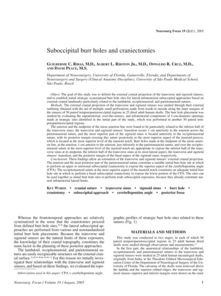
Fig. 1. Photographs of dried adult human skulls showing the external cranial surface (left) and the internal cranial surface (right).
surface of the skulls after multiple small perforations were made on (Fig. 2). The analysis of its distance to the superior and
its bone landmarks from inside to outside, and the main points to be inferior margins of the transverse sinus, and of the sinus
studied were identified. height at this point, led us to conclude that the asterion is
The second part of the study was done at the Anatomical Museum
of the Biomedical Sciences Institute of the University of São Paulo particularly related to the inferior half of the transverse
through observations and measurements obtained in 25 adult human sinus. The topography of the transverse sinus segment pos-
skulls cataloged according to their race, sex, and age (Table 1).The terior to the lambdoid and occipitomastoid sutures was
calvariae had already been removed in these skulls, and the points to evaluated through the disposition of the inion–asterion line
be studied were lightly marked with a pencil. in relation to the sinus (Fig. 3).
The burr hole study was conducted by evaluation of the posteri- The analysis of the distances to both margins from the
or fossa, the area over the sinuses, and supratentorial components of midpoint of the inion–asterion line, and the sinus height at
1-cm-diameter burr holes placed at strategic sites related to the cran-
ial sutures. The burr holes were plotted with the aid of a circular de- that point, showed that its position in relation to the trans-
vice adapted to a compass, and the measurements of the height of verse sinus is similar to the disposition of the asterion,
each burr hole’s topographic components provided the data to elab- being also related to the inferior aspect of the sinus sulcus
orate its topographic profile. (Fig. 4).
An extensive statistical analysis was done to compare the results The topography of the part of the transverse sinus ante-
among the different sides, sexes, and races.
RESULTS
Anatomical Relationships of the Lambdoid, Occipitomastoid,
and Parietomastoid Sutures With the Transverse and Sigmoid
Sinuses
Anatomical Relationships of the Lambdoid and Occipitomas-
toid Sutures With the Transverse Sinus. The relationships of
the lambdoid and occipitomastoid sutures with the trans-
verse sinus were evaluated through the asterion position
Fig. 2. Schematic drawing showing the variation of the asteri-
on’s position in relation to the transverse sinus (mean values, tak-
ing into account variations between races). Values in all schematic
TABLE 1 drawings except Fig. 3 are given in centimeters, and commas
Identification of the 25 catalogued skulls studied denote decimal points.
Feature Value
race
African-American 7
Caucasian 9
mixed 9
sex
F 11
M 14
age (yrs)
min 18.0
max 60.0
mean 33.9
Fig. 3. Schematic drawing showing the disposition of the
median 34.0
inion–asterion line in relation to the transverse sinus.
2 Neurosurg. Focus / Volume 19 / August, 2005
3. Suboccipital burr holes and craniectomies
Fig. 4. Schematic drawing showing variation in the position of Fig. 8. Schematic drawing showing variations in the position of
the midpoint of the inion–asterion line in relation to the transverse the occipitomastoid suture/sigmoid sinus posterior margin crossing
sinus (mean values, taking into account variations between races). point along the occipitomastoid suture (mean values, taking into
account variations between races).
rior to the lambdoid and occipitomastoid sutures was eval-
uated through analysis of the disposition of its most antero-
superior and anteroinferior points in relation to the asteri-
on. These points, which were equivalents to the superior
and inferior aspects, respectively, of the transverse sinus/
sigmoid sinus transition, presented the disposition depicted
in Fig. 5 in relation to the asterion.
Anatomical Relationships of the Parietomastoid Suture
With the Transverse Sinus. These relationships were initial-
ly evaluated through analysis of the transverse sinus’ most
anterosuperior and its most anteroinferior points in relation
to this suture. Whereas the position of the point corre-
sponding to the superior aspect of the sinuses’ transition
varied from 0.1 to 0.3 cm above the parietomastoid suture,
the point corresponding to the inferior aspect of the sinus-
Fig. 5. Schematic drawing showing the disposition of the most es’ transition was situated below the suture at a mean dis-
anterosuperior and anteroinferior points in the asterion and trans- tance of 0.8 cm, regardless of the race, sex, and side of the
verse sinus (T.S.; mean values according to race). skulls (Fig. 6).
The easily identifiable parietomastoid suture/squamous
suture meeting point was particularly studied in terms of its
position in relation to the superior margin of the transverse
sinus or to the middle fossa floor/tentorium, and was
shown to be related with this level (Fig. 7).
Anatomical Relationships of the Occipitomastoid Suture
With the Sigmoid Sinus. These relationships were evaluated
by studying the topography of the occipitomastoid suture/
sigmoid sinus posterior margin crossing point. The analy-
sis of the distances of this crossing point to the asterion and
to the jugular foramen identified its position along the oc-
cipitomastoid suture (Fig. 8).
Fig. 6. Schematic drawing showing variations in the position of This point was also shown to be closely related to the in-
the most anterosuperior and anteroinferior points in the transverse ion–mastoid tip line level and to the posterior aspect of the
sinus in relation to the parietomastoid suture (mean values, taking mastoid notch (Figs. 9 and 10).
into account variations between races).
Study of Burr Hole Sites Related to Lambdoid, Occipito-
mastoid, and Parietomastoid Sutures
The results presented here were determined through
evaluation of the 1-cm burr holes’ mean topographic pro-
files in six openings made in three different groups that
were studied for specific purposes.
Burr Holes at the Asterion and at the Midpoint of the In-
Fig. 7. Schematic drawing showing variations in the position of ion–Asterion Line. As shown in Fig. 11, these results prove
the parietomastoid suture/squamous suture meeting point in rela- that both burr holes are adequate to expose the transverse
tion to the transverse sinus superior margin/middle fossa floor level sinus, with the second one being more appropriate to ex-
(mean values, taking into account variations between races). pose both the sinus and the posterior fossa compartment.
Neurosurg. Focus / Volume 19 / August, 2005 3
4. G. C. Ribas, et al.
Fig. 9. Schematic drawing showing variations in the position of
the occipitomastoid suture/sigmoid sinus posterior margin crossing
point in relation to the level of the inion–mastoid tip line (mean val-
ues, taking into account variations between races).
Burr Holes Anterior to the Asterion. As shown in Fig. 12,
these two burr holes were also evaluated for their potential Fig. 11. Schematic drawings showing topographic profiles of
to expose the inferior aspect of the transverse sinus/sig- the 1-cm-diameter burr hole centered on the asterion (1), and cen-
moid sinus transition, and the latter proved to be more ap- tered on the midpoint of the inion–asterion line (2).
propriate for this purpose, yielding results not dependent
on race, sex, or side of the skull (Table 2).
Burr Holes Over the Occipitomastoid Suture. As shown in
Fig. 13, the comparison of their topographic profiles shows
that both burr hole sites are appropriate to expose the sig-
moid sinus posterior margin and the posterior fossa com-
partment.
DISCUSSION
The suboccipital approach to the CPA used nowadays is
still based on the unilateral craniectomy described by Dan-
dy8,9 in 1929 and in 1934, which was later made through a
straight lateral incision as proposed by Adson1 in 1941, and
then extended laterally and inferiorly as was already em-
phasized by Bucy7 in 1951. The microsurgical techniques
introduced by Rand and Kurze51 in 1965 enabled the devel-
opment of acoustic tumor exposure, particularly by drilling
of the posterior meatal wall.
Although they obtain very similar CPA exposures, the
most experienced neurosurgeons in this field perform their
craniectomies differently according to their own experi- Fig. 12. Schematic drawings showing topographic profiles of
the 1-cm-diameter burr hole with its superior base on the asterion
and on the parietomastoid suture (3), and centered 1 cm anteriorly
to the asterion and with its superior base on the parietomastoid
suture (4).
TABLE 2
Exposure of the inferior aspect
of the transverse sinus/sigmoid sinus transition*
Exposure (%)
Site Yes No
burr hole 3 34 66
Fig. 10. Schematic drawing showing variations in the position burr hole 4 84 16
of the occipitomastoid suture/sigmoid sinus posterior margin cross- * Exposure of the inferior aspect by the 1-cm burr hole with its superior
ing point in relation to the posterior and superior aspect of the mas- base on the asterion and on the parietomastoid suture, and by the 1-cm burr
toid notch (mean values, taking into account variations between hole centered 1 cm anteriorly to the asterion and with its superior base on
races). the parietomastoid suture.
4 Neurosurg. Focus / Volume 19 / August, 2005
5. Suboccipital burr holes and craniectomies
the parietomastoid suture; 3) the meeting point of the pari-
etomastoid and squamous sutures is located at the level of
the posterior part of the middle fossa floor; and 4) the
occipitomastoid suture and the crossing point of the sig-
moid sinus posterior margin are situated at the level of the
superior and posterior aspect of the mastoid notch, and also
correspond to the level of the intersection point of the
occipitomastoid suture with the imaginary line of the in-
ion–mastoid tip.
In the study of burr hole sites there were no significant
differences regarding the topographic profiles of the sexes
and sides of the skull. The main conclusions reached in this
part of the study were as follows: first, burr holes centered
at the asterion (1) and at the midpoint of the inion–asterion
line (2) are adequate to expose the transverse sinus; second,
burr holes with their superior base on the asterion and on
the parietomastoid suture (3) and centered 1 cm anterior to
the asterion with their superior base on the parietomastoid
suture (4) are sufficient to expose the inferior aspect of the
Fig. 13. Schematic drawings showing topographic profiles of transverse sinus next to the transverse sinus/sigmoid sinus
the 1-cm-diameter burr hole over the occipitomastoid suture at the transition, with the latter site more often involving the infe-
level of the posterior aspect of the mastoid notch (5), and centered
on the intersection point of the occipitomastoid suture/inion–mas- rior aspect of the sinuses’ transition; and third, burr holes
toid tip line (6). over the occipitomastoid suture at the posterior level of the
mastoid notch (5) and centered on the intersection point of
the occipitomastoid suture/inion–mastoid tip line (6) are
suitable to expose the posterior aspect of the sigmoid sinus
ence,37,38,44,45,52,53,55,56,58,59,67,68,76,77,79,80 placing the initial burr and the posterior fossa compartment.57
holes randomly or over nonspecific anatomical sites. The results of this study allowed the delineation of the
Malis37 placed his initial burr hole behind the occipito- transverse and the sigmoid sinuses externally based on the
mastoid suture, between the asterion and the posterior as- identification of external bone landmarks, and provided
pect of the mastoid notch, hence below the transverse sinus the groundwork for the part of the study relating to burr
and posterior to the sigmoid sinus according to our find- hole sites. That part of the study was conducted to estab-
ings. The lateral suboccipital craniotomy described by Ya¸- s lish systematized lateral suboccipital approaches to expose
argil and colleagues76,77,80 is done from “two infratentorial the superior aspect, the inferior aspect, and wide access to
burr holes located on the superior nuchal line and one su- the CPA.
pratentorial burr hole placed 2 to 3 centimeters above this
line.”76 Considering our results pertinent to the inion–aste- Delineation of the Transverse and Sigmoid Sinuses
rion line, which is higher than the superior nuchal line,6 and
the mean topographic profile of the burr hole centered 1 cm Allowing an error range of 0.5 cm, the results of this
above its midpoint, the highest burr hole proposed by Ya¸- s study enabled the transverse and sigmoid sinuses’ margins
argil might not be supratentorial in some circumstances. to be progressively traced on the skull’s outer surface from
Ojemann and Martuza46 described this approach as a “cran- the suture identification. Because the asterion is specifical-
iectomy that exposes the lateral two thirds of the cerebellar ly related to the inferior half of the transverse sinus, its iden-
hemisphere and the transverse and sigmoid sinuses medial tification, together with the knowledge of the sinus height
margins.” Samii and Draf58,59 and Rhoton52,53,56 also empha- at this point (1 cm), permit its inferior and superior margins
sized the importance of the lateral extension, without point- to be delineated at the level of the asterion. The occipito-
ing to specific landmarks for the burr holes and craniecto- mastoid suture and crossing point of the sigmoid sinus’
my placements. posterior margin can be identified along the suture at the
Our study was based on the assumption that there is a level of the most superior aspect of the mastoid notch, and/
precise relationship between the location and the volume of or at the level of this suture’s point of intersection with the
the venous sinuses and the markings on the skull, which imaginary line of the inion–mastoid tip, and allow the pos-
has already been confirmed by previous authors.43,73–75 The terior margin of the sigmoid sinus to be outlined. The
easy identification of these markings on the internal sur- knowledge of the relationship of the inion–asterion line and
face, and of the sutures on the external surface of the skull, its midpoint with the inferior aspect of the proximal portion
made this study feasible. The main conclusions reached of the transverse sinus permits its delineation, and by using
from the statistical analysis of the anatomical relationships parallelism the superior margins can then be completed
of the lambdoid, occipitomastoid, and parietomastoid su- (Fig. 14).
tures in the transverse and sigmoid sinuses part of the study
were as follows: 1) the asterion and the midpoint of the
inion–asterion line are particularly related to the inferior ASTERIONAL SUBOCCIPITAL CRANIECTOMY
half of the transverse sinus; 2) the superior and inferior A circumscribed craniectomy systematically started
points of the transverse and sigmoid sinus junction are sit- through a burr hole centered on the asterion, and extended
uated above and below, respectively, the posterior part of mainly along the parietomastoid suture is particularly ap-
Neurosurg. Focus / Volume 19 / August, 2005 5
6. G. C. Ribas, et al.
propriate for the approach to the upper portion of the CPA It is interesting to note that the Gray’s Anatomy text-
(Figs. 15 and 16), because it provides exposure of the infe- book,72 despite being strictly anatomical, describes the aste-
rior aspect of the distal portion of the transverse sinus and rion region as “a point for trephining over the transverse
its transition into the sigmoid sinus. The extension of the sinus.”
bone removal depends on the surgical plan and on the The burr hole centered 1 cm anteriorly to the asterion
experience of the surgeon. and with its superior base on the parietomastoid suture con-
The cranial approach required for posterior fossa neuro- stitutes another option to initiate an exposure of the upper
vascular decompression of the trigeminal nerve constitutes part of the angle, because this burr hole generally already
the prototype of a high lateral suboccipital craniectomy, be- exposes the inferior aspect of the transverse sinus/sigmoid
cause the cerebellum has to be predominantly retracted sinus transition. This burr hole site is approximately equiv-
along the tentorial aspect of its anterolateral margin30,41 due alent to the one located 3 to 5 mm below the posterior
to the trigeminal nerve/vascular relationships14,18,19,41,54 and extremity of the supramastoid crest proposed by Hakuba
to avoid stretching the facial nerve. and colleagues15–17 to expose the junction of the transverse,
Although varying in size and shape, the craniectomies sigmoid, and superior petrosal sinuses.
performed nowadays for this purpose are very similar to the According to our findings, the burr hole site located
one described by Dandy8 in 1929 when he proposed the “medially and inferiorly to the asterion” as proposed by Al-
partial section of the trigeminal sensory root for the treat- Mefty3 to expose the posterior fossa at the level of the
ment of tic douloureux, emphasizing the importance of the transverse sinus/sigmoid sinus junction may be too posteri-
inferior aspect of the transverse sinus/sigmoid sinus transi- or and inferior for its purpose. The initial burr hole for a
tion exposure to approach the fifth cranial nerve. Jannet- suboccipital craniectomy placed at the superior nuchal line/
ta,20,24,29,31,32 who popularized and further developed the neu- occipitomastoid suture intersection point as proposed by
rovascular decompression technique originally described Guthrie, et al.,13 might also be inferior to the transverse and
by Gardner and Miklos11 in 1959 and Rand and Kurze50 in posterior to the sigmoid sinus. Day, et al.,10 also described
1981, performed this procedure through a high suboccipi- the asterion over the transverse sinus and posterior to its
tal craniectomy that measured 1.5 to 2.3 ϫ 3 cm,21 expos- transition to sigmoid sinus.
ing the distal part of the transverse sinus. According to
Jannetta,31 the sigmoid sinus has to be exposed only in
dolichocephalic patients. Rhoton55,56 described his approach BASAL SUBOCCIPITAL CRANIECTOMY
for this purpose as a 4-cm suboccipital craniectomy, and
Fukushima did it through a 1- to 1.5-cm-diameter bone Because exposure of the proximal aspects of the facial
removal situated “above the superior nuchal line and below and vestibulocochlear nerves and related vessels36,39,
the asterion.” (Fukushima, personal communication, 1989)
41,48
requires the opening of the cerebellopontine and cere-
bellomedullary cisterns40 to allow retraction of the cerebel-
lum petrosal surface and flocculus, the suboccipital ap-
proach for neurovascular decompression of the facial nerve
is ideally made through a more basal craniectomy.4,5,22,23,25,33
For this purpose, and for the exploration of the vestibulo-
Fig. 14. Photograph showing delineation of the margins of the
transverse and sigmoid sinuses and their main points based on Fig. 15. Schematic drawing showing the area of bone removal
identification of the lambdoid, occipitomastoid, and parietomastoid from the asterion to expose the inferior aspect of the transverse
sutures. sinus/sigmoid sinus transition.
6 Neurosurg. Focus / Volume 19 / August, 2005
7. Suboccipital burr holes and craniectomies
Fig. 16. Photographs showing asterional suboccipital craniectomy steps (A–C) and microsurgical exposure (D) of the superior aspect of
the CPA.
cochlear, glossopharyngeal, and vagus nerves, Jannetta and approach described by Bremond, et al.,6 as a 3-cm craniec-
colleagues21–23,25–28,33,34 proposed a 2.5- to 4-cm-diameter ba- tomy located in the angle between the superior nuchal line
sal and lateral craniectomy. and a line tangential to the occipitomastoid margin, and to
A basal suboccipital craniectomy can be systematically the retrosigmoid approach proposed by Sterkers,66 which
started through a burr hole placed over the occipitomastoid consisted of a 3-cm bone removal situated between the
suture at the level of the most posterior aspect of the mas- superior and inferior nuchal lines.
toid notch, and/or at the intersection point of the occipi-
tomastoid suture/inion–mastoid process tip line, because
these burr holes already expose both the posterior fossa
compartment and the sigmoid sinus. The extension of the
craniectomy will also depend on each particular case, but
even very circumscribed bone removals started at this point
will provide an adequate exposure of the posterior aspect of
the internal meatus, of the facial–vestibulocochlear nerve
complex and related vessels, and of the glossopharyngeal,
vagus, and accessory nerves and related structures (Figs. 17
and 18). The surgeries for removal of small acoustic tumors
and for the aforementioned nerves’ neurovascular decom-
pression could be systematically done with such a crani-
ectomy.
Fukushima (personal communication, 1989) suggested
that this procedure be accomplished through a 1- to 1.5-cm-
diameter bone removal situated “one finger below the Fig. 17. Schematic drawing showing the initial burr hole site
superior nuchal line,” which is approximately equivalent to over the occipitomastoid suture at the level of the posterior aspect
the initial burr hole proposed here. The basal suboccipital of the mastoid notch and/or at the intersection point of the occipito-
craniectomy itself is equivalent to the “minima” posterior mastoid suture/inion–mastoid tip.
Neurosurg. Focus / Volume 19 / August, 2005 7
8. G. C. Ribas, et al.
Fig. 18. Photographs showing basal suboccipital
craniectomy steps (A–C), and microsurgical exposure
(D) of the inferior aspect of the CPA.
WIDE SUBOCCIPITAL CRANIECTOMY
More extensive suboccipital craniectomies, as are usual-
ly required for the surgical treatment of CPA tumors, could
be systematically initiated through the two burr holes just
mentioned for the upper and lower approaches to the CPA:
with its center on the asterion (1), and over the occipito-
mastoid suture at the most posterior aspect of the mastoid
notch (2), because they generally already expose the inferi-
or margin of the transverse sinus and the posterior margin
of the sigmoid sinus, which constitute the natural limits of
this approach. From these two initial burr holes the craniec-
tomy can be extended as much as necessary (Figs. 19–21).
Gardner, et al.,11 proposed extending a suboccipital craniec-
tomy inferiorly along the occipitomastoid suture for sur-
gery of glomus jugulare tumors, which is adequate consid-
ering the relationships of the inferior part of this suture with Fig. 19. Photograph showing the initial burr hole sites proposed
the sigmoid sinus. Descriptions of suboccipital bone re- for wide lateral suboccipital exposures.
8 Neurosurg. Focus / Volume 19 / August, 2005
9. Suboccipital burr holes and craniectomies
Fig. 20. Photographs showing a wide CPA craniectomy (A) and a microsurgical view (B) of the angle.
moval in more recent texts often relate the transverse and 7. Bucy PC: Surgical treatment of acoustic tumors. J Neurosurg
sigmoid sinuses with the asterion and with the occipito- 8:547–555, 1951
mastoid suture behind the mastoid process, respectively, in 8. Dandy WE: An operation for the cure of tic douloreux. Partial sec-
their illustrations, but usually without giving further ana- tion of the sensory root at the pons. Arch Surg 18:687–734, 1929
9. Dandy WE: Removal of cerebellopontine (acoustic) tumors
tomical details.2,3,64,65 through a unilateral approach. Arch Surg 29:337–344, 1934
10. Day JD, Kellogg JX, Tschabitscher M, et al: Surface and super-
CONCLUSIONS ficial surgical anatomy of the posterolateral cranial base: signi-
ficance for surgical planning and approach. Neurosurgery 38:
The asterion and the most posterior part of the parieto- 1079–1084, 1996
mastoid suture are related to the most lateral aspect of the 11. Gardner G, Robertson JT, Cocke EW, et al: Glomus jugulare
inferior margin of the transverse sinus and with the transi- tumors, in Schmidek HH, Sweet WH (eds): Operative Neu-
tion between the transverse and sigmoid sinuses, and hence rosurgical Techniques: Indications, Methods And Results.
New York: Grune & Stratton, 1982, Vol 1, pp 649–670
constitute proper sites at which to start and to delimit the 12. Gardner WJ, Miklos MV: Response of trigeminal neuralgia to
exposure of the superior aspect of the CPA. The occipito- “decompression” of sensory root. JAMA 170:1773–6, 1959
mastoid suture at the level of the mastoid notch is particu- 13. Guthrie BL, Ebersold MJ, Scheithauer BW: Neoplasms of the in-
larly related to the posterior margin of the sigmoid sinus, tracranial meninges, in Youmans JR (ed): Neurological Surgery,
and hence constitutes a proper initial burr hole site at which ed 3. Philadelphia: WB Saunders, 1990, Vol 5, pp 3250–3315
to perform a basal suboccipital craniectomy to expose the 14. Haines SJ, Jannetta PJ, Zorub DS: Microvascular relations of
lower portion of the CPA. Both can be used as initial burr the trigeminal nerve. An anatomical study with clinical correla-
hole sites to perform wide suboccipital exposures, because tion. J Neurosurg 52:381–386, 1980
they already constitute natural infratentorial lateral limits. 15. Hakuba A, Hashi K, Fujitani K, et al: Jugular foramen neurino-
mas. Surg Neurol 11:83–94, 1979
16. Hakuba A, Nishimura S, Inoue Y: Transpetrosal-transtentorial
References approach and its application in the therapy of retrochiasmatic
craniopharyngiomas. Surg Neurol 24:405–415, 1985
1. Adson AW: A straight lateral incision for unilateral suboccipi- 17. Hakuba A, Nishimura S, Jang BJ: A combined retroauricular
tal craniotomy. Surg Gynecol Obstet 72:99–100, 1941 and preauricular transpetrosal-transtentorial approach to clivus
2. Al-Mefty O: Petrosal approach to clival tumors, in Sekhar LN, meningeomas. Surg Neurol 30:108–116, 1988
Janecka IP (eds): Surgery of Cranial Base Tumors. New York: 18. Hardy DG, Peace DA, Rhoton AL Jr: Microsurgical anatomy of
Raven Press, 1993, pp 307–315 the superior cerebellar artery. Neurosurgery 6:10–28, 1980
3. Al-Mefty O: Surgery of the Cranial Base. Boston: Kluwer 19. Hardy DG, Rhoton AL Jr: Microsurgical relationships of the
Academic, 1989 superior cerebellar artery and the trigeminal nerve. J Neuro-
4. Almeida GM, Teixeira MJ, Salles AFY: Espasmo hemifacial, surg 49:669–678, 1978
tratamento microcirúrgico. Arq Bras Neurocirurg 1:89–100, 20. Jannetta PJ: Arterial compression of the trigeminal nerve at the
1982 pons in patients with trigeminal neuralgia. J Neurosurg 26:
5. Apfelbaum RI: Surgical management of disorders of the lower 159–162, 1967
cranial nerves, in Schmidek HH, Sweet WH (eds): Operative 21. Jannetta PJ: Cranial rhizopathies, in Youmans JR (ed): Neuro-
Neurosurgical Techniques: Indications, Methods And Re- logical Surgery, ed 2. Philadelphia: WB Saunders, 1982, Vol
sults. New York: Grune and Stratton, 1982, Vol 2, pp 1063–1082 6, pp 3771–3784
6. Bremond G, Garcin M, Magnan JI: Preservation of hearing in the 22. Jannetta PJ: Hemifacial spasm, in Samii M, Jannetta PJ (eds):
removal of acoustic neuroma, (“minima” posterior approach by The Cranial Nerves. Berlin: Springer-Verlag, 1981, pp 484–493
retrosigmoidal route). J Laryngol Otol 94:1199–1204, 1980 23. Jannetta PJ: Hemifacial spasm: microvascular decompression
Neurosurg. Focus / Volume 19 / August, 2005 9
10. G. C. Ribas, et al.
Fig. 21. A and B: Preoperative (A) and postoperative (B) magnetic resonance images of an acoustic tumor located at the right CPA and
resected in a 50-year-old man. C–H: Intraoperative photographs showing the anatomy and approaches. Intraoperative identification of the
asterion, lambdoid, occipitomastoid, and parietomastoid sutures, with the patient in the sitting position (C). Initial burrholes placed just ante-
riorly to the asterion to expose the transition between the transverse and sigmoid sinuses (1), and over the occipitomastoid suture just poste-
riorly to the mastoid process at the most posterior level of the mastoid notch (2), which lies just posteriorly to the posterior margin of the sig-
moid sinus (D). Wide suboccipital craniectomy site (E). Dural opening (F). Exposure of the tumor (G). Skull base view of the facial nerve
after removal of the tumor (H).
10 Neurosurg. Focus / Volume 19 / August, 2005
11. Suboccipital burr holes and craniectomies
of the VIIth nerve intracanially, in Symon L (ed): Operative 47. Oliveira E, Rhoton AL Jr, Peace D: Microsurgical anatomy of the
Surgery, Neurosurgery, ed 3. London: Butterworths, 1979, pp region of the foramen magnum. Surg Neurol 24:293–352, 1985
374–381 48. Ouaknine GE: Microsurgical anatomy of the arterial loops in
24. Jannetta PJ: Microsurgical approach to the trigeminal nerve for the ponto-cerebellar angle and the internal acoustic meatus, in
tic douloureux. Prog Neurol Surg 7:180–200, 1976 Samii M, Jannetta PJ (eds): The Cranial Nerves: Anatomy,
25. Jannetta PJ: Microsurgical exploration and decompression of Pathology, Pathophysiology, Diagnosis, Treatment. Berlin:
the facial nerve in hemifacial spasm. Curr Top Surg Res 2: Springer-Verlag, 1981, pp 378–390
217–220, 1970 49. Pernkopf E: Atlas of Topographical and Applied Human
26. Jannetta PJ: Neurovascular compression in cranial nerve and Anatomy. Baltimore: Urban & Schwarzenberg, 1980
systemic disease. Ann Surg 192:518–525, 1980 50. Rand RW: The Gardner neurovascular decompression operation
27. Jannetta PJ: Neurovascular cross-compression in patients with for trigeminal neuralgia. Acta Neurochir 58:161–166, 1981
hyperactive dysfunction symptoms of the eighth cranial nerve. 51. Rand RW, Kurze TL: Facial nerve preservation by posterior
Surg Forum 26:467–469, 1975 fossa transmeatal microdissection in total removal of acoustic
28. Jannetta PJ: Neurovascular cross-compression of the eighth cra- tumors. J Neurol Neurosurg Psychiatry 28:311–316, 1965
nial nerve in patients with vertigo and tinnitus, in Samii M, Jan- 52. Rhoton AL Jr: Microsurgical anatomy of acoustic neuromas.
netta PJ (eds): The Cranial Nerves. Berlin: Springer-Verlag, Neurol Res 6:3–21, 1984
1981, pp 552–555 53. Rhoton AL Jr: Microsurgical anatomy of the brainstem surface
29. Jannetta PJ: Treatment of trigeminal neuralgia by micro-opera- facing an acoustic neuroma. Surg Neurol 25:326–339, 1986
tive decompression, in Youmans JR (ed): Neurological Sur- 54. Rhoton AL Jr: Microsurgical anatomy of the posterior fossa cra-
gery, ed 2. Philadelphia: WB Saunders, 1982, Vol 6, pp nial nerves. Clin Neurosurg 26:398–462, 1979
3589–3603 55. Rhoton AL Jr: Microsurgical removal of acoustic neuromas.
30. Jannetta PJ: Treatment of trigeminal neuralgia by micro-opera- Surg Neurol 6:211–219, 1976
tive decompression, in Youmans JR (ed): Neurological Sur- 56. Rhoton AL Jr: Suboccipital—retrolabyrinthine removal of
gery, ed 3. Philadelphia: Saunders, 1990, Vol 6, pp 3928–3942 acoustic neuromas. J Fla Med Assoc 70:895–901, 1983
31. Jannetta PJ: Treatment of trigeminal neuralgia by suboccipital and 57. Ribas GC: Estudo das relações topográficas das suturas
transtentorial cranial operations. Clin Neurosurg 24:538–549, lambdóide, occipitomastóidea e parietomastóidea com os
1977 seios transverso e sigmóide, e de trepanações da região. São
32. Jannetta PJ: Vascular decompression in trigeminal neuralgia, in Paulo: Tese de Doutoramento, Faculdade de Medicina da Uni-
Samii M, Jannetta PJ (eds): The Cranial Nerves. Berlin: versidade de São Paulo, 1991
Springer-Verlag, 1981, pp 331–340 58. Samii M: Microsurgery of acoustic neurinomas with special
33. Jannetta PJ, Abbasy M, Maroon JC, et al: Etiology and defini- emphasis on preservation of seventh and eighth cranial nerves
tive treatment of hemifacial spasm. Operative techniques and and the scope of facial nerve grafting, in Rand RW (ed): Micro-
results in 47 patients. J Neurosurg 47:321–328, 1977 neurosurgery, ed 3. St. Louis: CV Mosby, 1985, pp 366–388
34. Jannetta PJ, Moller MB, Moller AR, et al: Neurosurgical treat- 59. Samii M, Draf W: Surgery of the Skull Base. An Interdis-
ment of vertigo by microvascular decompression of the eighth ciplinary Approach. Berlin: Springer-Verlag, 1989
cranial nerve. Clin Neurosurg 33:645–665, 1986 60. Seeger W: Differential Approaches in Microsurgery of the
35. Lang J: Inferior skull base anatomy, in Sekhar LN, Schramm Brain. Wien: Springer, 1985
VK Jr (eds): Tumors of the Cranial Base: Diagnosis and 61. Seeger W: Microsurgery of Cerebral Veins. Wien: Springer-
Treatment. Mt. Kisco: Futura, 1987, pp 461–529 Verlag, 1984
36. Lister JR, Rhoton AL Jr, Matsushima T, et al: Microsurgical 62. Seeger W: Microsurgery of the Brain: Anatomical and
anatomy of the posterior inferior cerebellar artery. Neurosur- Technical Principles. Wien: Springer-Verlag, 1980
gery 10:170–199, 1982 63. Seeger W: Microsurgery of the Cranial Base. Wien: Springer-
37. Malis LI: Acoustic Neuroma Surgery. Randolph: Codman and Verlag, 1983
Shurtleff, 1987 64. Sekhar LN, Tzortzidis F: Retrosigmoid approach to the cerebel-
38. Malis LI: Microsurgical treatment of acoustic neurinomas, in lopontine angle, in Sekhar LN, Oliveira E (eds): Cranial Micro-
Handa H (ed): Microneurosurgery. Tokyo: Igaku Shoin, surgery: Approaches and Techniques. Thieme: New York,
1975, pp 105–120 1999, pp. 352–360
39. Martin RG, Grant JL, Peace D, et al: Microsurgical relation- 65. Sen C, Sekhar LN: Extreme lateral transcondylar and tran-
ships of the anterior inferior cerebellar artery and the facial-ves- sjugular approaches, in Sekhar LN, Janecka IP (eds): Surgery
tibulocochlear nerve complex. Neurosurgery 6:483–507, 1980 Of Cranial Base Tumors. New York: Raven Press, 1993, pp
40. Matsuno H, Rhoton AL Jr, Peace D: Microsurgical anatomy of 389–411
the posterior fossa cisterns. Neurosurgery 23:58–80, 1988 66. Sterkers JM: Retro-sigmoid approach for preservation of hear-
41. Matsushima T, Rhoton AL Jr, Oliveira E, et al: Microsurgical ing in early acoustic neuroma surgery, in Samii M, Janetta PJ
anatomy of the veins of the posterior fossa. J Neurosurg 59: (eds): The Cranial Nerves: Anatomy, Pathology, Patho-
63–105, 1983 physiology, Diagnosis, Treatment. Berlin: Springer-Verlag,
42. McMinn RMH, Hutchings RT, Logan BM: Color Atlas of 1981, pp579–585
Head and Neck Anatomy. Chicago: Year Book Medical Publ, 67. Sugita K: Microneurosurgical Atlas. Berlin: Springer-Verlag,
1981 1985
43. Ogata M: [On the anatomical measurements of the posterior 68. Sugita K, Kobayashi S: Technical and instrumental improve-
fossa using dry skulls from neurosurgical standpoint.] No Shin- ments in the surgical treatment of acoustic neurinomas. J Neu-
kei Geka 12:717–723, 1984 (Jpn) rosurg 57:747–752, 1982
44. Ojemann RG: Microsurgical suboccipital approach to cerebel- 69. Testut L, Jacob O: Tratado de Anatomia Topográfica, ed 5.
lopontine angle tumors. Clin Neurosurg 25:461–479, 1978 Barcelona: Salvat, 1932
45. Ojemann RG, Crowell RC: Acoustic neuromas treated by micro- 70. Testut L, Latarjet A: Tratato de Anatomia Humana, ed 8.
surgical suboccipital operations. Prog Neurol Surg 9:337–373, Barcelona: Salvat, 1932
1978 71. Waddington M: Atlas of the Human Skull. Rutland: Academy
46. Ojemann RG, Martuza RL: Acoustic neuroma, in Youmans JR Books, 1981
(ed): Neurological Surgery, ed 3. Philadelphia: WB Saunders, 72. Williams PL, Warwick R (eds): Gray’s Anatomy, ed 36. Phil-
1990, Vol 5, pp 3316–3350 adelphia: Saunders, 1980
Neurosurg. Focus / Volume 19 / August, 2005 11
12. G. C. Ribas, et al.
73. Woodhall B: Anatomy of the cranial blood sinuses with partic- 79. Ya¸argil MG, Mortara RW, Curic M: Meningiomas of basal
s
ular reference to the lateral. Laryngoscope 49:966–1009, 1939 posterior cranial fossa. Adv Tech Stand Neurosurg 7:3–115,
74. Woodhall B: Variations of the cranial venous sinuses in the 1980
region of the torcular herophili. Arch Surg 33:297–314, 1936 80. Ya¸argil MG, Smith RD, Gasser JC: Microsurgical approach to
s
75. Woodhall B, Sedds AE: Cranial venous sinuses-correlations acoustic neurinomas. Adv Tech Stand Neurosurg 4:93–129,
between skull markings and roentgenograms of the occipital 1977
bone. Arch Surg 33:867–875, 1936
76. Ya¸argil MG: Microneurosurgery. Stuttgart: Georg Thieme,
s
1984
77. Ya¸argil MG, Fox JL: The microsurgical approach to acoustic
s Manuscript received June 9, 2005.
neurinomas. Surg Neurol 2:393–398, 1974 Accepted in final form July 18, 2005.
78. Ya¸argil MG, Fox JL, Ray MW: The operative approach to an-
s Address reprint requests to: Guilherme C. Ribas M.D., R Eduar-
eurysms of the anterior comunicating artery. Adv Tech Stand do Monteiro 567, Sao Paulo 05614-120 Brazil. email: guilherme@
Neurosurg 2:113–168, 1975 ribas.med.br.
12 Neurosurg. Focus / Volume 19 / August, 2005