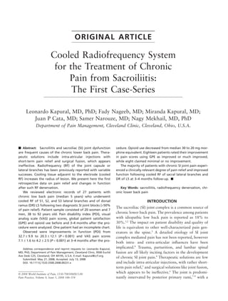
2. Radiofrequency Treatment for Sacroiliitis • 349
Table 1. Presented Here are General Characteristics of 26 Patients Treated with Cooled Radiofrequency of Their
Sacra Lateral Branches and Dorsal Ramus of L5 and Their Data on Pain Scores, Functional Capacity Change (PDI),
Patient Satisfaction, and Opioid Use
VAS (cm) PDI GPE Opioid MSO4 mg
No Age (Years) Sex Years of Pain Base After Base After Pain Function Satisfaction Before After
1 70 M 3 8 2 28 7 3 3 3 90 30
2 59 M 2 8 3 30 16 2 2 2 30 15
3 69 M 5 5 0 23 8 3 3 3 40 0
4 63 M 11 6 0 21 7 3 3 3 20 0
5 50 F 6 8 7 26 26 0 0 0 30 30
6 78 F 3 10 4 19 9 2 2 2 20 20
7 55 F 5 7 5 22 10 2 2 2 20 20
8 55 F 6 5 4 37 32 1 1 1 40 40
9 45 F 10 8 3 32 11 3 3 3 80 80
10 78 F 1 8 7 23 16 0 0 0 10 10
11 77 F 7 10 3 23 9 3 3 3 0 0
12 53 F 1 5 0 23 1 3 3 3 60 20
13 92 F 20 7 5 34 19 2 2 2 30 30
14 38 F 10 7 7 32 10 3 3 3 240 240
15 51 F 3 8 7 30 13 3 2 3 20 20
16 72 F 1 8 7 40 37 -2 -2 -2 0 0
17 44 F 1 10 3 31 17 3 3 3 30 15
18 82 F 2 8 10 29 27 0 0 0 0 0
19 57 M 3 6 2 59 23 3 3 3 250 200
20 56 F 17 8 8 46 46 0 0 0 10 10
21 75 F 6 8 4 32 22 2 2 2 140 165
22 53 F 11 5 2 46 23 3 3 3 120 40
23 58 F 5 5 5 38 38 0 0 0 0 0
24 52 F 2 5 5 32 32 0 0 0 60 60
25 44 F 10 5 5 43 42 2 2 2 40 40
26 55 M 10 6 2 50 28 3 2 2 15 15
Sex: M, male; F, female; VAS, visual analog scale; Base, baseline; PDI, pain disability index; GPE, global percieved effect; MSO4 mg, morphine opioid equivalent in mg.
major contribution from the lateral branches of S1 and applications in pain management, cooled RF electrodes
S2 and from the dorsal ramus (DR) of L5. Radiofre- rely on a closed loop fluid circuit consisting of sterile
quency (RF) denervation of the joint capsule or lateral water and a computerized peristaltic pump to regulate
branches has been performed using conventional RF cooling during RF delivery.
cannula either individually or in a bipolar arrange- In this study, we conducted an initial evaluation of
ment.4,7,10,11 Although preliminary results were largely the Pain Management SInergy System (Baylis Medical
positive, no gold standard for treatment has emerged Inc., Montreal, Canada) to treat chronic SI joint pain.
subsequently. Presented here are data on pain relief, functional capac-
The inherent challenge when treating SI joint pain ity and patient satisfaction from a 27-patient case-series.
with RF energy is the inconsistent location of targeted
lateral branch nerves.7,12 The development of a novel
cooled RF electrode to create larger lesions may provide METHODS
a means of overcoming this difficulty. By treating a After Institutional Review Board approval was
greater volume of the tissue lateral to the S1 through S3 obtained, chart review was conducted of 27 consecutive
sacral foramina, it intuitively follows that there is a patients with chronic SI joint mediated pain who were
greater likelihood of disrupting sacral lateral branches. treated using cooled RF denervation of the SI joint.
The use of electrode cooling to increase lesion size Patients were identified based on the computer-
was previously described for tumor ablation by Gold- generated list from our database of patients who under-
berg et al.13 and Lorentzen et al.15 Cooled electrodes went this procedure. To be included in the study, the
are capable of creating larger lesions compared with patient record had to contain all of the data shown in
noncooled ones because they remove heat from adjacent Table 1. Among the 27 charts reviewed, one had incom-
tissue and allow power delivery to be increased without plete data. A database was generated for the remaining
causing high impedance and tissue charring.14,15 For 26 patients (Table 1).
3. 350 • kapural et al.
Figure 2. Anterior-posterior fluoroscopic view of the sacral
bone. Note a 27G spinal needles placed within S1, S2, and S3
foramina serving as orienteer for the placement of radio-
frequency electrode introducer. Epsilon shaped ruler is used to
limit the area of heating away from the foraminal opening.
Visible is appropriately positioned radiofrequency electrode
lateral and somewhat inferior to the S1 foramina similar to the
position shown in schematic of the Figure 1.
Figure 1. Schematic describing electrode position during cooled
radiofrequency of S1 lateral branches. Epsilon ruler guides
appropriate distance of the electrode from the foramina. Three Because the posterior sacral foramina (PSFA) are diffi-
lesions are suggested at the lateral aspect of S1.
cult to identify with certainty on most patients, three
27-gauge 3.5-inch Quincke needles were placed into the
Patients who received RF denervation had pre- S1, S2, and S3 PSFA under anterior-posterior imaging to
viously failed to achieve adequate improvement with establish internal reference points. There were seven
comprehensive nonoperative treatments including reha- patients where such placement could not be achieved in
bilitation and medical therapies. Diagnosis was based one or two out of three sacral foramina. We did not use
on physical examination (axial pain below the L5 bowel enema/preparation before the procedure to
vertebrae).16–18 Diagnosis was confirmed by >50% improve sacral foramina visualization.
improvement in visual analog scale (VAS) pain scores Beginning at the S1 level, an introducer with stylet
following two fluoroscopically guided SI joint injections was inserted onto the bone endpoint of the posterior
using a 3 cc of local anesthetic bupivacaine 0.5% and sacrum. The distance between the introducer and the
40 mg triamcinolone. aperture of PSFA of S1 was measured using a circular
RF denervations were completed under minimal stainless steel ruler (Epsilon Ruler, Baylis Medical Inc,
sedation, anxiolysis (midazolam 1–2 mg) and infiltra- Montreal, Canada). The final position of the introducer
tion of local anesthetics. Patients communicated with was 8–10 mm from the lateral edge of PSFA (Figures 1
the attending staff physician throughout the procedure. and 2). A depth marker was fixed to the level of the skin
After placing the patient in the prone position with and the stylet removed. When inserted, the stylet
sterile site preparation and drape, the skin was infil- extends 6 mm beyond the tip of introducer. The RF
trated with 1% lidocaine over the desired target entry probe, which was subsequently inserted via same intro-
point. C-arm fluoroscopy was used to visualize the ducer, extends only 4 mm beyond the tip of introducer
sacrum by imaging through the L5/S1 disc space. (Figure 3a,b). Therefore, the final position of the probe
4. Radiofrequency Treatment for Sacroiliitis • 351
(a) (b)
Figure 3. Sequence photographs of the cooled radiofrequency introducers and electrode introduced through the skin within the
sacroiliac area. (a) Three 27G spinal needles and two electrode introducers are shown piercing the skin over the sacral area. Having
two introducers facilitated procedure itself, as they can be re-positioned under different angles to denervate a specific points lateral
to sacral foraminal openings while other electrode is in use. (b) Shown here (middle, black) is a cooled radiofrequency electrode
inserted via one of two introducers placed appropriately in the sacral area. The electrode is connected to the generator via electrical
cable (shown black) and tubing used for the electrode cooling by water pump (shown transparent). Two introducers are needed to
facilitate the procedure eg, while the ablation is conducted using electrode positioned via one of the introducers, other is being
repositioned under fluoroscopy.
tip should be approximately 2 mm from the surface of one or two procedures) 3–4 months after procedures.
the sacrum. Lateral fluoroscopy confirmed that the RF Comparisons were made using unpaired and paired
probe was not within sacral canal and impedance was t-tests and the graphs were generated using Sigma Plot
verified to be in the range of 100–500 ohms. If higher (Systat Software Inc, Chicago, IL) computer program.
impendence was observed, the electrode repositioning
was performed by first re-introducing stylet and slightly RESULTS
adjusting the location of the introducer. Once suitable A summary of absolute and relative VAS pain scores is
electrode location and impedance were achieved, the provided in Table 1. The mean VAS scores of the
heating protocol was initiated, delivering RF energy for patients in this study decreased from 7.1 1 1.6 to
2 minutes and 30 seconds. Target electrode temperature 4.2 1 2.5 (P < 0.001) at 3–4 months after procedures.
was 60°C. Once energy delivery was complete, the RF Functional capacity also improved significantly, with a
probe was removed, stylet replaced and introducer redi- change in PDI scores from 32.7 1 9.9 to 20.3 1 12.1
rected to the next target. Either two or three lesions (P < 0.001). A summary of all baseline and outcome
were created at each sacral level. Typically, these lesions data is presented in Tables 1 and 2.
were spaced about 1 cm apart from one another, creat- A benchmark for successful treatment of SI joint pain
ing a strip of lesioned tissue lateral to each foramina has been reported in the literature as a 50% reduction
(Figure 1). Only one skin entry point was made at each on VAS from baseline.7,10,19 For continuity among pub-
level. Multiple electrode placements could be achieved lished outcomes, we present data using similar metrics
at a given level by changing the angle of introducer from for successful response (Tables 2 and 3). At three to four
the same location at the skin. Those who required bilat- months following treatment, 13 of 26 patients (50%)
eral RF received contralateral RF within 1–2 weeks (10 had achieved the primary outcome of at least 50%
out of 27 patients). reduction in VAS pain scores. Four of the responders
Outcome tools included pain disability index (PDI), (15%) had over 75% pain relief and three patients
VAS pain scores, global perceived effect (GPE) for patient (12%) reported being completely pain free. The mean
satisfaction, and morphine (MSO4) mg equivalent reduction in pain scores (VAS) among responders was
opioids used. Data were collected at baseline (before the 5.2 +/-1.2. 15 patients (58%) experienced at least a
5. 352 • kapural et al.
Table 2. Summarized Outcome Measures for All of the Patients Treated Using Cooled Radiofrequency
All patients Responders Nonresponders
Outcome measure Mean SD Mean SD Mean SD P value
Visual analog scale for pain severity (0–10)
n (patients) 26 — 13 — 13 —
Pretreatment 7.1 1.6 7.3 1.9 6.8 1.3 0.49
3–4 months 4.2 2.6 2.2 1.4 6.3 1.7 0.0000008
Change 2.8 2.6 5.2 1.2 0.5 1.1
Pain Disability Index for physical functioning (0–70)
n (patients) 26 — 13 — 13 —
Pretreatment 32.7 9.9 32.1 12.3 33.2 7.4 0.77
3–4 months 20.3 12.1 13.9 8.2 26.8 12.3 0.004
Change 12.3 9.3 18.2 7.1 6.5 7.6
Opioid use and subsequent post-treatment reduction (mg MSO4 equivalent)
n (patients) 26 — 13 — 13 —
Pretreatment (median) 30 — 40 — 20 — 0.25
3–4 months 20 — 20 — 20 — 0.76
Change 10 — 20 — 0 —
Patient subjective rating (global perceived effect)
n (patients) 26 — 13 — 13 —
Pain 1.8 1.4 2.8 0.44 0.8 1.4
Function 1.7 1.4 2.7 0.48 0.8 1.5
Satisfaction 1.8 1.4 2.7 0.48 0.8 1.4
SD, standard deviation.
Table 3. Demographic, Clinical and Treatment Characteristics of Treated Patients Using Cooled RF Based on
Outcomes
Feature Responders (n = 13) Nonresponders (n = 13) All patients (n = 26)
Age 61 1 12 years 60 1 16 years 61 1 14 years (38–92)
Gender 6 male, 7 female 0 male, 13 female 6 male, 20 female
Years back pain 5.6 1 3.8 6.8 1 6.0 years 6.2 1 5.0 years
Opioid use (median in mg 40 mg 20 mg 30 mg
MSO4 equivalent)
Bilateral RF 6 patients 4 patients 10 patients
RF, radiofrequency; MSO4 mg, morphine opioid equivalent in mg.
2-point drop in VAS, which is considered clinically rel- formed (Table 3). The novel use of internally cooled
evant (Table 2).20,21 electrodes to create lesions along the posterior sacrum
Opioid use varied greatly among the patients, both did not result in any complications, and the procedure
prior to and following procedure (Tables 1 and 2). was generally well tolerated.
However, following treatment there was an observed
decrease from a median value of 30 mg morphine DISCUSSION
equivalent to 20 mg morphine equivalent. Among This case series of 47 procedures (uni- and bilateral)
responders, median opioid use decreased by 50%; from completed on 27 patients is the first to retrospectively
40 to 20 mg MSO4 equivalent (Table 2). observe the effects of novel cooled RF denervation of
Eighteen patients (67%) rated their improvement in sacral lateral branches and L5 DR on chronic SI joint
pain scores using GPE as improved or much improved, pain. At 3–4 month follow-up significant improvements
while eight patients (30%) claimed minimal or no in patient’s pain scores and ability to perform everyday
improvement. Similar ratings were observed for GPE functions were observed (Tables 1 and 3). All of the 13
related to daily activities and whether these patients responders (based on their pain scores) experienced
would recommend the procedure to others (Tables 1 more than 10 points improvement in function by PDI
and 2). We were not able to observe any difference in scores (Tables 1 and 2). Correspondingly, GPE among
procedure success related to patient’s age, years of responders was rated high (Table 2). Among the 13
chronic pain, or whether unilateral or bilateral RF per- nonresponders, only three patients experienced a >50%
6. Radiofrequency Treatment for Sacroiliitis • 353
improvement in physical disability. Average GPE among originating from the SI joint complex.7 Additional effi-
nonresponders was predictably low (Table 2). As cacy study is required to support this conclusion, and to
expected, there is a good correlation between positive further justify the additional cost of the equipment.
outcomes for pain severity on VAS and positive out- However, loss of functional capacity, long-term inactiv-
comes both PDI and GPE, respectively. ity and dissatisfaction with daily chronic pain might be
While many of the patient records demonstrated sig- associated with higher costs to both the individual suf-
nificant reduction in pain severity, there were an equal fering and the society.
number of patients who improved their pain scores 50%
or less. The observed inconsistency among outcomes
may be due to a number of factors including dener- REFERENCES
vation of some but not all of the communicating 1. Schwarzer AC, Aprill CN, Bogduk N. The sacroiliac
nociceptive branches, anterior innervation carrying non- joint in chronic low back pain. Spine. 1995;20:31–37.
ciceptive responses in some patients associated with 2. Maigne JY, Aivaliklis A, Pfefer F. Results of sacro-
underlying pathologies or undiagnosed concurrent pain iliac joint double block and value of sacroiliac pain pro-
generators. vocation tests in 54 patients with low back pain. Spine.
First, criteria of 50% pain relief twice following SI 1996;21:1889–1892.
joint injection used to confirm diagnosis of patients in 3. Irwin RW, Watson T, Minick RP, Ambrosius WT.
this study may contribute to the variability among Age, body mass index, and gender differences in sacroiliac
joint pathology. Am J Phys Med Rehabil. 2007;86:37–44.
patient outcomes. It should be noted that diagnostic
4. Cohen S. Sacroiliac joint pain: a comprehensive
criteria varies significantly among outcome studies on
review of anatomy, diagnosis, and treatment. Anesth Analg.
RF treatment for SI joint pain. This may preclude direct 2005;101:1440–1453.
comparison of outcomes until more rigorous and stand- 5. Hansen HC, McKenzie-Brown AM, Cohen S,
ardized criteria are adopted. Some of the previously Swicegood JR, Colson JD, Manchikanti L. Sacroiliac joint
published studies on techniques for SI joint denervation interventions: a systematic review. Pain Physician.
have used more rigorous selection criteria.6,7 2007;10:165–184.
Second, position of the sacral lateral branches varies 6. Cohen S, Hurley RW. The ability of diagnostic spinal
among individuals, not only in location related to pos- injections to predict surgical outcome. Anesth Analg. 2007;
terior foraminal openings, but also in depth and number 105:1756–1575.
of lateral branches present. Variability even exists 7. Yin W, Willard F, Carreiro J, Dreyfuss P Sensory
between nerve supply of left or right SI joint.7,12 There- stimulation-guided sacroiliac joint radiofrequency neurotomy:
technique based on neuroanatomy of the dorsal sacral plexus.
fore, it is likely that in some patients lateral branches
Spine. 2003;28:2419–2425.
were not effectively treated. Variability in nerve loca-
8. Fortin JD, Kissling RO, O’Connor BL, Vilensky JA.
tion, procedural technique, or lesion characteristics (size Sacroiliac joint innervation and pain. Am J Orthop. 1999;
and shape) may all have contributed to variable 28:68–90.
outcome. Study of thermal lesion characteristics would 9. Grob KR, Neuhuber WL, Kissling RO. Innervation
be useful to establish the most appropriate procedural of the sacroiliac joint of the human. Zeitschr Rheumatol.
technique for this novel technology. 1995;54:117–122.
Finally, as noted in the methods section, placement of 10. Ferrante FM, King LF, Roche EA, et al. Radiofre-
27G reference needles into the posterior foramina could quency sacroiliac joint denervation for sacroiliac syndrome.
not be achieved in all of the patients. Electrode place- Reg Anesth Pain Med. 2001;26: 137–142.
ment relies on visualization of bony landmarks and was 11. Burnham RS, Yasui Y. An alternate method of
generally more lateral in patients where individual radiofrequency neurotomy of the sacroiliac joint: a pilot study
of the effect on pain, function, and satisfaction. Reg Anesth
intraforaminal reference needles could not be placed to
Pain Med. 2007;32:12–19.
ensure minimal dissipation of the heat to the sacral
12. Willard F, Carreiro J, Manko W. The long posterior
nerve roots. However, this study could not detect any interosseous ligament and the sacrococcygeal plexus. Third
differences in outcomes related to the ability of place- interdisciplinary world congress on low back and pelvic pain,
ment of reference needles (data not shown). 1998.
This retrospective, uncontrolled case series suggests 13. Goldberg SN, Gazelle GS, Solbiati L, et al. Radio-
that more extensive sacral lateral branches denervation frequency issue ablation: increased lesion diameter with a
may be a feasible option for treatment of chronic pain perfusion electrode. Acad Radiol. 1996;3: 636–644.
7. 354 • kapural et al.
14. Watanabe I, Masaki R, Min N, et al. Cooled-tip 18. Slipman CW, Sterenfeld EB, Chou LH, Herzog R,
ablation results in increased radiofrequency power delivery Vresilovic E. The predictive value of provocative sacroiliac
and lesion size in the canine heart: importance of catheter-tip joint stress maneuvers in the diagnosis of sacroiliac joint syn-
temperature monitoring for prevention of popping and imped- drome. Arch Phys Med Rehabil. 1998;79:288–292.
ance rise. J Interv Card Electrophysiol. 2002;6:9–16. 19. Cohen SP, Abdi S. Lateral branch blocks as a treat-
15. Lorentzen T. A cooled needle electrode for radiofre- ment for sacroiliac joint pain: a pilot study. Reg Anesth Pain
quency tissue ablation: thermodynamic aspects of improved Med. 2003;28:113–119.
performance compared with conventional needle design. Acad 20. Farrar JT, Young JP, Jr, LaMoreaux L, et al. Clinical
Radiol. 1996;3:556–563. importance of changes in chronic pain intensity measured on
16. Fortin JD, Dwyer AP, West S, Pier J. Sacroiliac joint: an 11-point numerical pain rating scale. Pain. 2001;94:149–
pain referral maps upon applying a new injection/ 158.
arthrography technique. Part I: asymptomatic volunteers. 21. Hagg O, Fritzell P, Nordwall A. The clinical impor-
Spine. 1994;19:1475–1482. tance of changes in outcome scores after treatment for chronic
17. Dreyfuss P, Michaelsen M, Pauza K, McLarty J, low back pain. Eur Spine J. 2003;12:12–20.
Bogduk N. The value of medical history and physical exami-
nation in diagnosing sacroiliac joint pain. Spine. 1996;
21:2594–2602.