Short case...Spinal dural arteriovenous fistula withn congestive myelopathy
•
1 like•353 views

Short case...Spinal dural arteriovenous fistula with congestive myelopathy http://yassermetwally.com http://yassermetwally.net

Recommended
More Related Content
What's hot
What's hot (12)
Viewers also liked
Viewers also liked (20)
Similar to Short case...Spinal dural arteriovenous fistula withn congestive myelopathy
Similar to Short case...Spinal dural arteriovenous fistula withn congestive myelopathy (20)
More from Professor Yasser Metwally
More from Professor Yasser Metwally (20)
Recently uploaded
Recently uploaded (20)
Short case...Spinal dural arteriovenous fistula withn congestive myelopathy
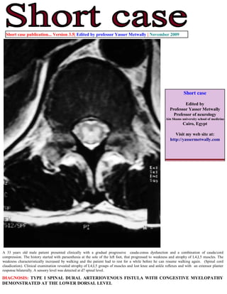
- 1. Short case publication... Version 3.5| Edited by professor Yasser Metwally | November 2009 Short case Edited by Professor Yasser Metwally Professor of neurology Ain Shams university school of medicine Cairo, Egypt Visit my web site at: http://yassermetwally.com A 33 years old male patient presented clinically with a gradual progressive cauda-conus dysfunction and a combination of cauda/cord compression. The history started with paraesthesia at the sole of the left foot, that progressed to weakness and atrophy of L4,L5 muscles. The weakness characteristically increased by walking and the patient had to rest for a while before he can resume walking again. (Spinal cord claudication). Clinical examination revealed atrophy of L4,L5 groups of muscles and lost knee and ankle reflexes and with an extensor planter response bilaterally. A sensory level was detected at d7 spinal level. DIAGNOSIS: TYPE I SPINAL DURAL ARTERIOVENOUS FISTULA WITH CONGESTIVE MYELOPATHY DEMONSTRATED AT THE LOWER DORSAL LEVEL
- 2. Figure 1. Precontrast MRI T1 images (A,B) and postcontrast MRI T1 image (C) at dorsal vertebrae D11,D12 showing mild precontrast T1 central hypointensity (A,B) with central bilateral contrast enhancement (C) Figure 2. MRI T2 images showing cord swelling at D11,D12 vertebrae with central hyperintensity taking the shape of the central grey matter
- 3. Figure 3. Precontrast MRI T1 image (A) and postcontrast MRI T1 images (B,C). Before contrast (A) the spinal cord is enlarged and hypointese. After contrast injestion, dilated enhanced perimedullary veins were observed on the surface of the spinal cord (B,C) Figure 4. MRI T2 imaging showing pencil-shape multisegmental central spinal cord hyperintensity extending from D6 to D12 vertebrae with cording swelling.
- 4. Figure 5. Lower dorsal type I dural spinal arteriovenous malformation SUMMARY The onset in middle age suggests that SDAVF is an acquired condition, in contrast to intradural ventral fistulas or AVMs, which are assumed to be congenital abnormalities An SDAVF is never located within the spinal parenchyma, in contrast to AVMs. Patients with SDAVF very rarely present with spinal haemorrhage in contrast to patients with AVMs. Associated vascular lesions are seen in AVMs, not in SDAVF. Intradural AVMs occur much more often in the cervical region than SDAVF (Rosenblum et al., 1987). The increased pressure causes the venous system to ‘arterialize’, that is, the walls of intramedullary veins become thickened and also tortuous. The radicular feeding artery is often a dural branch and in a minority, the medullary artery. The shunt results in venous hypertension in the spinal cord, because the intramedullary veins and the radicular vein share a common venous outflow. The reduced arteriovenous pressure gradient (with the resultant of venous hypertension) results initially in congestive myelopathy (cord edema, swelling and cord petechial hemorrhages) that is responsible for the spinal cord swelling and central T2 hyperintensity, T1 central hypointensity. The central T2 hyperintensity took the shape of the central grey matter denoting the cord edema is primarily located in the central grey matter in congestive myelopathy due to spinal arteriovenous fistula probably because the central grey matter has the highest blood supply and the highest venous return. On long term basis (and with persistence of venous hypertension) a decrease in tissue perfusion might occur and might eventually result in venous infarction and spinal cord atrophy. References 1. Metwally, MYM: Textbook of neurimaging, A CD-ROM publication, (Metwally, MYM editor) WEB-CD agency for electronic publishing, version 10.4a October 2009 Addendum A new version of short case is uploaded in my web site every week (every Saturday and remains available till Friday.) To download the current version follow the link "http://pdf.yassermetwally.com/short.pdf". You can download the long case version of this short case during the same week from: http://pdf.yassermetwally.com/case.pdf or visit web site: http://pdf.yassermetwally.com To download the software version of the publication (crow.exe) follow the link: http://neurology.yassermetwally.com/crow.zip At the end of each year, all the publications are compiled on a single CD-ROM, please contact the author to know more details. Screen resolution is better set at 1024*768 pixel screen area for optimum display For an archive of the previously reported cases go to www.yassermetwally.net, then under pages in the right panel, scroll down and click on the text entry "downloadable short cases in PDF format" Also to view a list of the previously published case records follow the following link (http://wordpress.com/tag/case- record/) or click on it if it appears as a link in your PDF reader