Case record...Focal midbrain glioma
•
1 like•923 views

Case record...Focal midbrain glioma http://yassermetwally.com http://yassermetwally.net

Recommended
Recommended
More Related Content
Viewers also liked
Viewers also liked (16)
More from Professor Yasser Metwally
More from Professor Yasser Metwally (20)
Recently uploaded
Recently uploaded (20)
Case record...Focal midbrain glioma
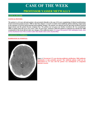
- 1. CASE OF THE WEEK PROFESSOR YASSER METWALLY CLINICAL PICTURE CLINICAL PICTURE: The patient is a 44 years old male patient, who presented clinically at the age of 10 years complaining of clinical manifestations of increased intracranial pressure. MRI at that time (Figures 1,2,3,4,5) revealed a focal midbrain glioma inducing compression of the aqueduct of Sylvius and producing hydrocephalic changes. The patient was shunted and the operation produced marked improvement and the patient became symptom free. The patient was not given any further treatment. He was examined by MRI at regular intervals (every two years). After 34 years (now...February 2010) the patient is symptom free and the last MRI examination of the brain did not show any changes of the midbrain tumor. (To inspect the patient's full radiological study, click on the attachment icon of the acrobat reader then double click on the attached file) RADIOLOGICAL FINDINGS RADIOLOGICAL FINDINGS: Figure 1. Precontrast CT scan showing midbrain calcification. Mild midbrain hypodensity is also probably present. The midbrain glioma could not be appreciated by CT scan and the patient was misdiagnosis as congenital aqueduct stenosis
- 2. Figure 2. Pre (A) and postcontrast (B) MRI T1 images showing enlargement of the midbrain, involving mainly the posterior part with a large irregular hypointensity on the precontrast image (A) involving the middle line area and extending from the interpeduncular area anteriorly to the periaqueductal area posteriorly, the tumor is apparently involving the medial parts of the midbrain. The aqueduct of Sylvius is compressed, pushed posteriorly. Dense patchy enhancement is observed on postcontrast image (B) and involved the linear hypointense zone observed on the precontrast image. The tumor is apparently sparing the crus cerebri and the lateral parts of the midbrain and its main bulk is located in the tectal plate posteriorly. Figure 3. Pre (A) and postcontrast (B) MRI images showing enlargement of the midbrain, involving mainly the posterior part. The aqueduct of Sylvius is compressed, pushed posteriorly and elongated (C). Dense patchy enhancement is observed on postcontrast image (B). Moderate hydrocephalic changes are also observed.
- 3. Figure 4. MRI T2 image (A) and FLAIR image (B). The tumor is hyperintense on the MRI T2, FLAIR images and involves the medial parts of the midbrain with selective sparing of the crus cerebri bilaterally and the lateral zones of the midbrain. The mainly part of the tumor is located posteriorly in the tectal plate and the tumor extends from the interpeduncular area anteriorly to the periaqueductal area posteriorly, Figure 5. The midbrain glioma DIAGNOSIS: DIAGNOSIS: DIFFUSE BRAIN STEM GLIOMA DISCUSSION DISCUSSION: Brainstem glioma, pilocytic astrocytoma and medulloblastoma are the most frequent infratentorial tumours in patients under 18 years4 representing 10-30% of brain tumours in children. They are usually infiltrative lesions and only a small number (dorsal exophytic) have a favourable prognosis. They occur mostly in childhood and adolescence (77% in ages below 20 years), representing 1% dos tumours in adults [5].
- 4. Midbrain tumours are a heterogeneous group of neoplasms with variable clinical and radiological features, relating with the location and tumour histology [6]. They occur in the tectal plate, tegmentum, invading the pons or cerebral aqueduct. Sometimes they represent midbrain invasion by tumours of adjacent regions, namely pineal and thalami6. Tectal gliomas are in majority low-grade astrocytomas, considered a "benign" sub-group of brain stem gliomas. They represent approximately 10% of brain stem gliomas in children7 and 6% of paediatric brain tumours surgically treated. Computhorized tomography (CT) reveals the hydrocephalous but may not be able to detect tectal plate tumours in up to 50% of patients. Calcifications are seen in 9-25% of cases [8]. Magnetic resonance imaging (MRI) is the chosen exam for diagnosis and follow up of tumours in this location. It allows a precise evaluation of the growth pattern and correct pre-operative diagnosis in most of cases6. Gadolinium enhancement, calcifications, cysts e exophytic nature are observed in both low and high-grade gliomas [8]. They are typically isointense in T1WI and iso or hiperintense in T2WI. Enhancement after endovenous contrast enclosures an undefined pathological significance. In the case of intrinsic tectal tumours, low-grade astrocytoma is the probable diagnosis. Differential diagnosis of exophytic tectal tumours includes pineal neoplasms, requiring histological verification [8]. Since biopsy is not performed upon many lesions, a precise statistic analysis is not possible. Midbrain gliomas are mainly astrocytomas (pilocytic astrocytoma, WHO II difuse astrocytoma, anaplastic astrocytoma, high- grade astrocytoma) but other lesions have been identified (oligodendroglioma e oligoastrocytoma, WHO II ependymoma, ganglioglioma, medulloblastoma, primitive neuroectodermal tumours, disembryoblastic neuroepithelial tumours, metastasis, melanoma, lipoma, cavernoma, abcess and periaqueductal gliosis). Many neurosurgeons perform a stereotactic biopsy to obtain histopathological confirmation of a low-grade tumour and only then the treatment is planned2. Clinical presentation with signs of raised intracranial pressure due to cerebral aqueduct compression resulting in supratentorial hydrocephalous is the most common clinical feature affecting all patients in some series [9]. Focal neurological findings are less frequent (as diplopia, visual field defects, nystagmus, Parinaud syndrome, seizures) and usually revert after correction of the hydrocephalous. It is not universally accepted that lesions with radiographic progression need to be treated. Paediatric tectal plate gliomas are usually low-grade tumours that can be managed conservatively even in the presence of radiographic enlargement, reserving radiotherapy and chemotherapy for clinical progression [3] which is described in 15-25% of cases. It is even more advantageous to observe these patients in order to avoid radiation therapy and chemotherapy induced neurodevelopmental and endocrinal injury to the developing brain [7]. The initial treatment is directed to correction of hydrocephalous. Ventricular-peritoneal shunt placement has good long-term results, if no dysfunction is verified. Third endoscopic ventriculostomy suppresses the need for shunt placement and a biopsy can be performed through an enlarged foramen of Monro. It allows resolution of signs and symptoms and the return of the ventricular system to its normal size. It is the procedure of choice for paediatric patients. Endoscopic aqueductoplasty with flexible systems (stent based or not) may be an option for some cases, but its long-term results are unknown. Due to its indolent course, open surgery is not usually indicated for low grade tumours. However, if a malignant, secondary or vascular lesion is clinically and radiographically suspected a microsurgical procedure should be performed. Simple stereotactic aspiration of cystic brain stem gliomas is not an effective treatment strategy, because they will frequently recur leading to progressive neurological deficit. When combined with stereotactical placement of intra-cyst catheters, intracavitary irradiation with radioactive solutions, external radiotherapy and chemotherapy, it may allow cyst control without permanent morbidity or mortality [10]. Resection or open biopsy of tumours in this location can be achieved by a supracerebellar-infratentorial or suboccipital- transtentorial approach, with the extent of removal being wider at the level of the superior colliculi and limited at the inferior colliculi due to high auditory risk [9]. Parinaud syndrome is one of the most frequent surgical complications. Auditory hallucinations and acoustic neglect syndrome can also occur. Despite of that, tectal plate region is a safer surgical field than the ventral midbrain. Early and middle-latency brain stem auditory evoked potentials should be used for functional brainstem evaluation [11] and definition of resection margins during tectal plate surgical procedures. Stereotactic radiosurgery can be employed on tumour progression but due to radiation side effects the dosage is limited. Optimal treatment of tectal plate gliomas is still to be determined. The role of different treatment modalities is unclear and universally accepted guidelines are still to be proposed. Serial neurological / clinical observations and MRI scans each 6-12 months is an option. Patients with well-differentiated brainstem gliomas may be cured by microsurgical resection [12]. Like in all high grade gliomas, resection of the tumour instead of biopsy, age equal or less than 60 years and a Karnofsky scale of 70 or greater are all correlated with better outcome.
- 5. Neuroanatomy based craniotomy for tumour resection is the mainstream of treatment currently available if it can be done safely, without further neurological deficits. However, surgical resection alone does not cure malignant brain tumours unless it is coupled with other treatment modalities addressed to the diffuse nature of these lesions, like chemotherapy and/or immunotherapy. In high-grade gliomas, partial resection may prolong survival and facilitate subsequent complementary therapeutics SUMMARY SUMMARY Adult brainstem gliomas are different from the childhood subtypes. Overall, brainstem gliomas are less aggressive in adults than in children. However, survival merely reflects the course of the most frequent subtype of tumours. Tectal gliomas fall under the grouping of childhood brainstem gliomas. They are typically low grade astrocytomas which expand from the tectal plate. Their expansion within the brainstem causes narrowing the aqueduct of Sylvius and causing obstructive hydrocephalus. They are slow growing and shunting is often the only required intervention for long term survival. Diffuse intrinsic low-grade brainstem glioma Interestingly, the most frequent type of of brainstem glioma in adults (representing 46% of the patients in this series) resembles the childhood diffuse gliomas of the pons in terms of clinical and radiological presentation but is radically different in course and survival. In both adults and children, the clinical picture is of a combination of cranial nerve and long tract signs. However, while the onset is rapid in children, the duration of symptoms is often long in adults. [13,14] In both children and adults, MRI at presentation reveals a diffuse infiltration of the pons, often increasing the size of the brainstem considerably. There is high signal on T2-weighted and low signal on T1-weighted images, which usually do not show contrast enhancement (100% in adults at diagnosis). It is worth noting that preferential location in the pons is less striking in adults than in children. When a biopsy is performed, which is far from routine practice in these diffuse intrinsic forms, a malignant glioma (grades III– IV) is found in many children, whereas a less aggressive histology is found the adults. [13,14] Malignant brainstem gliomas The other common tumour type identified in adult is clearly different from those discussed above. It occurs later than the diffuse, intrinsic, low-grade type and affects mainly older adults (most of them in their sixth decade). The clinical picture is characterized by the rapid onset of cranial nerve palsies and long tract signs leading to an early alteration in performance status. MRI reveals a brainstem mass that enhances after gadolinium infusion, often in a ring-like fashion. Contrast enhancement is a pejorative factor (particularly when the area of enhancement surrounded a low-signal area suggestive of necrosis) in contrast with children, in whom the prognostic value of contrast enhancement remains controversial. Pathologically, these tumours correspond to high-grade gliomas (grades III–IV) and median survival time is short (11.2 months) despite treatment with radiotherapy and chemotherapy. Thus, the clinical–radiological pattern, pathology and course closely resemble the common malignant supratentorial gliomas in adults and we suggest that this group be designated `malignant brainstem gliomas'. [13,14] Focal tectal gliomas Focal tectal gliomas represent the third type of adult brainstem glioma and constitute a small subgroup (8%) that also exists in children. The clinical picture is dominated by hydrocephalus. [13,14] Other types Other types of brainstem glioma can be observed in adults such as exophytic contrast-enhancing glioma arising from the floor of the fourth ventricle; this entity, which is associated with a good prognosis, is also well described in children (representing up to 10% of brainstem gliomas). A likely explanation for this discrepancy between the two age-groups is that most of the exophytic gliomas correspond to pilocytic astrocytoma, a very rare type of tumour in adults. [13,14] The brainstem is the second most frequent location of brain tumours after the optic pathways in patients with NF1. In contrast with children, in whom the course is usually very long, the tumour behaviour in adults with NF1 was much more aggressive, but larger series will be necessary to draw any conclusion on this point. [13,14]
- 6. Complications Except for locoregional progression, two main complications are observed during the course of adult brainstem gliomas, namely hydrocephalus and leptomeningeal dissemination. Hydrocephalus is observed in 20% of cases. Whereas some pontine tumours may have an important mass effect on the fourth ventricle, hydrocephalus is always associated with mesencephalic involvement and blockage of the CSF at the level of the sylvian aqueduct. Leptomeningeal dissemination occurred in 13% of cases and is the cause of a quarter of the deaths. This complication has also been reported with a high frequency in children. Close proximity of the tumour and CSF pathways could explain such an increased trend for leptomeningeal dissemination, but this remains to be demonstrated. [13,14] The role of biopsy Finally, this classification may help in the selection of patients for biopsy. In children, MRI has become the reference for the diagnosis of brainstem glioma and is used for the current classification of these tumours. MRI has replaced biopsy in the diagnosis of paediatric diffuse brainstem gliomas, for which most authors agree that anticancer treatments can be administered without pathological confirmation if the clinical course is rapid. However, we believe that biopsy is not useful in the diagnosis of intrinsic, diffuse, low-grade brainstem gliomas in adults when the clinical and radiological criteria described above are met. The issue is different in contrast-enhancing lesions because several reports have underlined the limits of MRI in differentiating tumours from infectious (e.g. tuberculomas) and inflammatory (sarcoidosis, Behciet's disease). [13,14] Addendum A new version of this PDF file (with a new case) is uploaded in my web site every week (every Saturday and remains available till Friday.) To download the current version follow the link "http://pdf.yassermetwally.com/case.pdf". You can also download the current version from my web site at "http://yassermetwally.com". To download the software version of the publication (crow.exe) follow the link: http://neurology.yassermetwally.com/crow.zip The case is also presented as a short case in PDF format, to download the short case follow the link: http://pdf.yassermetwally.com/short.pdf At the end of each year, all the publications are compiled on a single CD-ROM, please contact the author to know more details. Screen resolution is better set at 1024*768 pixel screen area for optimum display. Also to view a list of the previously published case records follow the following link (http://wordpress.com/tag/case- record/) or click on it if it appears as a link in your PDF reader To inspect the patient's full radiological study, click on the attachment icon of the acrobat reader then double click on the attached file. REFERENCES References 1. Lázaro BC, Landeiro JA. Tectal plate tumours. Arq Neuropsiquiatr 2006; 64:432-436. 2. Selvapaudian S, Rajshekhar V, Chandy MJ. Brainstem glioma: comparative study of clinico-radiological presentation, pathology and outcome in children and adults. Acta Neurochir (Wien) 1999;141:721-726; discussion 726-727. 3. Daniel CB, Christos G, Leslie JA, et al. Tectal gliomas: natural history of an indolent lesion in pediatric patients. Pediatr Neurosurg 2000;32:24-29. 4. Section of Pediatric Neurosurgery of the American Association of Neurological Surgeons (ed.). Pediatric neurosurgery. New York: Greene and Stratton, 1982. 5. Packer RJ, Nicholson HS, Vezina LG, et al. Brain stem gliomas. Neurosurg Clin N Am 1992;3:863-879. 6. Sun B, Wang CC, Wang J. MRI characteristics of midbrain tumours. Rev Neurol 1996;24:73-76. 7. Bowers DC, Georgiadis C, Burger PC, Melhem E, Cohen KJ. Tectal gliomas: radiographic progression does not mandate clinical intervention. Meeting abstract – 1999 ASCO Annual Meeting 8. Bognar L, Turjman F, Villanyi E, et al. Tectal plate gliomas. Part II: CT scans and MR imaging of tectal gliomas. Acta Neurochir 1994;127:48-54. 9. Lapras C, Bognar L, Turjman F, et al. Tectal plate gliomas. Part II: CT scans and MR imaging of tectal gliomas. Acta
- 7. Neurochir 1994;126:76-83. 10. Hood TW, McKeever PE. Stereotactic management of cystic gliomas of the brain stem. Neurosurgery 1989;24:373-378. 11. Fischer C, Bognar L, Turjman F, Villanyi E, Lapras C. Auditory early and middle-latency evoked potentials in patients with quadrigeminal plate tumours. Neurosurgery 1994;35:45-51. 12. Wang CC, Zhang JT, Liu AL. Surgical management of brain stem gliomas: a retrospective analysis of 311 cases. Zhongguo Yi Xue Ke Xue Yan Xue Bao 2005;27:7-12. 13. Metwally, MYM: Textbook of neuroimaging, A CD-ROM publication, (Metwally, MYM editor) WEB-CD agency for electronic publication, version 11.1a. January 2010 14. Metwally, MYM (2001): Brain stem glioma, A clinico-radiological study: A classification system with prognostic significance is suggested. Ain Shams medical journal, VOL. 51, NO. 10,11,12, pp 1085-1115 15. Metwally, MYM (2001): Brain stem glioma, A clinico-radiological study: A classification system with prognostic significance is suggested. Ain Shams medical journal, VOL. 51, NO. 10,11,12, pp 1085-1115 [Click to download in PDF format] 16. Case of the week...Brain stem glioma. [Click to download in PDF format]