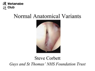
SlideShare a Scribd company logo
1 of 36
Normal Anatomical Variants


             Steve Corbett
Guys and St Thomas’ NHS Foundation Trust
Normal Anatomical Variants

•   Superior labrum / Biceps   •   Biceps
•   Anterosuperior Quadrant    •   AS head
•   AI Quadrant                •   Superior Cuff / Head
•   PI Quadrant                •   Posterior Cuff / Head
•   PS Quadrant                •   GHJ Surfaces
                               •   Bursa
Normal Anatomical Variants: Superior labrum/Biceps


                             • 15% loosely attached
                               meniscal type labrum

                             • 1-5mm width
Normal Anatomical Variants: Superior labrum/Biceps
Superior labrum/Biceps: SLAP
Normal Anatomical Variants: Superior labrum/Biceps


• Anatomy

    –   Type I 22%
    –   Type II 33%
    –   Type III 37%                     I           II
    –   Type IV 8%


                                         III              IV
Normal Anatomical Variants: Superior labrum/Biceps


• Vincula Biceps             • Bifid Biceps

   – Small strands of            – 1 part attached to cable
     mesentry                    – 2nd part attached to
   – Pass from biceps to           tubercle
     surrounding capsule
                             • Complete absence
Normal Anatomical Variants: Superior labrum


                             • 80% firmly attached

                             • 14-60% sublabral
                               foramen (Detrisac and
                               Johnson 20%
                               anatomic dissections)

                             • 6% Burford Complex
Normal Anatomical Variants: Superior labrum


                             • 6% Burford Complex

                                – Cord like MGHL

                                – No labral tissue ant/sup
                                  glenoid

                                – Surfaces smooth
Normal Anatomical Variants: Superior labrum


                             • 6% Burford Complex

                                – Cord like MGHL

                                – No labral tissue ant/sup
                                  glenoid

                                – Surfaces smooth
Normal Anatomical Variants: Superior labrum


                             • 6% Burford Complex

                                – Cord like MGHL

                                – No labral tissue ant/sup
                                  glenoid

                                – Surfaces smooth
Normal Anatomical Variants: Superior labrum


                             • Divides sup. 2/5 and inf.
                               3/5.
                             • Variable in depth
Normal Anatomical Variants: Subscapularis / SGHL


                             • Leading edge may be
                               split or bifid
                             • 3%

                             • SGHL present in
                               nearly 100%,
                               Occassionally frayed
Normal Anatomical Variants: MGHL


                           • Most variable of all
                             ligaments

                              – Variable origin

                              – 70% folded thickening
                                crossing subscapularis at
                                45º

                              – 20% cord like

                              – 10% thin veil or absent
Normal Anatomical Variants: Anterior Inferior Labrum


                              • 95% smooth
                                attachment

                              • 5% meniscoid
                                 – Probe can be inserted
                                   but labrum not
                                   detached
Normal Anatomical Variants: IGHL


                            • aIGHL

                               – Variable attachment to
                                 labrum

                               – Distinct superior band not
                                 always present (Defined by
                                 Turkel et al)

                               – May hypertrophy when
                                 MGHL absent
Normal Anatomical Variants: Inferior capsular recess


                               • Normally smooth

                               • Delicate synovial covering

                               • Small fenestrations

                               • Post. Sup. Band pIGHL
                                 not always well visualised
                                 (Schwartz et al)
Normal Anatomical Variants: Bare area


                              • Bare area
                                 – 2-3 mm
                                 – 2-3 cm
                                 – Frequent indentations,
                                   deep holes
                                 – Size varies with age
                                   (De Palma)
Normal Anatomical Variants: Bare area


                              • Must distinguish from
                                Hill Sachs
Normal Anatomical Variants: Superior cuff


                              • Layer of capsule and
                                synovium

                              • Rotator cable
Normal Anatomical Variants: Posterosuperior cuff


                              • May have
                                fenestrations in
                                superficial layers
Normal Anatomical Variants: Posterior labrum / Capsule


                              • 95% firmly attached

                              • 5% meniscoid, firmly
                                attached at periphery
Normal Anatomical Variants: Posterior labrum / Capsule


                              • Normal to have a deep
                                cleft in capsule
                                posterior to labrum
Thank you

More Related Content

Viewers also liked

Normal radiographic variants of immature skeleton
Normal radiographic variants of immature skeletonNormal radiographic variants of immature skeleton
Normal radiographic variants of immature skeletonRajeev Ks
 
K-oral.m-Normal anatomical-variants
K-oral.m-Normal anatomical-variantsK-oral.m-Normal anatomical-variants
K-oral.m-Normal anatomical-variantsYahya Almoussawy
 
Mayer rokitansky-küster-hauser syndrome
Mayer rokitansky-küster-hauser syndromeMayer rokitansky-küster-hauser syndrome
Mayer rokitansky-küster-hauser syndromeMATIAS FREITAS FH
 
Sistema musculoesqueletico
Sistema musculoesqueleticoSistema musculoesqueletico
Sistema musculoesqueleticoJonathan Trejo
 
Oral Cancer Screening
Oral Cancer Screening Oral Cancer Screening
Oral Cancer Screening Ghasaq Bakkal
 
Spinal trauma IMAGING
Spinal trauma  IMAGINGSpinal trauma  IMAGING
Spinal trauma IMAGINGSanal Kumar
 
Oral cancer screening
Oral cancer screeningOral cancer screening
Oral cancer screeningmrboy
 
Internal Carotid Artery and Normal Variants
Internal Carotid Artery and Normal VariantsInternal Carotid Artery and Normal Variants
Internal Carotid Artery and Normal VariantsMATIAS FREITAS FH
 
Radiological anatomy of kidney, ureter & bladder
Radiological anatomy of kidney, ureter & bladderRadiological anatomy of kidney, ureter & bladder
Radiological anatomy of kidney, ureter & bladderrajss007
 
Deep bite malocclusions-M. ABOULNASER- Orthodontist, BUA, USA. O. SANDID- Ort...
Deep bite malocclusions-M. ABOULNASER- Orthodontist, BUA, USA. O. SANDID- Ort...Deep bite malocclusions-M. ABOULNASER- Orthodontist, BUA, USA. O. SANDID- Ort...
Deep bite malocclusions-M. ABOULNASER- Orthodontist, BUA, USA. O. SANDID- Ort...Dr Mohamad ABOUALNASER -Orthodontist
 
Introduction oral medicine-primary and secondary lesions
Introduction oral medicine-primary and secondary lesionsIntroduction oral medicine-primary and secondary lesions
Introduction oral medicine-primary and secondary lesionsManali Rajvansh
 

Viewers also liked (20)

Pejkov H - AIMRADIAL 2014 - Anatomical variations
Pejkov H - AIMRADIAL 2014 - Anatomical variationsPejkov H - AIMRADIAL 2014 - Anatomical variations
Pejkov H - AIMRADIAL 2014 - Anatomical variations
 
Schussler JM and Rao SV 2014
Schussler JM and Rao SV 2014Schussler JM and Rao SV 2014
Schussler JM and Rao SV 2014
 
Gilchrist IC 201110
Gilchrist IC 201110Gilchrist IC 201110
Gilchrist IC 201110
 
Normal anatomy final
Normal anatomy finalNormal anatomy final
Normal anatomy final
 
Normal radiographic variants of immature skeleton
Normal radiographic variants of immature skeletonNormal radiographic variants of immature skeleton
Normal radiographic variants of immature skeleton
 
K-oral.m-Normal anatomical-variants
K-oral.m-Normal anatomical-variantsK-oral.m-Normal anatomical-variants
K-oral.m-Normal anatomical-variants
 
Tenotomy v tenodesis
Tenotomy v tenodesisTenotomy v tenodesis
Tenotomy v tenodesis
 
Tenotomy 2013 final
Tenotomy 2013 finalTenotomy 2013 final
Tenotomy 2013 final
 
Final, normal anatomy 2013
Final, normal anatomy 2013Final, normal anatomy 2013
Final, normal anatomy 2013
 
Gilchrist IC - Anatomy of radial and brachial arteries
Gilchrist IC - Anatomy of radial and brachial arteriesGilchrist IC - Anatomy of radial and brachial arteries
Gilchrist IC - Anatomy of radial and brachial arteries
 
Mayer rokitansky-küster-hauser syndrome
Mayer rokitansky-küster-hauser syndromeMayer rokitansky-küster-hauser syndrome
Mayer rokitansky-küster-hauser syndrome
 
Sistema musculoesqueletico
Sistema musculoesqueleticoSistema musculoesqueletico
Sistema musculoesqueletico
 
Oral Cancer Screening
Oral Cancer Screening Oral Cancer Screening
Oral Cancer Screening
 
Spinal trauma IMAGING
Spinal trauma  IMAGINGSpinal trauma  IMAGING
Spinal trauma IMAGING
 
Oral cancer screening
Oral cancer screeningOral cancer screening
Oral cancer screening
 
Internal Carotid Artery and Normal Variants
Internal Carotid Artery and Normal VariantsInternal Carotid Artery and Normal Variants
Internal Carotid Artery and Normal Variants
 
Radiological anatomy of kidney, ureter & bladder
Radiological anatomy of kidney, ureter & bladderRadiological anatomy of kidney, ureter & bladder
Radiological anatomy of kidney, ureter & bladder
 
Pdf- open-bite-malocclusion
Pdf- open-bite-malocclusionPdf- open-bite-malocclusion
Pdf- open-bite-malocclusion
 
Deep bite malocclusions-M. ABOULNASER- Orthodontist, BUA, USA. O. SANDID- Ort...
Deep bite malocclusions-M. ABOULNASER- Orthodontist, BUA, USA. O. SANDID- Ort...Deep bite malocclusions-M. ABOULNASER- Orthodontist, BUA, USA. O. SANDID- Ort...
Deep bite malocclusions-M. ABOULNASER- Orthodontist, BUA, USA. O. SANDID- Ort...
 
Introduction oral medicine-primary and secondary lesions
Introduction oral medicine-primary and secondary lesionsIntroduction oral medicine-primary and secondary lesions
Introduction oral medicine-primary and secondary lesions
 

Normal anatomical variants