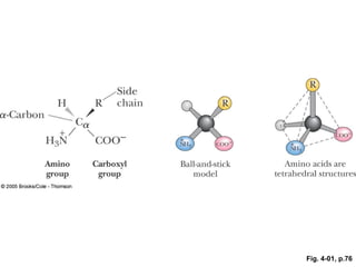
ANIMATED FIGURE 4.1 Anatomy of an amino acid. Except for proline and its derivatives, all of the amino acids commonly found in proteins possess this type of structure. See this figure animated at http://chemistry.brookscole.com/ggb3
ANIMATED FIGURE 4.2 The -COOH and -NH3 + groups of two amino acids can react with the resulting loss of a water molecule to form a covalent amide bond. (Illustration: Irving Geis. Rights owned by Howard Hughes Medical Institute. Not to be reproduced without permission.) See this figure animated at http://chemistry.brookscole.com/ggb3
ANIMATED FIGURE 4.2 The _-COOH and _-NH3 _ groups of two amino acids can react with the resulting loss of a water molecule to form a covalent amide bond. (Illustration: Irving Geis. Rights owned by Howard Hughes Medical Institute. Not to be reproduced without permission.) See this figure animated at http://chemistry.brookscole.com/ggb3
ANIMATED FIGURE 4.2 The _-COOH and _-NH3 _ groups of two amino acids can react with the resulting loss of a water molecule to form a covalent amide bond. (Illustration: Irving Geis. Rights owned by Howard Hughes Medical Institute. Not to be reproduced without permission.) See this figure animated at http://chemistry.brookscole.com/ggb3
FIGURE 4.3 The 20 amino acids that are the building blocks of most proteins can be classified as (a) nonpolar (hydrophobic); (b) polar, neutral; (c) acidic; or (d) basic. (Illustration: Irving Geis. Rights owned by Howard Hughes Medical Institute. Not to be produced without permission.)
FIGURE 4.3 The 20 amino acids that are the building blocks of most proteins can be classified as (a) nonpolar (hydrophobic); (b) polar, neutral; (c) acidic; or (d) basic. (Illustration: Irving Geis. Rights owned by Howard Hughes Medical Institute. Not to be produced without permission.)
(a) nonpolar (hydrophobic)
(a) nonpolar (hydrophobic)
(b) polar, neutral
(b) polar, neutral
(c) acidic
(d) basic
FIGURE 4.4 The structures of several amino acids that are less common but nevertheless found in certain proteins. Hydroxylysine and hydroxyproline are found in connective-tissue proteins, pyroglutamic acid is found in bacteriorhodopsin (a protein in Halobacterium halobium), and aminoadipic acid is found in proteins isolated from corn.
FIGURE 4.4 The structures of several amino acids that are less common but nevertheless found in certain proteins. Hydroxylysine and hydroxyproline are found in connective-tissue proteins, pyroglutamic acid is found in bacteriorhodopsin (a protein in Halobacterium halobium), and aminoadipic acid is found in proteins isolated from corn.
FIGURE 4.4 The structures of several amino acids that are less common but nevertheless found in certain proteins. Hydroxylysine and hydroxyproline are found in connective-tissue proteins, pyroglutamic acid is found in bacteriorhodopsin (a protein in Halobacterium halobium), and aminoadipic acid is found in proteins isolated from corn.
FIGURE 4.4 The structures of several amino acids that are less common but nevertheless found in certain proteins. Hydroxylysine and hydroxyproline are found in connective-tissue proteins, pyroglutamic acid is found in bacteriorhodopsin (a protein in Halobacterium halobium), and aminoadipic acid is found in proteins isolated from corn.
ANIMATED FIGURE 4.6 The ionic forms of the amino acids, shown without consideration of any ionizations on the side chain. The cationic form is the low pH form, and the titration of the cationic species with base yields the zwitterion and finally the anionic form. (Illustration: Irving Geis. Rights owned by Howard Hughes Medical Institute. Not to be reproduced without permission.) See this figure animated at http://chemistry.brookscole.com/ggb3
FIGURE 4.7 Titration of glycine, a simple amino acid. The isoelectric point, pI, the pH where glycine has a net charge of 0, can be calculated as (pK1 _ pK2)/2.
ACTIVE FIGURE 4.8 Titrations of glutamic acid and lysine. Test yourself on the concepts in this figure at http://chemistry.brookscole .com/ggb3
Titrations of glutamic acid and lysine.
Titrations of glutamic acid and lysine.
ACTIVE FIGURE 4.9 Typical reactions of the common amino acids (see text for details). Test yourself on the concepts in this figure at http://chemistry.brookscole. com/ggb3
Typical reactions of the common amino acids.
Typical reactions of the common amino acids.
Typical reactions of the common amino acids.
Typical reactions of the common amino acids.
Typical reactions of the common amino acids.
ANIMATED FIGURE 4.12 Enantiomeric molecules based on a chiral carbon atom. Enantiomers are nonsuperimposable mirror images of each other. See this figure animated at http://chemistry.brookscole .com/ggb3
ANIMATED FIGURE 4.13 The configuration of the common L-amino acids can be related to the configuration of L(_)-glyceraldehyde as shown. These drawings are known as Fischer projections. The horizontal lines of the Fischer projections are meant to indicate bonds coming out of the page from the central carbon, and vertical lines represent bonds extending behind the page from the central carbon atom. See this figure animated at http://chemistry. brookscole.com/ggb3
ANIMATED FIGURE 4.14 The stereoisomers of isoleucine and threonine. The structures at the far left are the naturally occurring isomers. See this figure animated at http://chemistry. brookscole.com/ggb3
The stereoisomers of isoleucine.
The stereoisomers of threonine.
FIGURE 4.15 The ultraviolet absorption spectra of the aromatic amino acids at pH 6. (From Wetlaufer, D. B., 1962. Ultraviolet spectra of proteins and amino acids. Advances in Protein Chemistry 17:303–390.