Call Girl Bangalore Nandini 7001305949 Independent Escort Service Bangalore
32 breast
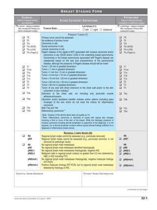
1. BREAST STAGING FORM
C L I NI C AL P AT HOL OG IC
Extent of disease before STAGE CATEGORY DEFINITIONS Extent of disease through
any treatment completion of definitive surgery
y clinical – staging completed L ATERALITY: y pathologic – staging completed
after neoadjuvant therapy but T UMOR S IZE : after neoadjuvant therapy AND
before subsequent surgery
left right bilateral subsequent surgery
PRIMARY TUMOR (T)
TX Primary tumor cannot be assessed TX
T0 No evidence of primary tumor T0
Tis Carcinoma in situ Tis
Tis (DCIS) Ductal carcinoma in situ Tis (DCIS)
Tis (LCIS) Lobular carcinoma in situ Tis (LCIS)
Tis (Paget’s) Paget’s disease of the nipple is NOT associated with invasive carcinoma and/or Tis (Paget’s)
carcinoma in situ (DCIS and/or LCIS) in the underlying breast parenchyma.
Carcinomas in the breast parenchyma associated with Paget's disease are
categorized based on the size and characteristics of the parenchymal
disease, although the presence of Paget's disease should still be noted
T1 Tumor £ 20 mm in greatest dimension T1
T1mi Tumor £ 1 mm in greatest dimension T1mi
T1a Tumor >1 mm but £ 5 mm in greatest dimension T1a
T1b Tumor > 5 mm but £ 10 mm in greatest dimension T1b
T1c Tumor >10 mm but £ 20 mm in greatest dimension T1c
T2 Tumor > 20 mm but £ 50 mm in greatest dimension T2
T3 Tumor > 50 mm in greatest dimension T3
T4 Tumor of any size with direct extension to the chest wall and/or to the skin T4
(ulceration or skin nodules)*
T4a Extension to the chest wall, not including only pectoralis muscle T4a
adherence/invasion
T4b Ulceration and/or ipsilateral satellite nodules and/or edema (including peau T4b
d'orange) of the skin which do not meet the criteria for inflammatory
carcinoma
T4c Both T4a and T4b T4c
T4d Inflammatory carcinoma** T4d
*Note: Invasion of the dermis alone does not qualify as T4.
**Note: Inflammatory carcinoma is restricted to cases with typical skin changes
involving a third or more of the skin of the breast. While the histologic presence of
invasive carcinoma invading dermal lymphatics is supportive of the diagnosis, it is not
required, nor is dermal lymphatic invasion without typical clinical findings sufficient for a
diagnosis of inflammatory breast cancer.
REGIONAL LYMPH NODES (N)
NX Regional lymph nodes cannot be assessed (e.g., previously removed) NX
pNX Regional lymph nodes cannot be assessed (e.g., previously removed, or not pNX*
removed for pathologic study)
N0 No regional lymph node metastases N0
pN0 No regional lymph node metastasis identified histologically pN0
pN0(i-) No regional lymph node metastases histologically, negative IHC pN0(i-)
pN0(i+) Malignant cells in regional lymph node(s) no greater than 0.2 mm (detected by pN0(i+)
H&E or IHC including ITC)
pN0(mol-) No regional lymph node metastases histologically, negative molecular findings pN0(mol-)
(RT-PCR)
pN0(mol+) Positive molecular findings (RT-PCR), but no regional lymph node metastases pN0(mol+)
detected by histology or IHC
HOSPITAL NAME /ADDRESS PATIENT NAME / INFORMATION
(continued on next page)
American Joint Committee on Cancer • 2010 32-1
2. BREAST STAGING FORM
N1 Metastases to movable ipsilateral level I, II axillary lymph node(s) N1
pN1 Micrometastases; or metastases in 1 to 3 axillary lymph nodes; and/or in pN1
internal mammary nodes with metastases detected by sentinel lymph node
biopsy but not clinically detected**
pN1mi Micrometastases (greater than 0.2 mm and/or more than 200 cells, but none pN1mi
greater than 2.0 mm)
pN1a Metastases in 1 to 3 axillary lymph nodes, at least one metastasis greater than pN1a
2.0 mm
pN1b Metastases in internal mammary nodes with micrometastases or pN1b
macrometastases detected by sentinel lymph node biopsy but not clinically
detected**
pN1c Metastases in 1 to 3 axillary lymph nodes and in internal mammary lymph pN1c
nodes with micrometastases or macrometastases detected by sentinel lymph
node biopsy but not clinically detected**
N2 Metastases in ipsilateral level I, II axillary lymph nodes that are clinically fixed
or matted; or in clinically detected* ipsilateral internal mammary nodes in the
absence of clinically evident axillary lymph node metastases
pN2 Metastases in 4 to 9 axillary lymph nodes; or in clinically detected*** internal pN2
mammary lymph nodes in the absence of axillary lymph node metastases
N2a Metastases in ipsilateral axillary lymph nodes fixed to one another (matted) or
to other structures
pN2a Metastases in 4 to 9 axillary lymph nodes (at least one tumor deposit greater pN2a
than 2.0 mm)
N2b Metastases only in clinically detected*** ipsilateral internal mammary nodes and
in the absence of clinically evident axillary lymph node metastases
pN2b Metastases in clinically detected*** internal mammary lymph nodes in the pN2b
absence of axillary lymph node metastases
N3 Metastases in ipsilateral infraclavicular (level III axillary) lymph node(s) with or
without level I, II axillary lymph node involvement; or in clinically detected*
ipsilateral internal mammary lymph node(s) with clinically evident level I, II
axillary lymph node metastases; or metastases in ipsilateral supraclavicular
lymph node(s) with or without axillary or internal mammary lymph node
involvement
pN3 Metastases in 10 or more axillary lymph nodes; or in infraclavicular (level III pN3
axillary) lymph nodes; or in clinically detected*** ipsilateral internal
mammary lymph nodes in the presence of 1 or more positive level I, II
axillary lymph nodes; or in more than 3 axillary lymph nodes and in internal
mammary lymph nodes with micrometastases or macrometastases detected
by sentinel lymph node biopsy but not clinically detected**; or in ipsilateral
supraclavicular lymph nodes
N3a Metastases in ipsilateral infraclavicular lymph node(s)
pN3a Metastases in 10 or more axillary lymph nodes (at least one tumor deposit pN3a
greater than 2.0 mm); or metastases to the infraclavicular (level III axillary
lymph) nodes
N3b Metastases in ipsilateral internal mammary lymph node(s) and axillary lymph
node(s)
pN3b Metastases in clinically detected*** ipsilateral internal mammary lymph nodes pN3b
in the presence of 1 or more positive axillary lymph nodes; or in more than 3
axillary lymph nodes and in internal mammary lymph nodes with
micrometastases or macrometastases detected by sentinel lymph node
biopsy but not clinically detected**
N3c Metastases in ipsilateral supraclavicular lymph node(s)
HOSPITAL NAME /ADDRESS PATIENT NAME / INFORMATION
(continued from previous page)
32-2 American Joint Committee on Cancer • 2010
3. BREAST STAGING FORM
pN3c Metastases in ipsilateral supraclavicular lymph nodes pN3c
*Classification is based on axillary lymph node dissection with or without sentinel lymph
node biopsy. Classification based solely on sentinel lymph node biopsy without subse-
quent axillary lymph node dissection is designated (sn) for “sentinel node,” for example, pN0(sn).
**Note: Not clinically detected is defined as not detected by imaging studies
(excluding lymphoscintigraphy) or not detected by clinical examination.
***Note: Clinically detected is defined as detected by imaging studies (excluding
lymphoscintigraphy) or by clinical examination and having characteristics highly
suspicious for malignancy or a presumed pathologic macrometastasis based on
fine needle aspiration biopsy with cytologic examination. Confirmation of clinically
detected metastatic disease by fine needle aspiration without excision biopsy is
designated with an (f) suffix, for example, cN3a(f). Excisional biopsy of a lymph node
or biopsy of a sentinel node, in the absence of assignment of a pT, is classified as
a clinical N, for example, cN1. Information regarding the confirmation of the nodal status
will be designated in sitespecific factors as clinical, fine needle aspiration, core biopsy,
or sentinel lymph node biopsy. Pathologic classification (pN) is used for excision or
sentinel lymph node biopsy only in conjunction with a pathologic T assignment.
Note: Isolated tumor cell clusters (ITC) are defined as small clusters of cells not
greater than 0.2 mm, or single tumor cells, or a cluster of fewer than 200 cells in
a single histologic cross-section. ITCs may be detected by routine histology or by
immunohistochemical (IHC) methods. Nodes containing only ITCs are excluded
from the total positive node count for purposes of N classification but should be
included in the total number of nodes evaluated
DISTANT METASTASIS (M)
M0 No clinical or radiographic evidence of distant metastases (no pathologic M0;
use clinical M to complete stage group)
cM0(i+) No clinical or radiographic evidence of distant metastases, but deposits of
molecularly or microscopically detected tumor cells in circulating blood,
bone marrow or other non-regional nodal tissue that are no larger than
0.2 mm in a patient without symptoms or signs of metastases
M1 Distant detectable metastases as determined by classic clinical and M1
radiographic means and/or histologically proven larger than 0.2 mm
HOSPITAL NAME /ADDRESS PATIENT NAME / INFORMATION
(continued on next page)
American Joint Committee on Cancer • 2010 32-3
4. BREAST STAGING FORM
ANATOMIC STAGE • PROGNOSTIC GROUPS
C LINICAL P ATHOLOGIC
GROUP T N M GROUP T N M
0 Tis N0 M0 0 Tis N0 M0
IA T1* N0 M0 IA T1* N0 M0
IB T0 N1mi M0 IB T0 N1mi M0
T1* N1mi M0 T1* N1mi M0
IIA T0 N1** M0 IIA T0 N1** M0
T1* N1** M0 T1* N1** M0
T2 N0 M0 T2 N0 M0
IIB T2 N1 M0 IIB T2 N1 M0
T3 N0 M0 T3 N0 M0
IIIA T0 N2 M0 IIIA T0 N2 M0
T1* N2 M0 T1* N2 M0
T2 N2 M0 T2 N2 M0
T3 N1 M0 T3 N1 M0
T3 N2 M0 T3 N2 M0
IIIB T4 N0 M0 IIIB T4 N0 M0
T4 N1 M0 T4 N1 M0
T4 N2 M0 T4 N2 M0
Stage IIIC Any T N3 M0 Stage IIIC Any T N3 M0
Stage IV Any T Any N M1 Stage IV Any T Any N M1
* T1 includes T1mi * T1 includes T1mi
** T0 and T1 tumors with nodal micrometastases only are excluded from Stage IIA ** T0 and T1 tumors with nodal micrometastases only are excluded from
and are classified Stage IB. Stage IIA and are classified Stage IB.
Stage unknown Stage unknown
PROGNOSTIC FACTORS (SITE-SPECIFIC FACTORS) General Notes:
For identification of special cases of
REQUIRED FOR STAGING: None
TNM or pTNM classifications, the "m"
CLINICALLY SIGNIFICANT: suffix and "y," "r," and "a" prefixes are
Paget’s disease: ___________________________________________________________ used. Although they do not affect the
stage grouping, they indicate cases
Tumor grade (Scarff-Bloom-Richardson system):__________________________________
needing separate analysis.
Estrogen receptor and test method (IHC, RT-PCR, other): ___________________________
m suffix indicates the presence of
Progesterone receptor and test method (IHC, RT-PCR, other): _______________________ multiple primary tumors in a single
site and is recorded in parentheses:
HER2 status and test method (IHC, FISH, CISH, RT-PCR, other): ____________________ pT(m)NM.
Method of lymph node assessment (e.g., clinical, fine needle aspiration; core biopsy; y prefix indicates those cases in
sentinel lymph node biopsy): ________________________________________________ which classification is performed
during or following initial multimodality
IHC of regional lymph nodes: _________________________________________________
therapy. The cTNM or pTNM
Molecular studies of regional lymph nodes: ______________________________________ category is identified by a "y" prefix.
The ycTNM or ypTNM categorizes
Distant metastases method of detection (clinical, radiographic, biopsy): ________________ the extent of tumor actually present at
Circulating Tumor Cells (CTC) and method of detection (RT-PCR, immunomagnetic the time of that examination. The "y"
categorization is not an estimate of
separation, other): ________________________________________________________ tumor prior to multimodality therapy.
Disseminated Tumor Cells (DTC; bone marrow micrometastases) and method of detection r prefix indicates a recurrent tumor
(RT-PCR, immunohistochemical, other): _______________________________________ when staged after a disease-free
interval, and is identified by the "r"
Multi-gene signature score: ___________________________________________________ prefix: rTNM.
Response to neoadjuvant therapy will be collected in the registry but does not affect the post- a prefix designates the stage
neoadjuvant stage: ________________________________________________________ determined at autopsy: aTNM.
HOSPITAL NAME /ADDRESS PATIENT NAME / INFORMATION
(continued from previous page)
32-4 American Joint Committee on Cancer • 2010
5. BREAST STAGING FORM
Histologic Grade (G) (also known as overall grade) General Notes (continued):
Grading system Grade surgical margins is data field
2 grade system Grade I or 1 recorded by registrars describing the
3 grade system Grade II or 2 surgical margins of the resected
primary site specimen as determined
4 grade system Grade III or 3 only by the pathology report.
No 2, 3, or 4 grade system is available Grade IV or 4 neoadjuvant treatment is radiation
therapy or systemic therapy
A DDITIONAL D ESCRIPTORS (consisting of chemotherapy,
Lymphatic Vessel Invasion (L) and Venous Invasion (V) have been combined into Lymph-Vascular hormone therapy, or immunotherapy)
Invasion (LVI) for collection by cancer registrars. The College of American Pathologist (CAP) Checklist administered prior to a definitive
should be used as the primary source. Other sources may be used in the absence of a Checklist. Priority surgical procedure. If the surgical
procedure is not performed, the
is given to positive results.
administered therapy no longer meets
Lymph-Vascular Invasion Not Present (absent)/Not Identified the definition of neoadjuvant therapy.
Lymph-Vascular Invasion Present/Identified
Not Applicable
Unknown/Indeterminate
Residual Tumor (R)
The absence or presence of residual tumor after treatment. In some cases treated with surgery and/or
with neoadjuvant therapy there will be residual tumor at the primary site after treatment because of
incomplete resection or local and regional disease that extends beyond the limit of ability of resection.
RX Presence of residual tumor cannot be assessed
R0 No residual tumor
R1 Microscopic residual tumor
R2 Macroscopic residual tumor
Clinical stage was used in treatment planning (describe):
National guidelines were used in treatment planning NCCN Other (describe):
Physician signature Date/Time
HOSPITAL NAME /ADDRESS PATIENT NAME / INFORMATION
(continued on next page)
American Joint Committee on Cancer • 2010 32-5
6. BREAST STAGING FORM
Illustration
Indicate on diagram primary
tumor and regional nodes
involved.
HOSPITAL NAME /ADDRESS PATIENT NAME / INFORMATION
(continued from previous page)
32-6 American Joint Committee on Cancer • 2010