The Most Attractive Hyderabad Call Girls Kothapet 𖠋 9332606886 𖠋 Will You Mis...
NCP-ANATOMY-1.docx
1. ASSESSME
NT
NSG.DIAGN
OSIS W/
SCIENTIFIC
BASIS
OBJECTIVE
OF CARE
NURSING
INTERVENTION
RATIONALE EVALUATION
S: No verbal
cues
O:
>Hemiplegia
> Altered
mental
status
>Restlessne
ss
NEEDS:
PHYSIOLO
GICAL
NEEDS
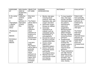
Ineffective
cerebral
tissue
perfusion r/t
interruption
of blood
flow.
Short term
goals:
The Patient
will be able to
display
decrease
signs of
ineffective
tissue
perfusion as
evidence by
gradual
improvement
of vital signs.
Long term
goals:
The Patient
will be able to
gradually
improve
tissue
perfusion as
evidence by
higher GCS
score and
increased
level of
Monitor vital signs,
including blood
pressure, heart rate,
respiratory rate, and
oxygen saturation.
Administer oxygen
therapy as ordered.
Administer
medications as
ordered, such as
antiplatelet agents,
thrombolytics, or
anticoagulants.
Elevate the head of
the bed to promote
cerebral blood flow.
Assess airway
patency and
respiratory pattern.
Perform GCS and
Frequently assess
and monitor
neurological status.
Maintain bed rest
and promote a quiet
and relaxing
environment.
To have baseline
data, Vital signs
provide valuable
information about
the patient’s
hemodynamic
status and
oxygenation.
Monitoring them
regularly helps
detect changes
that may indicate
worsening tissue
perfusion.
Supplemental
oxygen can help
increase the
oxygen supply to
the brain and
improve tissue
perfusion.
These medications
can help improve
blood flow to the
brain, prevent clot
formation, and
dissolve existing
clots, thereby
Patient shall
have gradually
improved tissue
perfusion as
evidenced by
gradual
improvement of
vital signs
The Patient
shall have
gradually
improved tissue
perfusion as
evidenced by
higher GCS
score and
increased level
of
consciousness.
2. consciousnes
s.
improving tissue
perfusion.
Elevating the head
of the bed can help
improve blood flow
to the brain,
reducing the risk of
further tissue
damage.
Clients with a
decreased level of
consciousness
should be
assessed to
ensure that they
are able to protect
their airways
The GCS
evaluates changes
in awareness
based on verbal,
sensorimotor, and
pupillary reflexes.
Restlessness and
assess also for
trends in the level
of consciousness
(LOC), the
potential for
increased ICP, and
helps determine
the location,
extent, and
3. progression of
damage.
CHAPTER IX
ANATOMY AND PHYSIOLOGY
The brain has a few regions. The most obvious is the cerebrum, which is divided into two cerebral hemispheres, each of
which has a cortex. The outer region is divided into four lobes including the frontal lobe, parietal lobe, temporal lobe and
the occipital lobe. The cerebellum which is located below and the brainstem which connects to the spinal cord. The right
4. cerebrum controls the muscles on the left side of your body and vice versa. The frontal lobe controls movement, and
executive functions, which is our ability to make decisions. The parietal lobe processes sensory information, which lets us
locate exactly where we are physically and guides movements in a three-dimensional space. The temporal lobe plays a
role in hearing, smell and memory, as well as visual recognition of faces and languages. The occipital lobe is primarily
responsible for vision. The cerebellum helps with muscle coordination and balance. The brainstem plays a vital role in
functions like heart rate, blood pressure, breathing, gastrointestinal function and consciousness.
The brain receives blood from the left and right carotid arteries, as well as the left and right vertebral arteries, which come
together to form the basilar artery. The internal carotid arteries turn into the left and right middle cerebral arteries which
serve the lateral portions of the frontal, parietal and temporal lobes of the brain. Each of the internal carotid arteries also
5. give off branches called the anterior cerebral arteries which serve the medical portion of the frontal and parietal lobes and
connect with one another whit a short little connecting blood vessels called the anterior communicating artery. Meanwhile,
the vertebral arteries and basilar arteries give off branches to supply the cerebellum and the brainstem.
In addition, the basilar artery divides to become the right and left posterior cerebral artery which mainly serves the
occipital lobe and some of the temporal lobe as well as the thalamus. Finally, the internal carotid arteries each give off a
branch called the posterior communicating artery which attaches to the posterior arteries on each side. So together, the
main arteries and the communicating arteries complete what is called the Circle of Willis. The Circle of Willis is the ring
where blood can circulate from one side to the other in case of a blockage. It also offers alternative ways for blood to get
around an obstructed vessel.
6. In general, the brain can get by on diminished blood flow, especially when it happens gradually because that allows
enough time for collateral circulation to develop, which is where a nearby vessel starts sending out branches of blood
vessels to serve an area that’s in need. But once the supply of blood flow is reduced to below the needs of the tissue it
causes tissue damage, which we call an ischemic stroke.
7. ASSESSMENT NSG.DIAGNOSIS
W/ SCIENTIFIC
BASIS
OBJECTIVE
OF CARE
NURSING
INTERVENTION
RATIONALE EVALUATION
S: No verbal cues
O:
>Ineffective or
absent cough.
> Abnormal
breath sounds
(crackles, rhonchi,
wheezes)
NEEDS:
PHYSIOLOGICAL
NEEDS
Ineffective airway
clearance related
to decreased
level of
consciousness
secondary to
CVA
After 8
hours of
nursing
intervention
the patient
will maintain
clear, open
airways as
evidenced
by the
absence of
abnormal
breath
sounds, a
respiratory
rate within
the normal
range and
maintaining
oxygen
saturation
levels within
the normal
range
(above
95%) during
rest and
activity, as
measured
Assess the
airway for
patency.
Auscultate
lungs for the
presence of
normal or
adventitious
breath sounds.
Assess
respirations.
Note quality,
rate, pattern,
depth, flaring
of nostrils,
dyspnea on
exertion,
evidence of
splinting, use
of accessory
muscles, and
position for
breathing.
Note the
presence of
sputum;
evaluate its
quality, color,
amount, odor,
Maintaining a
patent airway is
always the
priority ,
especially in
cases like
trauma, acute
neurological
decompensation,
or cardiac arrest.
Abnormal breath
sounds can be
heard as fluid
and mucus
accumulate. This
may indicate
ineffective airway
clearance.
A change in the
usual respiration
may mean
respiratory
compromise. An
increase in
respiratory rate
and rhythm may
be a
compensatory
response to
After 8 hours
of nursing
intervention
the patient will
maintain
clear, open
airways as
evidenced by
the absence
of abnormal
breath
sounds, a
respiratory
rate within the
normal range
and
maintaining
oxygen
saturation
levels within
the normal
range (above
95%) during
rest and
activity, as
measured by
pulse
oximetry.
8. by pulse
oximetry.
and
consistency.
Note for
changes in
HR, BP, and
temperature.
Asses for
clubbing of the
fingernails and
cyanosis.
Position the
client upright if
tolerated.
Regularly
check the
client’s
position to
prevent sliding
down in bed.
airway
obstruction.
The unusual
appearance of
secretions may
be a result of
infection,
bronchitis,
chronic smoking,
or other
conditions. A
discolored
sputum is a sign
of infection; an
odor may be
present.
Increased
breathing work
can lead to
tachycardia and
hypertension.
The heart pumps
faster to deliver
oxygenated
blood to vital
organs and
tissues in an
attempt to meet
the body’s
oxygen demand.
Clubbing of the
fingers appears
as sponginess of
9. the nail bed is a
sign of lung
disease.
Cyanosis, a
bluish coloring of
the skin, is a
very late
indicator of
hypoxia.
Upright position
limits abdominal
contents from
pushing upward
and inhibiting
lung expansion.
This position
promotes better
lung expansion
and improved air
exchange.
ASSESSMENT NSG.DIAGNOSIS
W/ SCIENTIFIC
BASIS
OBJECTIVE
OF CARE
NURSING
INTERVENTION
RATIONALE EVALUATION
S: No verbal cues
O:
> Tube feedings
>Reduced level of
consciousness
Risk of Aspiration
related to
impaired
swallowing,
decreased level
of consciousness,
and/or altered
gag reflex
secondary to
After 8
hours of
nursing
intervention
the patient
will be able
Maintain
patent
airway
Assess level of
consciousness
Auscultate
breath sounds
for
development
of crackles
and/or
Rhonchi.
The primary risk
factor of
aspiration is
decreased level
of
consciousness.
Aspiration of
small amounts
can occur
without coughing
After 8 hours
of nursing
intervention
the patient will
be able
Maintain
patent airway
as evidenced
by
10. >Inability to
maintain upright
body posture.
Need: Safety
Needs, Love and
belonging
cerebrovascular
accident (CVA)
as
evidenced
by
oxygen
saturation of
95-100%
and
Remain free
of
aspiration
with no
adventitious
breath
sounds
noted and
reduce the
risk of
Recurrence.
Educate the
family for the
need for
proper
Positioning.
or sudden onset
of respiratory
distress,
especially in
patients with
decreased levels
of
consciousness.
Crackle and
rhonchi that
occur suddenly
could be an
indicator of a
small amount of
aspiration.
Upright
positioning
decreases the
risk for
aspiration.
oxygen
saturation of
95-100% and
Remain free
of
aspiration with
no
adventitious
breath
sounds noted
and
reduce the
risk of
Recurrence.