Call Girls Service Pune Vaishnavi 9907093804 Short 1500 Night 6000 Best call ...
BĂNG HUYẾT SAU SINH - ĐẶT BÓNG CHÈN
1. 29
THE BALLOON INTERNAL UTERINE TAMPONADE
AS A DIAGNOSTIC TEST
S. Ferrazzani, L. Guariglia and C. Dell’Aquila
INTRODUCTION
During the last several years, a number of new
and simpler techniques have been developed in
the attempt to avoid major surgical procedures
for treatment of postpartum hemorrhage1–8.
Although a variety of surgical options have
been proposed to avoid hysterectomy including
uterine artery ligation, ovarian artery ligation,
internal iliac artery ligation, and B-Lynch Brace
suture9, a suitable conservative technique is still
lacking1 and all proposed options have risks as
well as advantages10.
In most cases, procedures are effective in
avoiding hysterectomy, but a delay carries a
poorer prognosis. Moreover, each of these tech-
niques entails a laparotomy and skilled person-
nel must perform the procedure. Rarely, major
complications follow radical surgery for post-
partum hemorrhage; these include loss of fertil-
ity, other morbidity and even maternal death11.
B-Lynch and colleagues12,13 used brace
sutures to compress the uterus without com-
promising major vessels. The advantage of the
B-Lynch procedure is that identification of spe-
cific blood vessels is not necessary, a process
which is often difficult. Although helpful during
Cesarean section, the B-Lynch procedure
requires a laparotomy and therefore may not
be ideal as the first approach in cases of
postpartum hemorrhage following vaginal
delivery14.
This chapter will focus on one of the recently
reported conservative measures to control hem-
orrhage – internal uterine tamponade. Although
uterine atony is the main indication for internal
uterine tamponade, this methodology is also
useful for postpartum hemorrhage arising from
placenta previa/accreta. The technique can be
easily carried out by doctors in training while
awaiting help from a senior colleague.
UTERINE PACKING
Control of postpartum hemorrhage by uterine
packing is not new15. For many years, uterine
packing with sterile gauze has been used in
the clinical management of severe postpartum
hemorrhage and as the last resort before hyster-
ectomy16. Because of the availability of better
uterotonic medications, this practice lost its
appeal, but reports on its successful use con-
tinued to appear17–19. Recently, some authors
raised concerns about concealed bleeding and
infection20; a newer technique, however, has
allayed some of these concerns21.
Uterovaginal packing may sometimes obviate
the need for surgery altogether. In cases of
deliveries complicated by postpartum hemor-
rhage, after excluding uterine rupture, genital
tract lacerations, and retained placental tissue,
efforts are directed toward contracting the
uterus by bimanual compression and uterotonic
agents. If these are not successful, one must
resort to surgical techniques. At this stage, an
alternative option to remember is uterovaginal
packing. Easy and quick to perform, it may be
used to control bleeding by tamponade effect
and stabilize the patient until a surgical
procedure is arranged.
Chapter 28 describes the technique of
uterine packing in more detail.
BALLOON TAMPONADE
Tamponade with different types of balloon
catheters used prior to surgery is a conservative
268
290
Z:Sapiens PublishingA5211 - Postpartum HemorrhageMake-upPostpartum Hemorrhage - Voucher Proofs #T.vp
30 August 2006 14:21:47
Color profile: Generic CMYK printer profile
Composite Default screen
2. procedure that is available before invasive
surgical techniques are needed1,3. Chapter 28
describes the various types of balloon catheters
that are available.
Balloon tamponade of the uterus is a recog-
nized procedure in those with massive and
intractable hemorrhage17,22–29.
The use of the Sengstaken–Blakemore eso-
phageal or gastric catheter is described in the
literature for the control of massive postpartum
hemorrhage due to an atonic uterus not
responding to oxytocics including prosta-
glandins3,17,23,30,31 or due to placenta accreta32.
Multiple Foley catheters in the case of a vaginal
delivery22,25 have also been used and even
rubber catheters fitted with a condom have
been used successfully to control postpartum
hemorrhage in undeveloped countries33. Uro-
logical fluid-filled catheters (300–500 ml)26,28,29
or of silicone balloons designed for tamponade
function27 also seem to be very effective, with
further possibilities in cases of hemorrhage after
Cesarean section for placenta previa/accreta.
Theoretical principle of action
The theoretical principle of the balloon
tamponade is that temporary and steady
mechanical compression of the bleeding surface
of the placental site can be performed while
waiting for the natural hemostatic mechanisms
of the blood to take effect. The balloon, inflated
inside the uterine cavity in order to stretch the
myometrial wall, can exert an intrauterine pres-
sure that overcomes the systemic arterial pres-
sure, resulting in cessation of the intrauterine
blood flow. Probably, a quite different mecha-
nism can be advocated for its efficacy in the case
of uterine atony. With separation of the pla-
centa, the many uterine arteries and veins that
carry blood to and from the placenta are severed
abruptly. Elsewhere in the body, hemostasis in
the absence of surgical ligation depends upon
intrinsic vasospasm and formation of blood
clots locally. At the placental implantation site,
the most important factors for achieving
hemostasis are contraction and retraction of the
myometrium in order to compress the vessels
and obliterate their lumens. Uterine atony
from any origin can prevent this physiological
mechanism, leading to massive hemorrhage.
The first therapeutic approach to this situa-
tion is mechanical stimulation by massage of the
uterus and then the use of uterotonic drugs. In
this case, the efficacy of the tamponade balloon
may derive from the mechanical stimulation of
myometrial contraction caused by the balloon’s
elasticity pressing against the myometrial wall.
The simultaneous and continuous stimulation
of myometrial contraction and the tamponade
effect on the open vessels, reached with the
contraction, explain its efficacy. However, the
uterus must be empty for the tamponade to
be successful.
In the presence of placenta accreta, the
balloon must be used with great caution, as a
failure or delay to control hemorrhage in such
patients could be catastrophic.
In the small series reported in the literature,
in which the different types of balloon catheter
were filled with various volumes, ranging from
30 to 500 ml, Seror and colleagues chose an
inflation volume of 250 ml, since this value
corresponds to the approximate volume of the
uterine cavity after delivery31.
BALLOON TAMPONADE AS A TEST
To date, there is no diagnostic test to identify
those patients with intractable hemorrhage who
will need surgery. Condous and colleagues3
proposed the use of an inflated Sengstaken–
Blakemore balloon catheter as a test to create
tamponade and identify patients who will or will
not need surgery (‘tamponade test’). When its
results are positive, the tamponade test not only
halts the blood loss and preserves the uterus,
but also gives an opportunity to reverse and
correct any consumptive coagulopathy. More
than 87% of their patients (14/16) with intracta-
ble postpartum hemorrhage responded to the
tamponade test3. More recently, Seror and col-
leagues reported that, in a series of 17 cases,
tamponade treatment prevented surgery in 88%
of patients31.
According to these clinical experiences, an
early use of the balloon catheter may reduce
the total blood loss, and it is probable that
any type of inflatable balloon with high fluid-
filling capacity could be used for the same
purpose.
269
The balloon internal uterine tamponade as a diagnostic test
291
Z:Sapiens PublishingA5211 - Postpartum HemorrhageMake-upPostpartum Hemorrhage - Voucher Proofs #T.vp
30 August 2006 14:21:47
Color profile: Generic CMYK printer profile
Composite Default screen
3. The experience at the Catholic University
of Rome, Italy
A longitudinal study is currently running in the
Obstetrics and Gynecology Department of the
Catholic University of Rome, Italy; it started in
January 2002 and the Institute review board
approved the study.
Patients and methods
In the period January 2002–August 2005,
10 773 patients delivered in our maternity
ward. During this period, there were 124
(1.15%) instances of postpartum hemorrhage.
Of these, 13 were considered critical and the
women underwent treatment by intrauterine
tamponade.
An atonic uterus caused postpartum hemor-
rhage in one case and placenta previa/accreta
was noted in 12 cases, of which two were
associated with uterine atony.
The mean age of the patients was 35 years
(26–39 years). The mean gestational age at
delivery was 36 weeks from the first day of
the last menstrual period (26 weeks and 5
days to 40 weeks and 1 day). Nine patients
were multiparous (69.2%). The mean parity
was 2.1.
Labor was spontaneous in three cases, and
stimulated with dinoprostone intravaginal gel
or oxytocin infusion in two cases. The mean
duration of labor was 6 h and 42 min (5–9 h).
Three patients had a vaginal delivery, and ten
had a Cesarean section (of which seven were
planned).
Routinely, the patients who delivered by the
vaginal route had prophylactic intramuscular
oxytocin/ergometrine in the third stage of labor
and all the patients who underwent Cesarean
section had intramyometrial and intravenous
oxytocin during/after the placenta was
delivered.
In the 13 cases of postpartum hemorrhage
considered in this study, patients were treated
with appropriate oxytocic agents and prosta-
glandin analogues (intravenous infusions
of oxytocin (40–100 U), intramyometrial
oxytocin (20 U), intramuscular ergometrine
(0.25–0.5 mg), and/or intravenous infusion of
sulprostone (500 mg)).
In the three patients delivering by the vaginal
route, an examination was performed under
regional or general anesthesia for retained tissue
and lacerations and, when necessary, retained
tissue or placenta was removed and lacerations
were sutured.
Coagulation studies were carried out simul-
taneously to exclude coagulopathy as the first or
the complimentary cause of the hemorrhage.
In those patients considered for the study
who showed no response to these measures, a
sterile hydrostatic (bladder distention) balloon
catheter size Ch. 16, 5.3 mm (Rüsch UK High
Wycombe, England) (Figure 1) was inserted
into the uterine cavity via the cervix. This was
achieved using minimal analgesia or regional
anesthetic. The insertion was facilitated by
grasping the anterior and lateral margins of the
cervix with sponge forceps and placing the bal-
loon into the uterine cavity with another sponge
forceps. The balloon catheter was then filled
with 120–300 ml of warm saline solution until
a contracted uterus was palpable through the
abdomen. Applying gentle traction at this stage
confirmed that the filled balloon was firmly
270
POSTPARTUM HEMORRHAGE
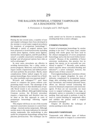
Figure 1 The Rüsch hydrostatic balloon catheter
292
Z:Sapiens PublishingA5211 - Postpartum HemorrhageMake-upPostpartum Hemorrhage - Voucher Proofs #T.vp
30 August 2006 14:21:49
Color profile: Generic CMYK printer profile
Composite Default screen
4. fixed in the uterine cavity. If no or minimal
bleeding was observed through the cervix,
laparotomy was avoided and a gauze packing
of the vagina was performed to avoid self-
expulsion of the balloon from the completely
dilated cervical os. If significant bleeding
continued through the cervix, the ‘tamponade
test’ had failed and laparotomy was performed.
In all the patients delivering by urgent or
planned Cesarean section, the problem of
abnormal insertion or suspicion of morbid
adhesion of the placenta was detected by ultra-
sound scan before surgery. The placenta was
delivered by firmly controlled cord traction, or
by manual removal if it was abnormally adherent
to the uterine wall. If severe bleeding persisted
despite a contracted uterus after local intramyo-
metrial and endovenous infusion of oxytocin
and prostaglandin analogues, the hydrostatic
balloon catheter previously described was
inserted intrabdominally, through the uterine
incision, into the cervical opening and through
the cervical canal by a sponge forceps, leaving
the balloon in the uterine cavity (Figure 2). The
balloon was then filled with 180–300 ml of
warm saline solution, using a 60 ml bladder
syringe. Tamponade was achieved by pulling
the distal extremity of the catheter shaft out of
the vagina. The uterine contraction over the
balloon was maintained, after the uterine clo-
sure, by a slow oxytocin infusion (20–40 U) that
was given over the next 24 h. A single-layer
closure of the uterine incision was performed,
taking care not to include the balloon in the
suture line. The Cesarean section was con-
cluded following the classical technique. Only
when the bleeding was adequately controlled
was the abdominal wall closed.
Those who responded to the balloon catheter
therapy were stabilized in the labor and delivery
unit for ongoing management. In all cases,
intravenous broad-spectrum antibiotics were
administered for at least the first 24 h. The bal-
loon catheter was left in situ until the next day.
During this time interval, blood transfusion and
coagulopathy correction were possible. Once
the above parameters were within acceptable
limits, the balloon catheter was slowly deflated
and withdrawn and the patient observed for any
active bleeding.
271
The balloon internal uterine tamponade as a diagnostic test
Figure 2 Intra-abdominal insertion of the hydrostatic balloon catheter into the uterine cavity
293
Z:Sapiens PublishingA5211 - Postpartum HemorrhageMake-upPostpartum Hemorrhage - Voucher Proofs #T.vp
30 August 2006 14:21:53
Color profile: Generic CMYK printer profile
Composite Default screen
5. Results
The ‘tamponade test’ was positive in 12 out of
13 cases and the hydrostatic catheter immedi-
ately arrested hemorrhage. In one case, tampon-
ade failed after 3 h and bleeding re-occurred. In
our series of 13 cases, the primary cause for
postpartum hemorrhage was bleeding at the
placental site alone (ten cases), uterine atony
associated with bleeding at the placental site
(two cases), and uterine atony with cervical
laceration and pre-eclampsia-associated dis-
seminated intravascular coagulation (one case).
Tables 1 and 2 provide details of the 13 cases.
Because, according to Benirschke and
Kaufmann34, the diagnosis of accreta cannot
be made when the placenta is not removed with
the uterus, in such a condition the diagnosis of
placenta accreta was based on clinical criteria
and consisted of the inability to remove it by
controlled cord traction because of a severe
adherence to the underlying myometrium and
failure to develop a cleavage plane between the
placenta and uterus.
Among the three patients delivering by the
vaginal route, two deserve a more detailed
description. One patient (case 5) had a pre-
delivery ultrasound diagnosis of marginal pla-
centa previa. The woman had a normal labor,
but soon after delivery of the placenta a profuse
hemorrhage began. The uterine cavity was
explored and the placenta accurately removed.
A 3-cm fragment of the placenta was lacking but
even a very vigorous examination of the uterine
cavity was unsuccessful in removing that frag-
ment. A catheter balloon inserted through the
vagina soon arrested the severe bleeding. The
patient was administered 2 units of blood and
the balloon was removed after 24 h. The patient
was discharged from the hospital 7 days later
and her progress was uneventful until 11 days,
when she was re-admitted to hospital for further
hemorrhage due to the expulsion of the placen-
tal fragment. An examination of the uterine cav-
ity resolved the case with no other intervention
or further blood transfusion.
Another patient (case 12) had a normal
vaginal delivery with no pre-delivery suspicion
of abnormal adherence of the placenta. After
delivery of the placenta, the lack of a 3-cm pla-
cental fragment was observed. The examination
of the uterine cavity was unsuccessful but, in the
absence of further bleeding, the patient was kept
under observation with no other intervention.
During the subsequent 24 h, sub-acute vaginal
bleeding was associated with a progressive fall of
the hematocrit level. A further examination of
the uterine cavity was planned, and the removal
of the retained placental fragment caused a
severe hemorrhage that was quickly stopped by
introducing a balloon catheter through the
vagina into the uterine cavity.
272
POSTPARTUM HEMORRHAGE
Case
Age
(years) Gravidity Parity
Gestation
(weeks)
Duration of
labor (h) Mode of delivery
1
2
3
4
5
6
7
8
9
10*
11
12
13
36
29
38
30
36
34
34
36
39
35
39
33
26
2
5
4
1
1
3
2
2
5
2
3
6
4
1
1
2
0
0
0
1
1
1
0
2
1
1
35
35
37
37
40
34
37
36
30
26
40
38
35
–
–
–
–
6
–
–
–
–
–
9
5
–
planned CS
planned CS
planned CS
planned CS
spontaneous labor
urgent CS
planned CS
planned CS
urgent CS
urgent CS
induced/oxytocin
induced/oxytocin
planned CS
CS, Cesarean section; *failed ‘tamponade test’
Table 1 Clinical details of patients with postpartum hemorrhage who underwent a balloon tamponade
294
Z:Sapiens PublishingA5211 - Postpartum HemorrhageMake-upPostpartum Hemorrhage - Voucher Proofs #T.vp
30 August 2006 14:21:53
Color profile: Generic CMYK printer profile
Composite Default screen
6. Among the ten cases resulting in Cesarean
section, one patient (case 13) showed at ultra-
sound scan high suspicion of morbid adhesion of
the placenta (accreta/increta) before the planned
Cesarean section for total placenta previa. Dur-
ing Cesarean section, in order to prevent severe
bleeding at delivery of the placenta by reducing
the blood flow to the uterus, a prophylactic
O’Leary suture35 was positioned around the
uterine arteries immediately after delivery of the
infant, with the placenta still in situ, using a
2-monofilament absorbable suture on a high-
curve needle. Subsequently, a bilateral utero-
ovarian vessel ligation was performed with a
1-monofilament absorbable suture, including the
broad ligament close under the tubal insertion to
273
The balloon internal uterine tamponade as a diagnostic test
Case Cause of bleeding
Estimated
blood loss
(ml)
Intrapartum
RBC and
FFP
Postpartum
RBC Medical treatment
Postpartum
hospital
admission
(days)
1 total placenta previa 1000 0 0 intramyometrial oxytocin,
oxytocin infusion
5
2 total placenta previa 3000 RBC 2 U 0 intramyometrial oxytocin,
oxytocin infusion
11
3 total placenta previa 1000 0 0 intramyometrial oxytocin,
oxytocin infusion
4
4 total placenta previa 1200 RBC 1 U RBC 2 U intramyometrial oxytocin,
oxytocin infusion
6
5 marginal placenta previa,
focally accreta
1200 0 RBC 2 U oxytocin infusion, i.m.
ergometrine
8
6 total placenta previa 1500 0 0 intramyometrial oxytocin,
oxytocin infusion
5
7 total placenta previa 1500 0 0 intramyometrial oxytocin,
oxytocin infusion
5
8 focal placenta accreta 1000 0 0 intramyometrial oxytocin,
oxytocin infusion
4
9 focal placenta accreta,
atony
3300 RBC 6 U,
FFP 6 U
0 intramyometrial oxytocin,
oxytocin infusion,
sulprostone infusion
5
10* marginal placenta previa,
focally accreta, atony
5000 RBC 9 U 0 intramyometrial oxytocin,
oxytocin, sulprostone
infusion
6
11 atony, cervico-isthmic tear
and DIC in pre-eclampsia
1100 0 RBC 2 U i.m. oxytocin, oxytocin
infusion
7
12 focal placenta accreta 1500 RBC 2 U RBC 2 U oxytocin and sulprostone
infusion, i.m. ergometrine
5
13 total placenta
previa/accreta
1100 0 0 intramyometrial oxytocin,
oxytocin infusion
5
RBC, red blood cell; FFP, fresh frozen plasma; DIC, disseminated intravascular coagulation; i.m., intra-
muscular; *failed ‘tamponade test’
Table 2 Cause of bleeding and medical treatment before tamponade procedure
295
Z:Sapiens PublishingA5211 - Postpartum HemorrhageMake-upPostpartum Hemorrhage - Voucher Proofs #T.vp
30 August 2006 14:21:53
Color profile: Generic CMYK printer profile
Composite Default screen
7. the uterus and the utero-ovarian ligament. The
placenta was found to extend across the internal
cervical os. The inability to remove it by firmly
controlled cord traction because of a severe
adherence to the underlying myometrium and to
develop a cleavage plane between the placenta
and uterus became the clinical confirmation of
placenta accreta34. Therefore, the placental tis-
sue was manually removed in fragments and the
placental site inspected. No myometrial defects
were found, as the adhesion was limited to the
myometrial layer. In order to control a persistent,
although moderate, bleeding from the placental
site which did not respond to pharmacological
uterotonic therapy, a hydrostatic balloon cathe-
ter was inserted through the uterine incision,
leaving the balloon in the uterine cavity as previ-
ously described. The patient did not need blood
transfusion and a 6-month follow-up by Doppler
ultrasound demonstrated regular reperfusion of
the uterus.
Conservative treatment with the balloon
catheter was unsuccessful in two cases and hys-
terectomy was performed (cases 9 and 10). In
case 9, the balloon catheter was inserted, after
Cesarean section was concluded, by the vaginal
route because of a persistent vaginal bleeding.
The ‘tamponade test’ was successful and the
patient was monitored for 3 h. However, the
patient then had a hemorrhage due to secondary
uterine atony not responding to oxytocics and
sulprostone infusion. Even further filling of the
balloon was unsuccessful and, soon after the
removal of the balloon, a large amount of blood
and clots were expelled from the cervical os, so
that urgent hysterectomy was mandatory. In
case 10, the ‘tamponade test’ failed and no
other surgical approach was attempted before
hysterectomy. The reason for the failure of the
‘tamponade test’ was uterine atony refractory to
any pharmacological treatment.
The 13 patients had a total estimated blood
loss of 23.4 liters. The lowest and highest
estimated blood losses experienced were 1 and
5 liters. A total of 28 U of blood and 6 U of
fresh frozen plasma were transfused.
Discussion
The effectiveness of the Rüsch urological hydro-
static balloon as a conservative procedure in the
therapy of postpartum hemorrhage has been
shown in two cases described by Johanson36 and
in four cases more recently reported28,29. How-
ever, its efficacy in severe postpartum hemor-
rhage needed to be evaluated in a larger series.
In the present provisional study, the insertion
of the Rüsch urological hydrostatic balloon in
patients with massive postpartum hemorrhage
was very successful and was associated with no
significant complications. The procedure failed
in only two cases. As opposed to the traditional
gauze uterine packing, the technique with the
balloon catheter provides immediate knowledge
of its effectiveness in controlling the postpartum
hemorrhage, so that subsequent surgery can be
expedited in failed cases.
If bleeding continues despite the insertion of
a balloon, the Rüsch urological hydrostatic bal-
loon gives less information than a Sengstaken–
Blakemore catheter, since bleeding is noted only
through the cervix but not from the uterine
fundal cavity. However, the Rüsch urological
hydrostatic balloon is simpler and cheaper than
the other. At the same time, its overturned
pear-shape better fits in the uterine cavity, with
probably less risk of self-expulsion. The uterus
must be empty for successful tamponade. If the
uterine cavity is completely empty and uterine
contraction sustained by adequate pharmaco-
logical assistance, there is probably no need for
monitoring bleeding from the uterine fundal
cavity. A larger series of cases will be necessary
to support this last opinion.
The Rüsch urological hydrostatic balloon
takes a few minutes to insert, is unlikely to cause
trauma and is easy to place with minimal or no
anesthesia, whereas its removal is painless and
simple. Whether the patient is going to bleed
after removal of the balloon is a general con-
cern, but this series demonstrates that there
were no cases of rebleeding after the planned
removal of the Rüsch urological hydrostatic
balloon. In case of rebleeding, it is possible to
replace the balloon while planning an oppor-
tune uterine arterial embolization in a patient
who is now in a stable condition36–39.
There were two cases of failure; atony was
the cause of failure and subsequent hysterec-
tomy in both. In these cases, an attempt to
mechanically favor uterine contraction by
applying a B-Lynch Brace suture of the uterus
274
POSTPARTUM HEMORRHAGE
296
Z:Sapiens PublishingA5211 - Postpartum HemorrhageMake-upPostpartum Hemorrhage - Voucher Proofs #T.vp
30 August 2006 14:21:53
Color profile: Generic CMYK printer profile
Composite Default screen
8. combined with an additional insertion in the
uterine cavity of a balloon catheter could possi-
bly have resolved the problem, with the com-
bined conservative approach already described
by Danso and Reginald40.
One of the difficulties in the management of
patients with intractable postpartum hemor-
rhage, not responding to uterotonic agents, is
the decision to perform a laparotomy and, in
case of Cesarean section, the decision to per-
form a hysterectomy. The delay can be cata-
strophic. In the present series, average blood
loss was considerably less than that of other
series recently reported3,31. In all the cases but
two, the risk of postpartum hemorrhage was
known in advance. When there is confidence
that the management of postpartum hemor-
rhage can be conservative, easy and effective, as
in the case of application of a balloon catheter,
there is no reason for a delay.
In conclusion, the safe, low-cost, and easy
procedure of utilizing a balloon catheter can
be applied in any situation of life-threatening
postpartum hemorrhage and avoids radical sur-
gery in patients so that reproductive capacity is
preserved.
References
1. Tamizian O, Arulkumaran S. The surgical
management of postpartum haemorrhage. Curr
Opin Obstet Gynecol 2001;13:127–31
2. Papp Z. Massive obstetric hemorrhage. J Perinat
Med 2003;31:408–14
3. Condous GS, Arulkumaran S, Symonds I,
Chapman R, Sinha A, Razvi K. The ‘tamponade
test’ in the management of massive postpartum
hemorrhage. Obstet Gynecol 2003;101:767–72
4. El-Refaey H, Rodeck C. Post-partum haemor-
rhage: definitions, medical and surgical manage-
ment. A time for change. Br Med Bull 2003;67:
205–17
5. Pahlavan P, Nezhat C, Nezhat C. Hemorrhage
in obstetrics and gynecology. Curr Opin Obstet
Gynecol 2001;13:419–29
6. Gielchiensky Y, Rojansky N, Fasoulitios SJ,
Ezra Y. Placenta accrete – summary of 10 years:
a survey of 310 cases. Placenta 2002;23:210–14
7. Shevell T, Malone FD. Management of obstetric
hemorrhage. Semin Perinatol 2003;27:86–104
8. Mousa HA, Walkinshaw S. Major postpartum
haemorrhage. Curr Opin Obstet Gynecol 2001;13:
595–603
9. Tamizian O, Arulkumaran S. The surgical man-
agement of post-partum haemorrhage. Best Pract
Res Clin Obstet Gynecol 2002;16:81–98
10. Drife J. Management of primary postpartum
haemorrhage. Br J Obstet Gynaecol 1997;104:
275–7
11. El-Hamamy E, B-Lynch C. A worldwide review
of the uses of the uterine compression suture
techniques as alternative to hysterectomy in the
management of severe post-partum haemor-
rhage. J Obstet Gynecol 2005;25:143–9
12. Vangsgaard K. ‘B-Lynch-suture’ in uterine
atony. Ugeshr Laeger 2000;162:3468
13. B-Lynch C, Coker A, Lawal AH, Abu J, Cowen
MJ. The B-Lynch surgical technique for the
control of massive postpartum haemorrhage: an
alternative to hysterectomy? Five cases reported.
Br J Obstet Gynaecol 1997;104:372–5
14. Allam MS, B-Lynch C. The B-Lynch and other
uterine compression suture techniques. Int J
Gynaecol Obstet 2005;89:236–41
15. Drucker M, Wallach RC. Uterine packing: a
re-appraisal. Mt Sinai J Med 1979;46:191–4
16. American College of Obstetrician and Gynecolo-
gists. Diagnosis and management of postpartum
hemorrhage. ACOG technical bulletin no. 143.
Washington, DC: American College of
Obstetricians and Gynecologists, 1990
17. Katesmark M, Brown R, Raju KS. Successful
use of a Sengstaken-Blakemore tube to control
massive postpartum haemorrhage. Br J Obstet
Gynaecol 1994;101:259–60
18. Kauff ND, Chelmow D, Kawada CY. Intracta-
ble bleeding managed with Foley catheter
tamponade after dilatation and evacuation. Am J
Obstet Gynecol 1995;173:957–8
19. Bagga R, Jain V, Kalra J, Chopra S, Gopalan S.
Uterovaginal packing with rolled gauze in post-
partum hemorrhage. Med Gen Med 2004;13:50
20. Hsu S, Rodgers B, Lele A, Yeh J. Use of packing
in obstetric hemorrhage of uterine origin. J
Reprod Med 2003;48:69–71
21. Roman AS, Rebarber A. Seven ways to control
postpartum hemorrhage. Contemp Obstet Gynecol
2003;48:34–53
22. De Loor JA, van Dam PA. Foley catheters
for uncontrollable obstetric or gynecologic
hemorrahage. Obstet Gynecol 1996;88:737
23. Chan C, Razvi K, Tham KF, Arulkumaran S.
The use of a Sengstaken-Blakemore tube to
control post-partum hemorrhage. Int J Gynaecol
Obstet 1997;58:251–2
24. Bakri YN. Uterine tamponade-drain for hemor-
rhage secondary to placenta previa-accreta. Int J
Gynaecol Obstet 1992;37:302–3
275
The balloon internal uterine tamponade as a diagnostic test
297
Z:Sapiens PublishingA5211 - Postpartum HemorrhageMake-upPostpartum Hemorrhage - Voucher Proofs #T.vp
30 August 2006 14:21:53
Color profile: Generic CMYK printer profile
Composite Default screen
9. 25. Marcovici I, Scoccia B. Postpartum hemorrhage
and intrauterine balloon tamponade: a report of
three cases. J Reprod Med 1999;44:122–6
26. Johanson R, Kumar M, Oberai M, Young P.
Management of massive postpartum haemor-
rhage: use of a hydrostatic balloon catheter to
avoid laparotomy. Br J Obstet Gynaecol 2001;
108:420–2
27. Bakri YN, Amri A, Abdul Jabbar F.
Tamponade-balloon for obstetrical bleeding. Int
J Gynaecol Obstet 2001;74:139–42
28. Ferrazzani S, Guariglia L, Caruso A. Therapy
and prevention of obstetric hemorrhage by
tamponade using a balloon catheter. Minerva
Ginecol 2004;56:481–4
29. Ferrazzani S, Guariglia L, Triunfo S, Caforio L,
Caruso A. Successful treatment of post-Cesarean
hemorrhage related to placenta praevia using
an intrauterine balloon. Two case reports. Fetal
Diagn Ther 2006;21:277–80
30. Condie RG, Buxton EJ, Payne ES. Successful
use of a Sengstaken-Blakemore tube to control
massive postpartum haemorrhage [letter]. Br J
Obstet Gynaecol 1994;101:1023–4
31. Seror J, Allouche C, Elhaik S. Use of
Sengstaken-Blakemore tube in massive post-
partum hemorrhage: a series of 17 cases. Acta
Obstet Gynecol Scand 2005:84:660–4
32. Frenzel D, Condous GS, Papageorghiou AT,
McWhinney NA. The use of the ‘tamponade
test’ to stop massive obstetric haemorrhage in
placenta accreta. Br J Obstet Gynaecol 2005;112:
676–7
33. Akhter S, Begum MR, Kabir Z, Rashid M, Laila
TR, Zabeen F. Use of a condom to control
massive postpartum hemorrhage. Med Gen Med
2003;5:38
34. Benirschke K, Kaufmann P, eds. Pathology of the
Human Placenta, 4th edn. New York: Springer,
2000:554
35. O’Leary JA. Uterine artery ligation in the control
of postcaesarean haemorrhage. J Reprod Med
1995;40:189–93
36. Mitty H, Sterling K, Alvarez M, Gendler R.
Obstetric haemorrhage: prophylactic and
emergency arterial catheterization and embolo-
therapy. Radiology 1993;188:183–7
37. Pelage JP, Le Dref O, Jacob D, Soyer P,
Herbreteau D, Rymer R. Selective arterial
embolization of the uterine arteries in the man-
agement of intractable post-partum hemorrhage.
Acta Obstet Gynecol Scand 1999;78:698–703
38. Corr P. Arterial embolization for haemorrhage in
the obstetric patient. Best Pract Res Clin Obstet
Gynecol 2001;4:557–61
39. Tourn G, Collet F, Seffert P, Veyret C. Place
of embolization of the uterine arteries in the
management of post-partum haemorrhage: a
study of 12 cases. Eur J Obstet Gynecol Reprod
Biol 2003;110:29–34
40. Danso D, Reginald P. Combined B-Lynch
suture with intrauterine balloon catheter tri-
umphs over massive postpartum haemorrhage.
Br J Obstet Gynaecol 2002;109:963
276
POSTPARTUM HEMORRHAGE
298
Z:Sapiens PublishingA5211 - Postpartum HemorrhageMake-upPostpartum Hemorrhage - Voucher Proofs #T.vp
04 September 2006 14:09:34
Color profile: Generic CMYK printer profile
Composite Default screen