The Integration of Critical Steps: Diagnosis, Treatment Planning, Workflow, and Teamwork in Anterior Restorations
•
17 likes•5,448 views

Simplicity, predictability, respecting biology, and minimally invasive treatment was the goal in this author's surgical treatment. Creating an ideal prosthetic environment and allowing the dental technician to create an implant restoration with ideal prosthetic soft tissue support and long-term stability was the author's purpose. This slide show displays the incredible teamwork by Dr. Eric Van Dooren and Mr. Murilo Calgaro has they work together through the critical steps of an anterior implant restoration.

Recommended
More Related Content
Viewers also liked
Viewers also liked (10)
More from theaacd
More from theaacd (12)
Recently uploaded
Recently uploaded (20)
The Integration of Critical Steps: Diagnosis, Treatment Planning, Workflow, and Teamwork in Anterior Restorations
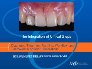
- 1. Diagnosis, Treatment Planning, Workflow, and Teamwork in Anterior Restorations Eric Van Dooren, DDS and Murilo Calgaro, CDT Images are © , All Rights Reserved, Dr. Eric Van Dooren The Integration of Critical Steps
- 2. This slide show presents a case regarding a 20-year-old male patient with the goal being to correct the esthetic problems in the anterior region. Soft tissue health and hygiene were acceptable, and no pockets were present. The patient presented a thin biotype with high scalloped gingival margins (which made the authors more cautious for any surgical soft tissue procedure). A 1-mm overjet and a 1-mm overbite with a mandibular range of motion within normal limits were observed. The periapical x-ray exhibited enough space to place a narrow platform implant in the lateral right incisor position. Silicone impressions were taken (Virtual, Ivoclar Vivadent; Schaan, Liechtenstein) and models were poured. A diagnostic wax-up was used to treatment plan the case. Diagnosis and proper treatment planning would ultimately be the guarantees for treatment success. Adapted from the full article, which originally appeared in the AACD’s Journal of Cosmetic Dentistry, Spring 2011, Volume 27, No. 1 issue. For more information, visit www.AACD.com. © 2011-2013 American Academy of Cosmetic Dentistry, All Rights Reserved
- 3. Agenesia of the left lateral incisor and microdontia with very conical tooth shape covered with an old composite were at the root of the esthetic problems. A Maryland bridge for replacement of #7, with metal retainers on both central incisor and canine, was placed by the referring dentist after completion of the orthodontic treatment.
- 4. The pretreatment periapical x-ray exhibited enough space to place a narrow platform implant in the lateral right incisor position.
- 5. A narrow-platform Branemark (Nobel Biocare, Göteborg, Sweden) implant was placed and a connective tissue graft was harvested from the maxillary tuberosity and placed in a buccal pouch. Grafts taken from the retromolar area are dense and fibrous, and exhibit long-term three-dimensional stability with minimal long- term volumetric changes. Care was taken to maximize soft tissue thickness around the implant. Seralene 6/0 sutures (American Dental Systems; Vaterstetten, Germany) were placed to secure the graft into position. A 3-mm NP healing abutment was placed at the day of surgery and healing was uneventful.
- 6. A narrow- platform, open-tray impression coping was secured and retraction cord placed. A final impression (Virtual Light and Heavy Body, Ivoclar Vivadent) was taken and a silicone key was placed to evaluate the preparation.
- 7. In most of the author’s anterior delayed- loading implant restorations, no attempt is made to develop the ideal gingival contour with individualized healing abutments or impression copings. Shown right is the final model with soft tissue mask. A high-quality full-contour esthetic and functional wax-up was made. It was important at that time to incorporate the ideal form and ideal three- dimensional gingival tooth position in the wax-up and draw the ideal buccal crown contour according to the wax-up.
- 8. The concept is to stay 0.5 mm away from the line drawn on the buccal aspect of the implant restoration. A round, coarse diamond bur is used to reshape the transmucosal space. The final dimensions of the transmucosal space are related to the silcone index and wax-up and the abutment margin positioned in the sulcus.
- 9. The perfect implant restoration would be a restoration where the abutment occupies 90% of the transmucosal space and where the crown would only emerge in the last 1 mm or 10% of the transmucosal space. Abutment 90% Crown 10%
- 10. The abutment should have a concave transmucosal profile to allow for connective tissue thickness in this critical zone. This will act as a mechanical barrier and protect the bone from the oral environment.
- 11. Layering of the porcelain (IPS e.max Ceram). Fabrication of a lithium disilicate coping (IPS e.max Press LT). The use of magnification both in the clinical and laboratory environment are mandatory. Precision relates to biology and soft tissue health in a direct way.
- 12. In this particular case, trying in the second bake, it became apparent that the prosthetic soft tissue support was insufficient and, consequently, the gingival scallop was flat. Minor modifications are necessary to improve color, form, and especially prosthetic soft tissue support. The final soft tissue profile and position around the implant restoration will depend on the amount of prosthetic pressure and the prosthetic contour.
- 13. Allowing for a steeper distal angle and a softer mesial angle. Adding low-fusion porcelain during the last bake on the disto-gingival portion of the restoration allowed us to create a high scalloped gingival design, modify the zenith position, and relocate the gingival culminating point.
- 14. Functional adjustments were made to restore the canine guidance, a composite was applied to the right upper and lower canines. The 12-month postoperative x-ray revealed a satisfactory bone remodeling.
- 15. Final esthetic and functional result. Acknowledgments The AACD thanks Dr. Eric Van Dooren (Belgium) and Murilo Calgaro, CDT (Brazil) for this slideshow.
- 16. To read the details of this case or to receive the quarterly, peer-reviewed Journal of Cosmetic Dentistry, become an AACD member at www.AACD.com/join