![Volume 2 | Issue 1
ajsccr.org 2
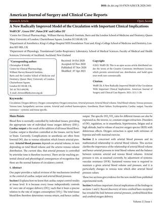
Figure 1. The uppermost traces are of cardiac output (CO), the middle traces heart rate (HR) and the lower traces stroke volume (SV). On the left (A) recordings were made
in intact dogs, on the right (B) following recovery after cardiac autonomic denervation. After Donald and Shepherd (5) with permission (American Journal of Physiology
– legacy collection).
SECTION 1
2. Matching Tissue Oxygen Supply to The Rate of Oxygen
Consumption is The Top Priority of The Circulation
2.1. Origins of the Utilization of Oxygen
Energy is an essential feature of living things, required not only for
movement, cerebration, digestion etc. but also to sustain molecular
organization within cells. Muscular activity and varying activity in
other tissues, such as brain, heart and gut, and their maintenance,
all require provision of energy. The earliest living things were single
cells and for them energy was obtained, in the main, from molec-
ular interactions which did not utilize oxygen (anaerobic respira-
tion, e.g. methanogens, which react hydrogen with carbon dioxide
making methane as a waste product).
Energy yield was less than can be obtained from interaction with
oxygen which is, nevertheless, highly toxic, from its ready conver-
sion to biochemically dangerous free radicals. Eventually certain
bacteria evolved with the ability to utilize oxygen in energy gen-
erating reactions which largely avoided free radical release [1, 2].
Multicellular organisms were initiated 600 million years ago when
an oxygen capable bacterium was incorporated into a second type
of unicellular organism – a member of the archaea [1, 3]. The ox-
ygen capable bacteria in these new combined organisms became
mitochondria. Multicellularity became possible for the new com-
bined organisms. Their evolution included branches where, with
increasing size, circulation evolved such that oxygen could be
transported from the atmosphere to the tissues. Hence, evolution
sub-served circulatory delivery of oxygen, appropriate to oxygen
consumption by the mitochondria. In this section evidence is given
for the precision regulation of blood flow to provide the appropri-
ate oxygen supply to major tissues. These include, specifically,
skeletal muscle, heart and brain, and evidence for whole body oxy-
gen supply, provided by appropriate cardiac output, the total of the
individually regulated tissue blood flows.
2.2. Cardiac Output in Exercise is Independent of Cardiac In-
nervation
Total circulatory blood flow is the sum of blood flows from every
tissue. All blood flow converges on the heart. “The heart puts out
what it receives” [4]. Hence, whole body blood flow becomes the
Cardiac Output (CO). Blood flow to skeletal muscle increases in
exercise, resulting in the increase in cardiac output. The fact that
tissues control their own blood flow is well illustrated by the ex-
perimental work of Donald and Shepherd [5]. Conscious mongrel
dogs were instrumented for continuous measurement of cardiac
function and Figure 1 shows the cardiac output resulting from 4
minutes exercise. On the left (A) is the result where the dogs exer-
cised with intact cardiac innervation. On the right (B) is shown the
result after recovery from full cardiac autonomic ablation (method
of Cooper et al, [6]. Although the response after denervation was
less regular the cardiac output increase during exercise was basi-
cally the same as in the normal dogs. The difference was that the
heart rate increase had largely been lost. Since the skeletal muscle
blood flow had increased to the same extent, but with longer filling
periods, stroke volume was correspondingly increased (lower re-
cord in B). These findings are consistent with the local regulation of
increased blood flow through the exercising skeletal muscle during
exercise, and of the appropriate rate of oxygen supply at the site of
the increased rate of oxygen consumption.](data:image/gif;base64,R0lGODlhAQABAIAAAAAAAP///yH5BAEAAAAALAAAAAABAAEAAAIBRAA7)
Recommended
Recommended
More Related Content
Similar to A New Radically Improved Model of the Circulation with Important Clinical Implications
Similar to A New Radically Improved Model of the Circulation with Important Clinical Implications (11)
More from suppubs1pubs1
More from suppubs1pubs1 (20)
Recently uploaded
Recently uploaded (20)
A New Radically Improved Model of the Circulation with Important Clinical Implications
- 1. American Journal of Surgery and Clinical Case Reports Research Article Open Access A New Radically Improved Model of the Circulation with Important Clinical Implications Wolff CB1* , Green DW2 , Paton JFR3 and Collier DJ1 1 Centre for Clinical Pharmacology, William Harvey Research Institute, Barts and the London School of Medicine and Dentistry, Queen Mary University of London, Charterhouse Square, London EC1M 6B, UK 2 Department of Anesthetics, King's College Hospital NHS Foundation Trust and, King's College School of Medicine and Dentistry, Lon- don SE5 9RS, UK 3 Department of Physiology, Translational Cardio-Respiratory Laboratory, School of Medical Sciences, Faculty of Medical and Health Sciences, University of Auckland, Auckland, New Zealand *Corresponding author: Christopher B Wolff, Centre for Clinical Pharmacology, William Harvey Research Institute, Barts and the London School of Medicine and Dentistry, Queen Mary University of London, Charterhouse Square, London EC1M 6B, UK, Tel: 44 7813 694190, E-mail: chriswolff@doctors.org.uk Received: 16 Oct 2020 Accepted: 02 Nov 2020 Published: 07 Nov 2020 Modified : 07 Apr 2021 Copyright: ©2021 Wolff CB. This is an open access article distributed un- der the terms of the Creative Commons Attribution License, which permits unrestricted use, distribution, and build upon your work non-commercially. Citation: Wolff CB, A New Radically Improved Model of the Circulation With Important Clinical Implications. American Journal of Surgery and Clinical Case Reports. 2021; 2(1): 1-28. Keywords: Circulation; Oxygen delivery; Oxygen consumption; Oxygen extraction; Arterial pressure; Arterial blood volume; Total blood volume; Venous pressure; Venous tone; Sympathetic nervous system; Arterial and cerebral baroreceptors; Anesthesia; Heart failure; Erythropoietin; Cardiac output; Vascular resistance – systemic and tissue specific. Main Points Blood flow is normally controlled by individual tissues, providing the appropriate rate of individual tissue oxygen delivery (DO2 ). Cardiac output is the result of the addition of all tissue blood flows. Cardiac output is therefore controlled at the tissues, not by heart or brain. Currently, Complications in anesthesia are often from lowered DO2 due to venous relaxation which lowers arterial pres- sure. Arterial blood pressure depends on arterial volume, in turn depending on total blood volume and the arterio-venous volume distribution. The current idea, that arterial pressure is controlled by arteriolar tone is therefore incorrect. There are far reaching po- tential clinical and physiological consequences of recognition that these are the normal features of circulatory control. 1. Abstract Our paper provides a radical revision of the mechanisms involved in the control of cardiac output and arterial blood pressure. Section 1 Explains how we know that cardiac output control resides at tissue level. It is because every tissue independently controls its’ own rate of oxygen delivery (DO2 ) such that it bears a precise relation to the rate of oxygen consumption (VO2 ). The total tissue blood flow therefore determines venous return, and hence cardiac output. The specific DO2 :VO2 ratio for different tissues can also be expressed as, the inverse, i.e. constant oxygen extraction. Disorders of DO2 regulation, as in anaesthesia, hypertension, fatigue and at high altitude, lead to release of reactive oxygen species (ROS) with deleterious effects. Oxygen extraction is upset with extremes of hypoxia and with maximal exercise. Section 2 is concerned with arterial blood pressure and its mathematical relationship to arterial blood volume. This section clarifies the importance of the relationship of arterial blood volume and hence arterial pressure to total blood volume and the balance between systemic arterial and venous blood volumes. Arterial pressure is not, as assumed currently, by adjustment of systemic vascular resistance (SVR). Sustained venous tone is required to maintain a given arterial blood volume and pressure, and it is particularly changes in venous tone which alter arterial blood pressure values. These two sections give evidence for the new model from published experimental work. Section 3 outlines important clinical implications of the findings in sections 1 and 2. Recent discovery of intra-cerebral baro-reception has revealed the link between arterial pressure, cerebral blood flow and cerebral oxygen delivery. Volume 2 | Issue 1 DOI: dx.doi.org/10.47829/AJSCCR.2020.2601
- 2. Volume 2 | Issue 1 ajsccr.org 2 Figure 1. The uppermost traces are of cardiac output (CO), the middle traces heart rate (HR) and the lower traces stroke volume (SV). On the left (A) recordings were made in intact dogs, on the right (B) following recovery after cardiac autonomic denervation. After Donald and Shepherd (5) with permission (American Journal of Physiology – legacy collection). SECTION 1 2. Matching Tissue Oxygen Supply to The Rate of Oxygen Consumption is The Top Priority of The Circulation 2.1. Origins of the Utilization of Oxygen Energy is an essential feature of living things, required not only for movement, cerebration, digestion etc. but also to sustain molecular organization within cells. Muscular activity and varying activity in other tissues, such as brain, heart and gut, and their maintenance, all require provision of energy. The earliest living things were single cells and for them energy was obtained, in the main, from molec- ular interactions which did not utilize oxygen (anaerobic respira- tion, e.g. methanogens, which react hydrogen with carbon dioxide making methane as a waste product). Energy yield was less than can be obtained from interaction with oxygen which is, nevertheless, highly toxic, from its ready conver- sion to biochemically dangerous free radicals. Eventually certain bacteria evolved with the ability to utilize oxygen in energy gen- erating reactions which largely avoided free radical release [1, 2]. Multicellular organisms were initiated 600 million years ago when an oxygen capable bacterium was incorporated into a second type of unicellular organism – a member of the archaea [1, 3]. The ox- ygen capable bacteria in these new combined organisms became mitochondria. Multicellularity became possible for the new com- bined organisms. Their evolution included branches where, with increasing size, circulation evolved such that oxygen could be transported from the atmosphere to the tissues. Hence, evolution sub-served circulatory delivery of oxygen, appropriate to oxygen consumption by the mitochondria. In this section evidence is given for the precision regulation of blood flow to provide the appropri- ate oxygen supply to major tissues. These include, specifically, skeletal muscle, heart and brain, and evidence for whole body oxy- gen supply, provided by appropriate cardiac output, the total of the individually regulated tissue blood flows. 2.2. Cardiac Output in Exercise is Independent of Cardiac In- nervation Total circulatory blood flow is the sum of blood flows from every tissue. All blood flow converges on the heart. “The heart puts out what it receives” [4]. Hence, whole body blood flow becomes the Cardiac Output (CO). Blood flow to skeletal muscle increases in exercise, resulting in the increase in cardiac output. The fact that tissues control their own blood flow is well illustrated by the ex- perimental work of Donald and Shepherd [5]. Conscious mongrel dogs were instrumented for continuous measurement of cardiac function and Figure 1 shows the cardiac output resulting from 4 minutes exercise. On the left (A) is the result where the dogs exer- cised with intact cardiac innervation. On the right (B) is shown the result after recovery from full cardiac autonomic ablation (method of Cooper et al, [6]. Although the response after denervation was less regular the cardiac output increase during exercise was basi- cally the same as in the normal dogs. The difference was that the heart rate increase had largely been lost. Since the skeletal muscle blood flow had increased to the same extent, but with longer filling periods, stroke volume was correspondingly increased (lower re- cord in B). These findings are consistent with the local regulation of increased blood flow through the exercising skeletal muscle during exercise, and of the appropriate rate of oxygen supply at the site of the increased rate of oxygen consumption.
- 3. Volume 2 | Issue 1 ajsccr.org 3 Figure 2. The figure (after Rowell, (8)) shows that there is a very similar relationship between CO and VO2 for normal subjects and athletes. The departure from the com- mon line only occurs with disease (here, mitral stenosis). 2.3. Changes in Cardiac Output Occur in Proportion to Chang- es in Oxygen Consumption With increasing work rates, and corresponding increases in the rate of oxygen consumption (VO2 ), normal subjects increase their cardiac output in direct proportion to the increase in VO2 [7, 8]. This is illustrated in Figure 2. Normally active subjects and athletes increase CO with the same linear relationship to VO2 . For athletes the line continues above and beyond the maximum for non-ath- letes (up to around 4 litres a minute to 5 l min-1 or more – 20 times the resting value). With heart disease there may be failure to proceed up the normal exercise line – mitral stenosis in the figure. The proportional increase in CO means there is also proportional increase in DO2 . Hence, DO2 increases in proportion to increases in VO2 . Hence exercising muscle DO2 (DO2 m) also increases in direct pro- portion to the increase in VO2 . In the normal subject the increase in VO2 is presumably skeletal muscle VO2 (VO2 m) so for normal exercise there is a constant DO2m :VO2m ratio. This also means that the inverse, the rate of oxygen extraction (VO2m /DO2m ), is constant, under normal circumstances. Whether constant oxygen extraction of exercising muscle is also the case with low arterial oxygen con- tent (CaO2 ) was investigated by Wolff, [9]. This followed a period of informal investigations concerning the validity or otherwise of CO measurement which showed that DO2 , calculated from arterial oxygen content (CaO2 ) times CO values given in the literature, of- ten gave supposed DO2 values less than measured VO2 ; an obvious error. Guyton et al [7] pointed out that accurate measurement of CO could be obtained from the direct Fick method or dye dilution. Also, that other methods gave values often only 2/3rd correct values. The formal study [9] analyzed results from two papers [10, 11] where supine exercise enabled study of a limited muscle group (thigh extensor) and included measurement of exercising muscle venous oxygen content (CvO2 ) via a retrograde femoral venous catheter. CO was measured by dye dilution and gave resting values above assumed norms in the literature of 5 litres per minute see (Table 1). One of the two studies [10] included rest, exercise at 30 watts (w) as well as maximum and half maximum intensity. This sequence was completed at both normal CaO2 and low CaO2 (from normo-volaemic anaemia). In the second study, [11] resting and 30w exercise were undertaken with normal CaO2 and with 3 low CaO2 conditions: anaemia, hypoxia and combined anaemia and hypoxia. The results from the two studies suggested constancy of exercising muscle oxygen extraction (or the inverse – DO2 /VO2 ). It is shown here that this important finding can be more strong- ly supported by further examination, presented here. The findings and derivation are given in two tables (Table 1 measured values and Table 2 derived values), making the data available to the reader for full assessment. The importance of the findings is that they demon- strate conclusively that cardiac output, at least for exercise, is deter- mined at individual tissue level (skeletal muscle, in this case), and is simply the sum of the tissue blood flows converging as the input to the heart; also, that the individual tissue blood flows sustain the rate of DO2 appropriate to the ongoing VO2 . Here, we consider the evidence for exercising skeletal muscle. We then show that appro- priate DO2 is also provided for cardiac and cerebral tissues in the face of hypoxia and metabolic rate change. 2.4. The Rate of Oxygen Delivery to Sub-Maximally Exercising Muscle is Sustained With Moderate Anaemia and Hypoxia These comprehensive measurements in Table 1 enable calcula- tion of the oxygen consumption by the exercising skeletal mus- cle (VO2 m), and its blood flow (Qm) and rate of oxygen delivery (DO2 m). The oxygen consumption is taken to be the excess during exercise above the resting value. The exercising skeletal muscle
- 4. Volume 2 | Issue 1 ajsccr.org 4 blood flow (derived from the Fick equation), is VO2 m divided by the arterio-venous oxygen content difference (Δa-vO2 ). Although the authors of the papers made measurements of limb blood flow which correlate with those from the Fick equation, the latter have been utilized here since it is only them which are compatible with all the measured and derived variables. Table 1: Measurements from two studies (10, 11) mentioned above. MEASURED Watts VO2 CaO2 CvO2 CO 10 Rest Normal O2 0 0.349 0.1901 0.0965 6.27 Ex 30 0.801 0.1905 0.0647 10.25 Ex 73 1.425 0.191 0.0565 14.73 Exmax 143 2.753 0.2025 0.0528 20.35 Rest Anaemia 0 0.411 0.1511 0.0666 7.72 Ex 30 0.89 0.1531 0.0502 12.05 Ex 55 1.188 0.1538 0.0489 13.69 Exmax 118 2.322 0.1623 0.0407 21.3 11 Rest Normal O2 0 0.35 0.1903 0.0958 Ex 30 0.84 0.1903 0.0639 10.2 Rest Anaemia 0 0.4 0.1507 0.0668 Ex 30 0.88 0.1513 0.0495 12 Rest Hypoxia 0 0.3 0.1624 0.0792 Ex 30 0.88 0.1507 0.0508 11 Rest Anaem&Hypox 0 0.38 0.131 0.0604 Ex 30 0.91 0.1147 0.0316 12.6 VO2 and CO are in litres per minute, CaO2 and CvO2 are expressed as volumes of oxygen per unit volume of blood (i.e. in the same units). Figure 3, on the left, shows a schematic for exercise with normal oxygenation; the middle section shows the relationship of exercis- ing muscle blood flow (Qm) to VO2 m for both the normal and the anaemic (normo-volaemic) subjects. There is greater blood flow where CaO2 is reduced. On the right DO2 m (DO2 for muscle) is plotted for both the anaemic and the normally oxygenated sub- jects. Since these two plots follow the same trend, there is confir- mation that, at least for the given degree of anaemia, DO2 m values are sustained over this range of VO2 m at the same values as for the normally oxygenated subjects. In other words, there is exercising muscle tissue compensation for the low CaO2 , as a result of a tissue mediated increase in Qm. The mechanism for this precision adjust- ment is, at present, unknown. DO2 m and Qm were calculated from the variables in Table 1 and appear with further derived variables in Table 2. On derivation of variables in Table 2. VO2 m, exercising muscle VO2 , is derived by subtraction of the rest value from the exercise value (Ex-rest); DO2 m is Qm times CaO2 ; Δa-vO2 is the arterio-venous oxygen content difference. Qm, mus- cle blood flow, is derived as VO2 m/Δa-vO2 (from the Fick equa- tion), Em% is the percentage oxygen extraction, here derived from the ratio of Δa-vO2 to CaO2 . Oxygen extraction is also VO2 m/ DO2 m and gives the same answer since it is (Qm x Δa-vO2 )/(Qm x CaO2 ) and Qm cancels out. We are now in a position to examine the relationship of the oxygen extraction, expressed as a percentage (E%), to arterial oxygen con- tent (left hand panel of Figure 4, and to the work rate (watts – mid- dle panel). The inverse – DO2m /VO2m is shown (right hand panel) for 30 watt exercise plotted against CaO2 . Figure 3: The left panel shows a schematic of the normal relationship between CO and VO2 , with assumptions as to exercising muscle oxygen consumption (VO2m ) and blood flow (Qm). For normal oxygenation the exercising blood flow (Qm) is as shown. With low CaO2 there may be greater muscle blood flow. The middle panel shows the exercising muscle blood flow (Qm) plotted against exercising muscle oxygen consumption (VO2m ). Qm is greater for low CaO2 than for normal CaO2 . As shown in the right hand panel, this excess blood flow compensates for low CaO2 and sustains the same DO2m as for normal oxygenation.
- 5. Volume 2 | Issue 1 ajsccr.org 5 Table 2: Derivation of variables in table 2 DERIVED (Ex-rest) ΔVO2/Δa-v Qm*CaO2 (Ca-Cv)/Ca Watts VO2m CaO2 Δa-vO2 Qm DO2m Em% 0 Normal 0.1901 0.0936 49.2 30 0.452 0.1905 0.1258 3.59 0.684 66 73 1.076 0.191 0.1345 8 1.528 70.4 143 2.404 0.2025 0.1497 16.06 3.252 73.9 0 Anaemia 0.1511 0.0845 55.9 30 0.479 0.1531 0.1029 4.66 0.713 67.2 55 0.777 0.1538 0.1049 7.41 1.139 68.2 118 1.911 0.1623 0.1216 15.72 2.551 74.9 0 Normal 0.1903 0.0945 49.7 30 0.49 0.1903 0.1264 3.88 0.738 66.4 0 Anaemia 0.1507 0.0839 55.7 30 0.48 0.1513 0.1018 4.72 0.713 67.3 0 Hypoxia 0.1624 0.0832 51.2 30 0.58 0.1507 0.0999 5.81 0.875 66.3 0 An&Hypox 0.131 0.0706 53.9 30 0.53 0.1147 0.0831 6.38 0.732 72.4 The illustrated results confirm tissue conservation of the preferred rate of oxygen extraction (E% 2/3rds for skeletal muscle) in the face of lowered arterial oxygen content at least for modest exercise (30 watts) and values close to this for higher work rates. So these relationships for exercising muscle blood flow and oxygen delivery convincingly demonstrate the primary role of this tissue in its own blood flow determination. The accuracy of the DO2 :VO2 matching is consistent with the point made by Lane [1] that an excess of DO2 endangers cellular biochemistry since it functions as dangerous free radical either itself or causes release of free radicals. The idea that too little arrival of oxygen is dangerous is common knowledge, as it is known to give rise to ischaemia with adverse consequences. Precision matching of delivery to usage therefore prevents severe consequences. 2.4.1. Comment The persistent, constant, DO2 :VO2 ratio (1.5 +/- 0.01 sd) also represents a constant rate of oxygen extraction (here 2/3rds) for sub maximal exercise. At exercise intensities above the anaero- bic threshold there is increased oxygen extraction, as shown for maximal exercise in the left hand panel of Figure 4. For maximal exercise DO2 then falls short of keeping pace with VO2 with the associated ischaemia and excess lactate release. Figure 4: In the left hand panel we see there is successive reduction in CaO2 from normal (around 19 ml/100ml) down to the very low level (around 11.5 ml/100ml) where anaemia and hypoxia are combined. Oxygen extraction (E%) at that lowest CaO2 is increased above the near constant levels for submaximal exercise (blue points). For maximal exercise E% is increased (triangles). The E% values for rest (maroon) are well below those for submaximal exercise, presumably reflecting mainly the E% value for bone. In the middle panel, E% versus work rate, the six values for 30 watt exercise show the same value – they are plotted in an array around 30 watts at 1 unit apart to show the individual points. For half maximum values E% is slightly raised as is the point for the lowest CaO2 , ‘o’ point. In the right hand panel the inverse of oxygen extraction – DO2m /VO2m – is shown for 30 watts exercise, plotted against CaO2 . This illustrates the constancy of the muscle DO2 :VO2 balance. Mean DO2 /VO2 for muscle is 1.50 (+/- 0.01 sd) Skeletal muscle is one of three major organs considered here which show constancy of oxygen extraction. The other two – car- diac muscle and brain – have different, chosen, constant oxygen extraction values – heart 62.3% and brain 33.3%. For the kidney, oxygen extraction is also constant with changing VO2 where CaO2 is normal, but DO2 is unchanged in hypoxia and anaemia, where the consequent reduction in venous oxygenation stimulates eryth- ropoietin production. 2.5. Oxygen extraction for Cardiac Muscle is Normally Sus- tained With Both Hypoxia and Changes in Metabolic Rate For cardiac muscle oxygen extraction is constant and runs at a little over 60% (Figure 5) corresponding with a DO2 :VO2 ratio of 1.6 [12]. The data in the figure were calculated from the results of the experimental work of Martinez et al [13]. The experiment consist- ed of hypoxic challenges in instrumented conscious dogs 10 days after instrumentation. The upper left hand panel of the figure shows blood flows with for different ranges of CaO2 : normal and with 3 progressively lower values (from lowered inspiratory oxygen). VO2 and DO2 shown against CaO2 are shown in the upper right hand panel, calculated from given CaO2 and CvO2 . When oxygen ex- traction (E%) is calculated for each level of CaO2 it is within 0.05% of the mean (62.30) for the upper 3 levels of CaO2 ; only the lowest CaO2 gives a small but significantly higher E% value, just over 64%.
- 6. Volume 2 | Issue 1 ajsccr.org 6 Figure 5: Right ventricular oxygenation under normal and hypoxic conditions in instrumented conscious dogs. The data came from Martinez et al (13) where arterial and right coronary artery venous blood were sampled and right coronary artery blood flow measured. The upper two and left lower panels (A, B and C) show plots of measurements and derivations from the normal data, while the lower right hand plot (D) shows, oxygen extraction, above, with NO blocked, open rectangles (□). The original unblocked plot is included, below, for comparison (closed circles). For interpretation see text. After Wolff (12). No permission required (Elsevier correspondence) The increase in blood flow accompanying lowered CaO2 was partly a result of cardiac hypoxia but also resulted from increased work of the heart. Cardiac work was increased as a result of increased blood flow to the other tissues, sustaining their normal DO2 . Hence, despite the combination of hypoxia and increased cardiac work rate cardiac oxygen extraction remained remarkably con- stant, lower left panel of Figure 5, except for the most severe hy- poxia. This illustrates the exquisite cardiac tissue control of oxygen delivery. Oxygen extraction lost its constancy when NO synthesis was blocked with nitroarginine (L-NNA panel D). Precise mainte- nance of oxygen extraction, again illustrates the tissue control of blood flow such that DO2 is sustained at the correct value for a specific tissue. Arterial blood flow and oxygen consumption were given in this study. It is of interest that calculation of oxygen extraction from CaO2 and CvO2 enables its independent enumeration. This is use- ful where there are difficulties in ensuring accuracy in the mea- surement of blood flow and oxygen consumption, since the correct value for oxygen extraction is still available. The third major tissue to be considered here is the brain, with constancy of oxygen ex- traction illustrated during exposure to the hypoxia of moderately high altitude. 2.6. Maintenance of Cerebral Oxygen Delivery With Sustained Hypoxia at Moderately High Altitude (3810 M, 12500 Feet) Six human subjects in the classic study of Severinghaus et al [14]. Provided cerebral blood flow and arterial oxygen data at sea level and following ascent to 3810 m (12,500 feet). The subjects arrived from sea level after around 8 hours ascent then stayed either for 3 or for 5 days. Again, the evidence here shows that cerebral oxygen delivery, at least for this modest altitude, is sustained at the sea level value during acclimatization to the hypoxia of altitude, despite the considerable changes in CaO2 . Values derived from the paper of Severinghaus et al: [14] were ana- lyzed to give the mean changes in arterial oxygen saturation (SaO2 ) and Cerebral Blood Flow (CBF) shown in Figure 6 [12]. The mean values of Cerebral Blood Flow (CBF) are also shown (on the right). It is apparent that the CBF values have an inverse pattern compati- ble with constant cerebral oxygen delivery. Constancy of cerebral DO2 is confirmed in the face of hypoxic change, as illustrated in Figure 7. Values of DO2 are shown coded separately for individuals on the left. On the right of the figure all points are plotted uncoded to enable regression analysis. Regres- sion shows no significant trend. Only data from five subjects was analyzed as there was no Hb value for one of the six. The constant DO2 , at the normal sea level value during acclimati- zation, illustrates the maintenance of a constant DO2 to VO2 ratio in the face of changes in CaO2 over five days at altitude. Mean ce- rebral oxygen extraction here is 34% so that, close to 3 ml DO2 are delivered per ml VO2 . The evolution of cerebral oxygen utilization is well described in a review by Bailey [15]. The review stresses the importance of sus- taining the constant ratio of oxygen delivery to oxygen consump- tion for the brain. As mentioned, both excess and insufficient DO2 give rise to excess free radicals – Reactive Oxygen Species (ROS). This reinforces the importance of the specific control of individual tissue blood flow such that DO2 is sustained at the appropriate ra- tio to VO2 . This, of course, amounts to sustaining constant oxygen extraction.
- 7. Volume 2 | Issue 1 ajsccr.org 7 Figure 6: This shows that, on average, there is a near 20% fall in SaO2 at 8 hrs (from 96% to 79%) then (steady recovery to 88% at 3 days and 90% at 5 days. The opposite changes in CBF, shown on the right, show an initial rise to over 25% above the sea level value, then gradual decline over 5 days to around 13% above the sea level value. (12) Here we have considered average values, which come from an earlier analysis (12) of data from Severinghaus et al. (14) Figure 7 shows individual DO2 values. Figure 7: Cerebral DO2 at sea level, at around 8 hours, 3 days and five days at 3810 m (12,500 feet) for individual subjects. On the left values are plotted shown with different symbols for individual subjects. On the right a regression line is fitted through the values for all five subjects. It is clear that DO2 was sustained, with maintenance of the sea level value during the five days acclimatization despite changing arterial oxygenation. This third major tissue confirms the exceedingly accurate control by individual tissues of blood flow to sustain the individual tissue appropriate DO2 relative to its rate of oxygen consumption. This confirms the fact that blood flow in the circulation is regulated at each individual tissue. It is apparent that control from neither heart or brain could render these precision rates of oxygen flux to indi- vidual tissues. 2.7. DO2 for Splanchnic and Renal Circulations Splanchnic auto-regulation, sub serving metabolic rate changes is seen following meals, where secretion, absorption and smooth muscle contraction are accompanied by increased blood flow ac- companying the increases in local VO2 . Renal blood flow priority also sub-serves DO2 requirements but, in the face of lowered CaO2 (hypoxic or anaemic), blood flow is unchanged, resulting in renal ischaemia [16]. At altitude this is illustrated in (Figure 8) by the increased renal oxygen extraction at 2400 m (7874 feet), whereas oxygen extraction is constant for muscle, liver and brain for this altitude. Renal ischaemia stimulates erythropoietin production sub-serving the oxygenation interests of the body as a whole. Note the lowered oxygen extraction at the highest altitude (5050 m, 16570 feet). Figure 8: Oxygen extraction is plotted against height (left) and SaO2 (right). (16) The original expedition data are from Beasley et al [17]. Figure re-plotted from original data
- 8. Volume 2 | Issue 1 ajsccr.org 8 2.8. Summary of Section 1 Evolution has enabled exquisitely accurate adjustment of organ, and within organ, input resistance such that there is an appropriate rate of oxygen arrival in the arterial blood. The reasons for the dif- fering tissue specific values for oxygen extraction (or for DO2 :VO2 ratio) is unknown, but the precision is extraordinary. Accepting this feature of circulatory control is important, simplifying many circulatory control features and avoiding assessment errors. Tis- sue regulation of blood flow means that blood pressure is not con- trolled by the relationship between flow, resistance and pressure. Control of arteriolar resistance for individual organs is dominated by tissue adjustment. Although there is sympathetic nervous in- nervation of arterioles, tissue adjustment via alteration of arteriolar resistance is managed around the ongoing Mean Arterial Pressure (MAP), commonly referred to as auto-regulation. This means there must be constant levels of arteriolar sympathetic activity to avoid interference in the accuracy of DO2 :VO2 tissue matching. It also means that control of arterial blood pressure cannot be via alter- ations in arteriolar resistance, especially in view of the fact that sys- temic vascular resistance is the combined effect of the accumulat- ed values for all tissues. In order to resolve the problem of arterial pressure control, independent of arteriolar resistance adjustment, the second section shows that arterial blood pressure is determined by the total blood volume and its distribution between arterial and venous compartments. Once accepted that tissues normally determine their own con- stancy of oxygen extraction the phenomenon requires research to find out the mechanism of such precise adjustment. This should be greatly facilitated by the discovery of the “pathway that directly signals oxygen levels in cells, and its major components”, described in a recent paper [18]. SECTION 2 3. Mechanical Features of the Circulation Which Under- pin Arterial Blood Pressure and its Changes 3.1. Introduction This section explains why the current idea that arterial blood pres- sure and its changes depend on systemic vascular resistance and its changes, requires radical revision. Also, that this revision explains paradoxes inherent in the current accounts of arterial blood pres- sure regulation. Guyton et al [7], studying circulatory control, recognized the pri- mary role of tissues in blood flow control and pointed out that “it is often thought that nervous control of arterial pressure is effected almost entirely by changing the resistance to blood flow through the small tissue blood vessels. However, an increase in resistance would decrease blood flow through the tissues. Therefore, when a tissue demands additional quantities of blood flow because of its local needs, vasoconstriction of its arterioles to maintain the arte- rial pressure would be very detrimental to the tissue’s own needs.” They then say “Fortunately, the local vasodilating effect caused by increased metabolism usually prevails over the vasoconstricting effect of the nervous signals, and the net result is decreased vascu- lar resistance rather than increased resistance.” There is therefore a problem, as to how arterial pressure can be adjusted dynamical- ly without resort to arteriolar resistance control and with blood flow determination primarily residing at tissue level. Although a solution was offered it was not dynamic, nor did it overcome the fundamental problem faced by attempting an explanation simply based on the Ohms law like relationship between cardiac output, systemic vascular resistance and mean arterial pressure difference from venous pressure. And the problem has been with us ever since. Section 1 confirms that the primary blood flow and DO2 reg- ulatory action occurs at arterioles, led by the tissues. As explained, changes in metabolic rate (as evidenced by oxygen consumption, VO2 ) of individual organs are accompanied by precise changes in blood flow, such that DO2 is increased or decreased in the same proportion as VO2 . For exercise there is an upper limit above which oxygen extraction is increased. In the usual range of metabolic rate (less than maximal exercise) the blood flow adjustments sustain the particular organ’s preferred DO2 to VO2 ratio (or its inverse – oxygen extraction). This preferred ratio (or, the inverse, oxygen extraction) is also sustained where arterial pressure changes and, where arterial oxygen content changes, so long as the changes are not too great. When arterial pressure changes, without a change in VO2 , each local organ’s arteriolar resistance changes in proportion to the pressure change – the well-known phenomenon of auto-reg- ulation. Again, this is tissue mediated, with the resistance change therefore being a consequence of the pressure change via tissue action. There must therefore be a reason for a change in pressure which is not dependent on SVR change, since that is already deter- mined as the integration of the actions of multiple tissues. The new model shows there can be arterial pressure change independently of sympathetic stimulation of arteriolar resistance. The changes in arterial blood pressure are specifically due to changes in arterial volume. This is enlarged upon later in this section. It is important to realize that changes in blood flow can be a result of changes in peripheral resistance, especially at arteriolar level. When arterial pressure does not change, despite changes in VO2 , say for example in mild-moderate exercise in normal subjects [19], the extra DO2 is delivered entirely as a result of an increase in blood flow to the tissues, and this is due to tissue mediated reduction of SVR (mainly the resistance fall of the exercising muscle). By analo- gy, the variable flow of air supplying the pipes during performance in an ancient hydraulic organ, was sustained by a wind-chest kept at constant pressure by a water chamber. The Hydraulis was de- signed by Ctesibius of Alexandria (circa. 300 BC), the earliest de- scription of any wind powered device [20]. This illustrates that blood-flow regulation by the tissues does not require a change in
- 9. Volume 2 | Issue 1 ajsccr.org 9 the mean arterial pressure; it may, and often does, result simply from adjustment of resistance. The argument, concerning arterial blood volume, in the present section is offered as the solution to the problem of arterial pressure control independent of SVR and blood flow. Since Guyton et al [7] put forward the problem subsequent authors have made conflicting assertions despite making or quoting many useful measurements. As a representative example of the problems involved in the interpretation of good data, a review of Safar and London [21] contains a multitude of examples. They assume for example ‘a role of capacitance vessels on the regulation of cardiac output’; ‘filling pressure of the heart is of paramount importance for the understanding of normal cardiac output…..’; ‘…increase in central venous pressure in hypertension ……accounted for by a decrease in right ventricular performance’; ‘changes in cardiac output induced by volume expansion’; ‘the contribution of vascular capacitance to the control of cardiac output’; and ‘the only possibil- ity of maintaining a normal CO in essential hypertension is to have an increased MCFP’ (Mean Circulatory Filling Pressure); ‘control of mean circulatory pressure and filling pressure of the heart so as to ensure adequacy of the circulation especially of the cardiac out- put.’ All these statements conflict with the correct comment, made at the end of their review, ‘Unlike the resistance vessels, which al- ter their calibre in response to metabolic changes, the capacitance vessels are regulated through sympathetic nerve stimulation.’ Many measurements quoted in the paper support the hypothesis put for- ward in the present paper. Here we point out that pressure change involves arterial volume change due to venous volume change in the opposite direction. More recently, Guyenet’s article on the sympathetic control of ar- terial blood pressure [22], while giving comprehensive informa- tion about cerebral connections and baro-reflex phenomena, gives statements which, again conflict with the idea that arteriolar resis- tance is controlled by tissues in relation to metabolism, also com- pensating for pressure change (auto-regulation) and for changes in arterial oxygen content (CaO2 ). For example ‘…neural control of the circulation is primarily designed to regulate blood volume and blood flow (cardiac output and its apportionment) at the expense of BP’. Also, ‘BP is a function of vascular resistance and cardiac out- put……two variables that are controlled by the autonomic nervous system.’ ‘cardiac output is controlled by three variables: end-dia- stolic volume, myocardial contractility and heart rate.’ Again, these are not compatible with tissue control of blood flow and DO2 . In a lecture given by Fink [23] consideration given to venous ca- pacitance and compliance is supportive of the present hypothe- sis. However, to quote: ‘The hallmark hemodynamic change in established hypertension clearly is increased vascular resistance’. This has been taken to mean that the raised pressure results from a sympathetically mediated increase in arteriolar resistance. This, again conflicts with the dominant role of the tissues in the control of blood flow. Once we realise that tissue resistance follows chang- es in metabolic rate, pressure and CaO2 , it becomes a truism that SVR change is normally secondary to pressure regulation, not the primary driver – correlate not cause. There are very many articles with interpretations of data, attempt- ing to explain arterial pressure regulation which conflict with the tissue control of blood flow and DO2 . We will show that while the tissues independently control blood flow, under normal circum- stances, the simple means by which arterial pressure is adjusted also acts independently. Published data is entirely compatible with the mechanism proposed here for independent arterial pressure control. Regulation of arterial pressure is volume based and de- pends on total blood volume and the venous to arterial distribu- tion which is largely determined by sympathetically driven venous tone. Once understood the new insight opens the flood gates for research likely to improve understanding of both normal and dis- ordered circulatory physiology. The following section considers the proposed underlying mechanisms in some detail. 3.2. Argument Blood vessel compliances determine the ways in which the internal pressures are related to the contained volumes. With different com- pliance values in arteries and veins the total blood volume is parti- tioned with a much larger venous than arterial volume. This is true in life at mean systemic pressure and even after death at zero pres- sure. In life the heart continuously pumps blood into the arterial side of the circulation and, facing arteriolar (peripheral) resistance, generates a pressure according to the resulting arterial volume. On reaching the venous system, the volume is much larger [24] and compliance much higher and adjustable. Venous compliance ad- justment occurs particularly from sympathetic activity acting on venous wall musculature. Venous blood flow continues to the heart with a much lower driving pressure. Normally, this ‘venous return’ becomes the output into the arterial system. This cardiac pump- ing action then sustains the arterial volume and hence the arterial blood pressure. The equilibrium state, between arteries and veins, depends both on total blood volume and the arterial and venous compliance proper- ties. Venous wall tone is effectively the inverse of compliance (dp/ dv rather than dv/dp). An increase in venous wall tone will mean there is a new compliance ratio between veins and arteries – a re- duction in vein: artery compliance ratio, or increase in venous to arterial tone. Where the change is an increase in venous wall tone it will result in a new volume equilibrium, with a reduction in venous and an increase in arterial volume. The increased arterial volume means there is an increase in arterial pressure. This occurs simply while circulation continues such that CO is unaffected – there is a volume shift between compartments (venous and arterial) rather than an increase in blood flow. The reverse, a reduction in venous
- 10. Volume 2 | Issue 1 ajsccr.org 10 wall tone will displace a volume from the arterial to the venous compartment; the basis for a reduction in arterial pressure (be- cause of the reduction in arterial volume). This concept of a vol- ume displacement, without any change in blood flow, is unfamiliar to most people but is an essential feature of the regulatory process. A reminder, although arterial blood pressure, at a given fixed value of SVR will depend on the force of contraction of the heart, pre- cise control by individual tissue components of the peripheral re- sistance normally determine the specific blood flow appropriate to the metabolic rate of each tissue (section 1). Any change in Mean Arterial Pressure (MAP) will have been caused by a change in ve- nous volume in the opposite direction. Each tissue will normally respond to such a pressure change with a proportional change in resistance, such that blood flow remains at the appropriate level - the basis of auto- regulation of blood flow [25]. The total of all the individual tissue blood flows provides the venous return, which then becomes cardiac output. Hence, cardiac output is controlled by the tissues and resides at all the individual tissues, despite the fact that the energy required to maintain blood flow and hydro- static pressure is provided by cardiac action. It is therefore the com- bined action of the tissues rather than the heart which determine cardiac output. To return to arterial blood pressure, in particular the mean value (MAP), it is apparent that the total blood volume is one important determinant, while the second is the volume distri- bution ratio between arteries and veins. Since, the distribution de- pends largely on venous tone the precise value of arterial pressure at any one time depends both on blood volume and venous tone, mainly adjustable by sympathetic innervation (specifically on the venous side of the circulation). The volume relationships determin- ing arterial pressure and its variations mean they are independent of the control of blood flow. To repeat, arterial pressure is normally independent of arterial blood flow. With blood flow adjusted by the tissues, sustaining a precise DO2 to VO2 ratio, changes in MAP are usually accompanied by parallel, tissue induced, changes in arteri- olar resistance for the majority of body tissues. This has meant that experiments where venous tone is increased are accompanied by increased SVR, which, to date, has been assumed to be mediated by the same agent. However, the increase in SVR will normally be tis- sue mediated, compensating for the increased arterial blood pres- sure, thereby sustaining the original blood flow (auto-regulation). Hence, there is no role, under most normal circumstances, for a change in the activity of the sympathetic innervation of arterioles. The radical change in emphasis in arterial pressure control from ar- teriolar resistance to venous tone (and total blood volume) means that there is no longer an expectation that there will be arteriolar sympathetic involvement in arterial pressure regulation. The ba- sic blood pressure control mechanism is therefore fundamentally different from current thinking. Arteriolar sympathetic innerva- tion may well be present to cater for emergency situations. It seems likely that there is normally a ‘tonic’ level of arteriolar sympathetic activity – a subject worthy of further investigation. Considerations of cerebral blood flow and auto-regulation help to illustrate the factors involved. Tests of auto-regulation have utilized induced increases in arterial pressure, which we can now attribute to changes in venous wall tone. Cerebral blood flow is unchanged over a wide range of arterial pressure, only rising with excessive pressure or falling with extremely low pressure. When the cere- bral sympathetic supply is removed there is no change in the range where cerebral blood flow remains unchanged. However, when some extra sympathetic stimulation is added, there is an extension to a higher pressure level before the rise in blood flow occurs. So, for cerebral auto-regulation no sympathetic activity changes are involved normally. Furthermore, “Neither surgical division nor electrical stimulation of the cervical sympathetic nerves influenc- es cerebral blood flow under normal conditions” [26]. There is an important role, however, for the sympathetic innervation of arte- rioles, which has been documented for cerebral vessels. Where a sudden extreme rise in pressure occurs it will be met by (arteriolar) vaso-spasm mediated by the sympathetic, protecting cerebral tis- sue from damage. An example of auto-regulation, applicable to the whole body, is the tissue adjustment of blood flow, which sustains normal resting car- diac output in patients with established hypertension, despite the raised Mean Arterial Pressure (MAP) [27]. Since this is the basis of auto-regulation it is clear that the Systemic Vascular Resistance (SVR) must have changed as a result of tissue action. It may be that auto-regulation has been recognized for individual organs but has not been recognized as being near universal and therefore ap- plicable to SVR and sustained cardiac output. It is therefore the response of the majority of tissues to raised pressure which leads to raised SVR – contrary to current thinking. The kidney also exhibits auto-regulation of blood flow in relation to arterial pressure changes and modifies blood flow in relation to changes in metabolic rate [28]. However, unlike other tissues, renal blood flow does not respond to reduction in the amount of oxygen carried in the arterial blood supply – (arterial oxygen content - CaO2 ). When arterial oxygen content is lowered, either from anae- mia or low arterial oxygen saturation (SaO2 ) there is no change in renal blood flow. This results in tissue ischaemia stimulating eryth- ropoietin production. Since, the result of existent venous tone will also depend on total blood volume there is room for differing pairs of values – venous tone and blood volume – for a given arterial pressure. The venous tone and its changes act well for rapid arterial pressure change though the sustained value of venous tone will also be relevant for longer term pressures. Normally, total blood volume changes are necessarily longer term. Furthermore, the venous tone, while under rapid dynamic control via the sympathetic innervation, is
- 11. Volume 2 | Issue 1 ajsccr.org 11 under influences from endocrine factors affecting endothelial re- ceptors. Examples are catecholamines, angiotensin 2 and endothe- lin 1 [29]. Conversely venoconstriction by both Endothelin-1 and the Endothelin-B receptor agonist S6c are markedly attenuated by endothelial ET-B mediated production of vasodilator substances prostacyclin and NO [29, 30] It is a simple fact of physics that there can be no change in arte- rial pressure without a change in arterial volume [31, 32]. There is a simple saturating exponential relationship between function- al arterial volume and pressure, approximating: V = 250(1 – e(- 0.0092P)) which is shown on the left of Figure 9. The data describ- ing the relationship originated in the work of Remington et al [31] and is part of an algorithm, utilized commercially (PulseCO, LiD- CO, PLC, UK) to derive stroke volume from arterial blood pressure [33]. The inverse equation: P = -108.7 x ln(1 – V/250), giving the plot on the right of Figure 9, has been used in a model to derive pu- tative arterial pressure waveforms from imposed stroke volumes. The commercial algorithm then returns values close to the original stroke volumes [32]. This direct relationship (arterial pressure ver- sus arterial volume) shows how movement of a volume of blood into or out of the arterial system will change the pressure. It also illustrates the great increase in arterial wall stiffness seen at the higher pressures, when the high pressure and reduced compliance oppose further volume input. Figure 9: Arterial pressure and volume. On the left functional arterial volume V, derived from arterial pressure P, is V = 250(1 – e(-0.0092P)); while on the right pressure is derived from the inverse equation, P = -108.7 x ln(1 – V/250). On the left we see arterial compartment volumes, above relaxation volume, corresponding to arterial pres- sures. On the right we see the arterial pressures resulting from those volumes. The exponential, derived by Wolff from lookup tables utilized by Band et al (33) in an early commercial monitor, fitted the original data of Remington et al. 3.2.1. A Second Reminder of The Basis of Arterial Pressure and its Changes. Changes in arterial volume, and hence changes in arterial pressure, can only come from exchange of a volume of blood to or from the rest of the circulation, in particular the systemic venous com- partment. Changes in mean arterial pressure result from changes in venous tone mediated by venous smooth muscle sympathetic innervation. This transfers blood from one compartment to the other. Since the arterial pressures are mathematically related to the arterial volume there can only be arterial pressure changes where venous volume changes occur (in the opposite direction). Although venous tone has been measured in a variety of situations [34, 35] its specific relationship to arterial pressure requires formal study. Theoretically, there will be minimum venous tone (maxi- mum compliance) when arterial pressure is zero (at relaxation vol- ume). Higher venous tone values will underpin the redistribution from veins to arteries required to sustain normal arterial pressures. Certainly Sharpey-Schafer [34, 35] demonstrated increased venous tone and arterial pressure following catecholamine administration. This means that rapid arterial pressure change must have meant similarly rapid changes in venous volume. Longer term adjustment of total blood volume on the basis of ‘pressure-natriuresis’ may, hypothetically, restore normal pressure in the face of persisting venous tone change; this, because it results in blood volume ad- justment. Total blood volume in hypertension is either normal or reduced and there is an associated reduction in the effectiveness of pressure natriuresis [36]. When sympathetic nervous innerva- tion of the venous wall musculature changes, for example when it reduces venous wall compliance (increases venous tone), there will be a small increase in venous return to the heart. The heart puts out what it receives, imparting the required energy to pass on any increased return as output. However, this volume increment is retained in the arterial compartment by the extra total system- ic pressure thereby increasing arterial volume and hence pressure. The sympathetic innervation responsible for pressure change is the venous sympathetic supply; [37] modulated, more slowly, by endo- thelial mediators such as Endothelin-1 [29]. The relative volumes of arteries, capillaries and veins are illustrated in (Figure 10).
- 12. Volume 2 | Issue 1 ajsccr.org 12 Figure10: The arterial and venous volumes differ in a ratio of approximately 1:5. The diagram gives typically quoted values of compartment percentages of the total volume. Venous compliance is far greater than arterial, often quoted as 30 to 1. But venous compliance varies with venous sympathetic ac- tivity because of the resulting changes in venous wall tension (or venous tone – the inverse of compliance). The question arises as to the extent to which short term arterial pressure changes depend on venous tone/compliance changes. Changes in the compliance or stiffness of arteries, in an individual, at least in the short term (as distinct from veins) is thought to be principally dependent on existing Mean Arterial Pressure (MAP) and the structural properties of the arterial walls. There is thought to be little if any dynamic effect of the sympathetic nervous sys- tem on individual “arterial stiffness; it is otherwise independent of sympathetic nervous activity” [38]. Hence, arterial pressures are intimately dependant on the state of venous compliance/tone. If we are to account for the sympathetic innervation of arterioles the tissue control will be operating normally only if the effect of arteri- olar sympathetic activity is constant. If MAP increases without any change in resistance CO will increase in proportion to the MAP increase. With stress induced increases in arterial pressure there may be similar increases in SVR preventing the increase in CO. This is the classic mechanism of auto-regulation. We have reached the point where it is apparent that there are sepa- rate causes of blood pressure (total blood volume and venous wall tone) and blood flow (tissue action on arteriolar resistance). The physical causes of blood flow include the upstream pressure and the local arteriolar resistance which is largely regulated by the tis- sues. It is therefore likely that pressure control includes keeping the sympathetic influence on arteriolar resistance constant, thereby enabling tissue regulation to exert changes which specifically serve DO2 regulation. We see from this argument that: 1. Arterial pressure depends on total blood volume and venous tone. 2. Arterial pressure changes result from venous volume changes mainly mediated in the short term by the venous sympathetic sup- ply. 3. Changes in arteriolar resistance are mainly mediated by tissue responses to metabolic rate change, alterations in MAP and in ar- terial oxygen content. All changes act to sustain appropriate DO2 . 4. The sympathetic supply to arterioles normally remains steady in the face of the precise tissue auto-regulatory action on arterioles. 5. Small changes in venous tone and volume generate large changes in arterial pressure since the small new volume enters a volume (arterial) much smaller than venous. The necessary energy to sus- tain arterial pressure comes from cardiac action. 6. Control of blood flow is a distributed function involving the re- sponses of individual tissues, whereas arterial pressure control is universal and mediated by reflexes and regulation of blood volume and its distribution. 7. The realisation that short term arterial blood pressure control involves venous volume change, and that this combines with total blood volume in the longer term, means that blood pressure con- trol is, for the most part, independent of blood flow control (see later). A schematic (Figure 11) illustrates the short and long term mechanistic basis for arterial blood pressure regulation. 3.3. Supportive Evidence Studies of the behavior of the venous system require measurement of its volume, pressure and the compliance or tone of its walls. An additional measurement is the Mean Systemic Filling Pressure (MSFP), obtained by pressure measurement after equilibration fol- lowing cessation of cardiac action. “It is an indicator of how full the circulatory system is (i.e. the volume of blood in the system compared to the capacity of the system), and is influenced by the volume of circulating blood and the smooth muscle tone in the walls of the venous system” [39]. The various approaches to mea- surement of venous volume and pressure are outlined in detail in a large review [40] in which measurement results are also quot- ed. The results given include changes of arterial pressure and SVR in the same direction, and this has been interpreted as action of the sympathetic on both veins and arterioles. To date it has been
- 13. Volume 2 | Issue 1 ajsccr.org 13 assumed that the correlation between MAP and SVR meant that SVR changes caused MAP changes. However, as explained, MAP changes come about from venous tone changes. As explained ear- lier, the correlation with SVR comes about from compensatory tis- sue adjustment of SVR preventing blood flow change. Hence, cau- sality is in the opposite direction – blood pressure change results in SVR change rather than the other way round – a complete reversal of current thinking. Figure 11: Volume relationships to arterial pressure. On the left the smoothed rectangles represent venous volume, the thin ovals the arterial volume. The upper pair rep- resents larger venous and smaller arterial volume meaning a lower arterial pressure. Below there has been loss of a small venous volume (small circle) and transfer to the arterial compartment which is now larger. The larger arterial volume will mean a higher arterial pressure. On the right the total volume is representative of systemic venous and arterial volumes together. Now the alteration of volume is to or from the exterior. The larger total volume (lower area) will mean higher pressure so long as the relative arterial/venous balance is appropriate (around 1 to 5). 3.3.1. Studies Concerning the Venous System in the Review of Catherine Pang This large review [40] examines the physical basis of venous com- pliance (or its functional inverse – tone). Methods of measurement are outlined first, then aspects of the sympathetic nervous system, the baroreceptor reflex and drugs with autonomic related effects. Venous function in disease is also considered. Measurements made with the various techniques confirm the major role of sympathet- ic activity. Increased sympathetic activity is shown repeatedly to be accompanied by increased MAP and SVR. Again, it is assumed that this means the increased SVR is driven by the increased sym- pathetic activity or sympathomimetic drugs. The accompanying increased venous tone is found to cause reduction in venous vol- ume and increased venous pressure. Also, there is the assertion that these increases in venous tone drive some blood from the venous side of the circulation to the arteries but the assumption is then made that this is generating increased blood flow. The phenomena quoted and measurements made are compatible with transfer of venous blood into the arterial system, as a result of venous volume reduction. However, the transfer is only between compartments, not a change in blood flow. The interpretation in the review, is that the increase in SVR is sym- pathetic driven and is the cause of the rise in pressure; this ignores the fact that the studies have shown reduction in venous volume which is necessarily the basis of the increase in arterial volume and hence the increase in arterial pressure. This means there must be a reason, after the event, for the increase in SVR. As described above there are strong reasons for concluding that it is the tissue response, preventing the increase in pressure from an inappropriate increase in blood flow – the basis for the increase in SVR. Examples From The Paper: Including Baro-Receptor Function And Cerebral Vascular Reactivity. Quotes from Pang [40] 1.…stimulation of the carotid sinus nerve reduced arterial pres- sure, peripheral vascular resistance…
- 14. Volume 2 | Issue 1 ajsccr.org 14 2….infusion of adrenaline (1.2 μg/kg/min) increased arterial pres- sure and resistance concurrently… As a result the author concludes there is sympathetic drive to both veins and arterioles: “These studies show that the carotid sinus baroreflex system regulates systemic resistance and capacitance vessels in the same direction…”. These results are consistent with the new theory, but most of the experimental work quoted fails to leave tissue regulation in place, because of experimentally keeping cardiac output or limb blood flow constant. 3. For example Deschamps and Magder [41] investigated whether splanchnic venous volume reduction induced by carotid sinus hy- potension (to 50 mm Hg) in dogs was due to sympathetic activa- tion. To accomplish this, phentolamine (an α-adrenoceptor antag- onist, 0.2 mg/kg) was given with a resultant increase in splanchnic volume (5.4 + 3.7 sd ml/kg). Further confirmation with the addi- tion of hexamethonium (a ganglionic blocker, 5 mg/kg) increased splanchnic volume further (to 10.2 + 2.5 ml/kg). However, their experiment involved diversion of superior and upper and lower in- ferior vena-caval blood to a reservoir. From there blood was taken to give a pre-determined venous return and hence a fixed cardiac output. The arteriolar resistance control by the tissues was thereby bypassed. 4. Several further experimental results with carotid sinus nerve stimulation or inhibition are given, all utilising artificially fixed levels of cardiac output. Low carotid sinus pressure reflexly raised systemic arterial pressure and high carotid sinus pressure lowered systemic pressure. The SVR values, obtained went in the same di- rection as arterial pressure. The same problem, as in the paper of Deschamps and Magder [41] was that the fixing of cardiac output (CO) prevented tissue control of blood flow. The fact that the SVR changes parallel the arterial pressure changes does not mean that it is reasonable to attribute the SVR changes to the arteriolar sympathetic innervation for two reasons: 1. The experimental work quoted in 3, and 4, above prevented nor- mal tissue effects on arteriolar resistance, and 2. The calculated SVR values depended on seeing a rise in pressure while forcing a fixed blood flow. This would mean that the calculat- ed resistance would change in precise proportion to the change in value of the mean arterial pressure and not represent the behavior of the arterioles. In other words the calculated SVR, under condi- tions of fixed blood flow, is a mathematical but non-physiological value. These experiments with carotid sinus pressure changes and fixed blood flow only demonstrate sympathetic involvement in the pres- sure change part of the response (via venous action). On the other hand a pharmacological example (5. below) illus- trates pressure and SVR changes consistent with tissue compen- sation, where there is a parallel change of pressure and SVR rather than sympathetic action on arterioles. 5. “In sedated intact dogs given atropine (antimuscarinic) to con- trol HR, a low (6 – 10 μg/kg/min) and a high (11 – 15 μg/kg/min) dose of methoxamine, which caused a 50% and 100% increase in arterial pressure and arterial resistance, respectively, dose-de- pendently increased the MCFP, but did not alter CO” [42]. In this example CO was not fixed by the operators, the increased MCFP means there was increased venous pressure causing the in- crease in arterial pressure by means of a venous to arterial volume shift. The tissue response could, and presumably did, operate, com- pensating for the CO driving effect of the increased pressure, with resulting increased SVR (auto-regulation again). Hence, CO was unchanged. These notes on re-interpretation of the findings quoted by Pang include the major misinterpretation, in the paper, of the venous control illustrated. “The major function of the capacitance vessel is to regulate (maintain) venous return and, therefore, CO.” In sum- mary, the apparent sympathetic effect on arterioles, is either from sustaining constancy of CO, or failing to realize the tissue role in the SVR changes in response to changes in arterial blood pressure. 3.3.2. Baro-Receptor Function Before and After Two Types of Carotid Endarterectomy Demirel et al [43] have compared the two major forms of carotid endarterectomy, both of which include removal of atheroma. One (older) manoeuvre often referred to as standard carotid endarter- ectomy (s-CEA) or conventional (C-CEA) utilizes a longitudinal arteriotomy at the carotid bifurcation, removal of the atheroma and closure of the artery occasionally with a patch. The carotid si- nus nerve (CSN) remains intact. The second (newer) method uti- lizes eversion of the vessel as part of the repair and includes carotid sinus nerve section. The acronym e-CEA or E-CEA is usually used to clarify that this method is used. Prior to operation many pa- tients were hypertensive. The study compared 27 C-CEA and 37 E-CEA patients pre- and post- operation – at 24 and 72 hours. For C-CEA patients carotid endarterectomy resulted in increased Ba- ro-Receptor Sensitivity (BRS) and a reduction in arterial pressure (both significantly) at both 24 and 72 hours post-operatively. For E-CEA carotid endarterectomy resulted in a highly significant fall in BRS and the arterial pressure fall was small (yet significant) at 24 hours but there was no significant change at 72 hours. The reduced BRS for E-CEA was from loss of the ipsilateral sinus nerve input as a result of nerve section. The authors point out that “ … increases in carotid bulb diameter from patch angioplasty after C-CEA may result in increased wall tension at the same intraluminal arterial pressure. Under preservation of the CSN, an increased BRS after plaque removal results in an increase of the CSN activity, followed by lowered HR, and decreased blood pressure. ” That the improve- ment in haemodynamics following C-CEA with intact carotid si-
- 15. Volume 2 | Issue 1 ajsccr.org 15 nus nerve may last long term is supported by the study of Hirschl et al [44]. “ … Improvement in receptor sensitivity was associated with a 5-year reduction in the absolute level and lability of blood pres- sure.” Pang’s article [40] states that Angel James and colleagues demon- strated that the manoeuvre (carotid endarterectomy) improved carotid baroreflex sensitivity as a result of increased arterial pul- satility, due to improved local arterial compliance. However, the paper, by Angell James, [45] actually shows that for their 11 carotid endarterectomy patients the vessels were stiffer and that increases in Carotid Sinus Nerve (CSN) activity correlated with increases in carotid arterial diameter. “The operative procedure was found to change the mechanical properties of the arterial wall, there being an overall increase in diameter and reduction in distensibility,…..”. For 6 of the patients, in whom arterial pressure and CSN activity were both measured they obtained the result shown in (Table 3). These findings and earlier quoted animal studies offer unique sup- port for the inverse relationship between baro-receptor firing rate in the sinus nerve and the level of arterial blood pressure. One of the authors of the present paper (DWG) has also noticed the high frequency of hypertension in patients requiring carotid endarter- ectomy [46]. With carotid endarterectomy (the older version with intact sinus nerve – (s- or C-CEA) arterial pressure was normalised in a high proportion of patients following operation. The beneficial effect of this version of carotid endarterectomy is confirmed by the study of Vachev et al [47]. Although the paper is in Russian, the ma- jor findings are given in the English translation of the abstract. 105 patients had carotid endarterectomy (C-CEA). Prior to operation all had more than 70% stenosis of the carotid bifurcation. Ninety two (87.6%) had presented with resistant hypertension. 43.8% (46 patients) were classified, prior to operation as having grade III hy- pertension. The incidence of hypertension fell postoperatively to 5.8% (6 patients) in the remote postoperative period. These results are consistent with improvement of baro-reflex sensitivity, though no information appeared on BRS in the abstract. A further surprise is that the two types of endarterectomy men- tioned above may well have been used in many studies where the universal finding is of loss of baro-receptor sensitivity [48]. Fur- thermore there are those who anaesthetise the sinus nerve to pre- vent hypertension during the operation. The realization that im- provement in arterial pressure can be achieved from carotid arteri- Table 3: Changes in CSN discharge and arterial blood pressure following C-CEA Number of patients 3 2 1 CSN discharge change + - 0 Arterial pressure change - + 0 ‘+’ signifies an increase; ‘-‘ signifies a decrease; ‘0’ signifies no change al endarterectomy, where the sinus nerve remains intact, suggests there could be considerable therapeutic benefit from adopting the particular way in which the older CEA is conducted. Careful an- atomical studies of the variation in location of the baro-receptor have been published recently [49]. This could well help optimise CEA methodology and lead to improved arterial pressure control in these patients. 3.3.3. Further Supportive Evidence a) The consistent finding during anaesthetic induction of a fall in arterial blood pressure has recently been attributed to a drug in- duced reduction in venous tone [50, 51]. The reasoning given is that with induction of anesthesia an increase occurs in the vari- ation of stroke volume and pulse pressure indicating volume re- sponsiveness due to venous relaxation and not loss of volume. For this state administration of fluids improves pressure and helps to restore reduced blood flow. Since, there has usually been no loss of actual blood volume; the apparent shortage has been attribut- ed to venous wall relaxation, so that some arterial blood has been transferred to the venous side of the circulation. This is supported by the fact that venous tone can be largely supported by the action of a low maintenance dose of phenylephrine introduced prior to and continued during induction. The improved venous tone helps to sustain arterial pressure and cardiac output to near normal val- ues. Hence, there is the advantage that the phenylephrine dosage reduces, or eliminates, the need for fluid supplementation [51, 52]. There is little if any effect of low dose phenylephrine on arteriolar resistance. The consistent fall in arterial pressure and cardiac out- put together on induction of anesthesia occurs without alteration of SVR. This then is an important illustration of the fact that it is not SVR change which causes arterial pressure change, it is venous tone and hence arterial volume change which, at least in the short term, causes arterial pressure change. b) The loss of some of the arterial blood volume to the veins is an unfortunate consequence of routine anaesthetic induction. How- ever, intentional re-distribution of blood volume from the arteri- al to the venous compartment has been introduced recently as an effective way of reducing arterial blood pressure, in patients with refractory hypertension. Use of the ROX coupler for hypertension [53] involves insertion of a 4mm diameter connection between the femoral artery and femoral vein. Blood flow of around 1 litre per minute through this arteriovenous connection has been recorded, with consequent reductions in arterial volume and shift in the ar- terial/venous volume relationship; i.e. an increase in venous vol- ume. Arterial pressure is reduced very rapidly by this manoeuvre, as soon as the connection is established on the catheter lab table, although the extent of this reduction may be later modified. The paper of Lobo et al [54] shows, nevertheless, that useful pressure reduction persists for at least 6 months. c) The work of Sharpey-Shafer [34] on venous tone (Figure 12)
- 16. Volume 2 | Issue 1 ajsccr.org 16 illustrates the increase seen with removal of blood from the cir- culation. With moderate haemorrhage, the fact that there is little or no fall in arterial pressure is compatible with the transfer of ve- nous blood into the arterial compartment. This arises from the ba- ro-reflex response to a reduced rate of rise of the arterial pressure waveform, which reduces the baro-receptor reflex, causing an in- crease in sympathetically mediated venous tone, from contraction of muscle fibres in the walls. In the same study, adrenaline, given intravenously also caused ve- nous constriction. He also showed that the postural hypotensive effect of ganglion blocking drugs was predominantly due to venous rather than arteriolar dilatation. These studies illustrate the fact that arterial pressure depends on total blood volume and its distribution between arterial and ve- nous compartments. Figure 12: Venous contraction increasing venous tone, with a large venesection in a normal subject:. The increased venous tone with haemorrhage will transfer venous volume, via the heart, to the arterial compartment, compensating for the loss of blood, thereby sustaining arterial pressure. From: Sharpey-Schafer with permission: copy- right of BMJ Publishing Group Ltd 3.3.4. Venous Sympathetic Innervation, Venous Tone, Arterial Pressure Changes and Independent Tissue Autoregulation a) The idea that venous tone and arterial pressure will go together is supported by the finding that “venous tone is increased via auto- nomic effector systems during the developmental stages of sponta- neous hypertension [55]”. b) Studies of King and Fink [56] linked salt, angiotensin II, Mean Arterial Pressure (MAP) and raised Mean Circulatory Filling Pres- sure (MCFP). Measurements were made on four groups of con- scious rats, fed a normal (0.4%) or high (2%) NaCl diet to study the effects of chronic, low dose, angiotensin II on HR and MAP (their Figure 1), blood volume and MCFP (their Figure 2). Angiotensin II was infused for 14 days in both low and high salt diet rats. Two control groups, on normal or high NaCl diets, functioned as con- trols. In Figure 13 data has been plotted for those on high NaCl receiving angiotensin II (150 ng per minute) for 14 days after a 3 day control period. So, Figure 13 shows the control and test values for MCFP and MAP in the rats on the high NaCl diet. The rise in MCFP is consistent with veno-constriction. The increase in MAP is consistent with the resultant transfer of venous volume into the arterial compartment. The way in which veno-constric- tion is mediated is not clear. Certainly, the result in rats on low dose NaCl showed no consistent change in MCFP or MAP despite receiving the same regime of continuous low dose angiotensin II. The result on high NaCl is strongly supportive of the fundamental dependency of arterial blood pressure on arterial blood volume. The precise measurements made in this paper included total circu- latory blood volume. There was no change in total blood volume to account for the arterial pressure change, so the whole change was from venous to arterial re-distribution. An important assertion in this paper’s introduction requires con- sideration. "Increases in splanchnic sympathetic nervous system activity cause a translocation of blood toward the heart, increasing cardiac diastolic filling and cardiac output.” As pointed out earlier, the increase in venous tone means that the venous to arterial ratio has increased. As circulation continues there is a redistribution of blood volume – reduction on the venous side and augmentation on the arterial side. The larger volume in the arteries means there is a higher arterial pressure. Blood flow, normally, remains unchanged, so there is no increase in CO – yet another illustration of auto-reg- ulation [25].
- 17. Volume 2 | Issue 1 ajsccr.org 17 Figure 13: Measurements of MAP and MCFP made on conscious rats taking a high salt diet and infused with low dose angiotensin II (see text). The data comes from a paper by King and Fink (56). The upper panel is a histogram plot of values prior to infusion followed by 14 days on angiotensin II. It shows increases of both MAP and MCFP. A regression line calculation through these values is shown in the lower panel. The strong correlation is compatible with a causal relation between MCFP and MAP. c) An example of maintenance of appropriate constant blood flow in the face of increased arterial pressure is provided by the response to a three minute autonomic testing manoeuvre. From supine the subject sits up and then has to maintain a straight back and keep his/her legs straight at right angles to his/her back. The manoeuvre is stressful and causes a progressive increase in arterial pressure Figure 14. The initial transient peak in CO is due to the use of back muscles during the effort of rising to the straight back position from supine, with the associated peak in VO2 . Increased metabolic rate continues, at a more moderate level, but there is a steady, pro- gressive, increase in MAP. CO remains at an increased, near steady value, appropriate to the steady, increased metabolic rate, despite the concurrent increase in MAP. Further (MAP driven) increase in CO, is prevented by the progressive increase in (SVR). Figure 14 illustrates circulatory variables during this ‘sit up’ manoeuvre in a normal subject. This increase in SVR, after the ‘transient’ (brief peak in CO, dip in SVR etc) parallels the subsequent increase in MAP. This prevents futher increase in CO, otherwise expected from the MAP increase. This again is simply ‘auto-regulation’ mediated by the tissues. The Sit Up manoeuvre, described here, has been found to be a valuable addition to a set of other manoeuvres utilized to assess autonomic function [57]. d. The usual assumption to date has been that arterial pressure changes are caused by a rise in SVR whereas it is apparent that changes in SVR result from a tissue response to changes in pres- sure. In Figure 14 the record of the ‘Sit Up’ manoeuvre showed MAP and SVR rising together. Since the causal relation for the apparent SVR-pressure correlation turns out to be opposite to the assumption to date, it is of interest to see whether MAP and SVR can behave differently. In Figure 15 we see the result of a second manoeuvre where the subject is stressed by a prolonged period sustaining a hand grip, squeezed at half their preceding maximum (brief attempt). MAP rises here but, in this case, SVR does not rise. As a consequence CO increases as a result of the increase in MAP.
- 18. Volume 2 | Issue 1 ajsccr.org 18 Figure 14: The ‘sit up’ manoeuvre – the subject rising from supine to a sitting up position, holding their back straight at right angles to straight horizontal legs. This gives rise, initially, to a large increase in metabolic rate and the initial peak in CO (red trace). For the rest of the manoeuvre CO remains near constant with a more moderately increase metabolic rate. Following the initial transient, MAP (red line in black BP record) and SVR (green) rise in parallel; the increase in SVR opposes the effect of MAP on CO. The heart rate increase is concomitant with the increased CO, not causal. This trace is derived from a recording using LiDCOView Pro (LiDCO PLC, UK). Figure 15: The record shows the changes which occur during sustained squeezing hand grip of half the maximum obtained (briefly) earlier. Despite the steady increase in mean arterial pressure (MAP), SVR does not follow it. Now CO increases while SVR barely changes during the manoeuvre – completed at the event mark (blue). 3.3.5. Evidence from Measurement of Venous and Total Blood Volume Although hypertension is by definition abnormal, studies showing changes which are associated with excess arterial pressure, support the notion that arterial pressure is related to venous pressure, ve- nous wall tone and inversely related to venous volume. There were already several important such measurements quoted in the review of Safar and London [21]. Forearm venous tone had been shown to be increased in hypertension [their Ref 4]. Total blood volume had been shown to be normal or reduced in hypertension – a figure of 12% reduction in severe hypertension – all of which (reduction) must be venous – since arterial volume is expanded at higher arte- rial pressure [58]. These authors refer to three listed studies, show- ing higher central venous pressures, in patients with sustained essential hypertension, than were present in age and sex matched normal subjects. Since intrathoracic blood volume has been found
- 19. Volume 2 | Issue 1 ajsccr.org 19 to be normal in essential hypertension, the predominant and sig- nificant reduction is in the ‘peripheral’ (systemic) portion of the venous volume. It is the reduced venous volume and increased ve- nous pressure from increased venous wall tone which sustains the excess distribution of circulatory volume in the arteries, thereby sustaining hypertension. 3.4. Excess Fixed Cerebral Vascular Resistance and Hyperten- sion The normal adjustment of blood flow to a given tissue is, as point- ed out in section 1, by control of arteriolar resistance. For cerebral blood flow this mechanism can fail, if there is sufficient abnormal resistance upstream of the tissue arterioles. If this occurs acutely it may result in inadequate local arterial pressure between the fixed upstream resistance and the arterioles. Auto-regulation of local blood flow will fail acutely generating cerebral tissue ischaemia. It has recently been shown that, under these circumstances, the consequent cerebral ischaemia causes stimulation of sympathetic output mediated by astrocytes. This is a new finding, where the mechanism is effectively an intra-cerebral baro-receptor [59]. The resulting systemic hypertension will give adequate arterial pressure between the fixed resistance and cerebral tissue arterioles enabling auto-regulation to sustain normal cerebral blood flow. Discovery of this phenomenon is the recent result of studies undertaken since an original hypothesis was put forward by John Dickinson, that narrowing of brain stem cerebral arteries from atheroma, re- sulting in local ischaemia, could be the basis of hypertension. He had found a strong correlation between vertebral artery resistance and the ante-mortem blood pressure in cadavers [60]. He provid- ed supportive evidence, in that the cerebral respiratory quotient in hypertensive patients and normal subjects differed; for normal subjects the cerebral respiratory quotient was 0.98 to 0.99 whereas for hypertensive subjects it was significantly lower, being close to 0.91 [61]. The fact that brainstem hypo-perfusion and ischaemia were linked with hypertension in man and animals led to the suggestion that the excess vascular resistance and hypertension were causally linked, with, as yet, no idea of the likely mechanism; though it seemed inconsistent with the background theory of orthodox baro-recep- tor function [62]. Brainstem hypoxia has since been confirmed in the spontaneously hypertensive rat, also known to have narrowed vertebral and basilar arteries [63, 64]. Increased cerebral vascular resistance occurs in more patients with hypertension than in con- trols. This has been especially related to congenital vascular anom- alies which include vertebral artery hypoplasia and incompleteness of the posterior vessels of the circle of Willis. These patients also exhibit a higher incidence of lacunar type cerebral infarcts [65]. Patients with coarctation of the aorta also exhibit these cerebral vascular anomalies and are hypertensive in 20 to 30% of cases af- ter coarctation repair [66]. Here the situation is more complicated, because the arterial pressure above the coarctation (narrowing) is high pre-operatively, with ordinary arterial pressures in main sys- temic vessels beyond the coarctation. The simplest argument is that the hypertension in the pre-coarctation segment of the aorta is due to the fixed resistance offered by the associated cerebral vascular anomalies. When the coarctation is repaired these fixed resistances are still in place. The mechanism of persistence of inadequate pres- sure between the fixed resistance and arterioles gives the ischaemic stimulus to astrocytes, with sympathetic nerve stimulation result- ing in hypertension. Mechanism operating where an abnormal fixed resistance is pres- ent in the cerebral vascular pathway. The proposed mechanism is illustrated functionally in Figure 16 Figure 16: Diagramatic illustration of hypothesised percentage changes in cerebral perfusion values of mean arterial pressure, resistance and cerebral blood flow (CBF) during passage of blood en-route from cerebral input vessels to cerebral venous outflow. Normally the arterioles regulate cerebral vascular resistance (Rcer) at the arte- rioles thereby sustaining appropriate CBF in the face of normal arterial pressures (in the auto-regulatory range). This is illustrated in the left panel with an event mark at the timing/distance along the vascular pathway indicating the location of arteriolar resistance. The middle panel shows the putative immediate result of a fixed upstream resistance. The new left hand event mark indicates the timing/distance along the cerebral vascular pathway of the new fixed vascular resistance. The pressure drop from this fixed resistance results in a pressure between the fixed resistance and arterioles which is below the auto-regulation range, Hence, CBF will be inadequate with an ischaemic stimulus to astrocytes. The right hand panel shows the expected longer term result re-instating normal CBF by means of the hypertensive increase in systemic arterial blood pressure, resulting from extra sympathetic stimulation of venous tone. Key: MAP maroon, Rcer orange/brown, CO blue. The event marks are each shown as a red triangle. Where both resistance and pressure (MAP) are the same the colour code for MAP (maroon) dominates. The acute phase of the introduction of a fixed cerebral upstream resistance is included here and is likely to be applicable to the acute onset of stroke. However, the end (adapted) result with associated systemic arterial hypertension will have resulted chronically, where the upstream resistance has been in place long term. In the acute phase, with normal arterial pressure the upstream resistance will cause an acute fall in local MAP (between here and the arterioles). The low MAP and high cerebral vascular resistance will cause local
- 20. Volume 2 | Issue 1 ajsccr.org 20 MAP, on the way to the arterioles, to fall to levels below the normal auto-regulatory range, with consequent reduction in CBF. The re- sultant ischaemia stimulates astrocytes which in turn gives rise to increased venous tone from increase in sympathetic activity. The result is systemic hypertension. 3.5. Summary – Section 2 Arterial blood pressure depends on arterial blood volume and compliance. Total blood volume and venous tone are the major determinants of arterial volume. The analysis and quoted measure- ments establish that the sympathetic nervous innervation of the ve- nous system is the major determinant of the distribution of blood volume between the venous and arterial compartments, and hence determines arterial blood pressure. This also means that changes in arterial blood pressure are particu- larly influenced by changes in venous sympathetic activity. The property of tissues, i.e. that they determine the blood flow re- quired to sustain appropriate oxygen delivery, means that tissue metabolic rate is the main determinant of arteriolar resistance. It means that blood flow and arterial pressure control are largely independent, and that there is probably constant but unchanging activity of the sympathetic innervation of arterioles under normal circumstances, and within moderate levels of physical activity. Recent work shows that high venous tone, and hence arterial pres- sure, can result from the presence of abnormal, fixed, additional cerebral resistance, from atheroma or stroke. The fixed resistance is on the cerebral vascular pathway to the tissues, and can be re- ferred to as ‘upstream’ of normal arteriolar resistance. Ischaemia of astrocytes, resulting in stimulation of the sympathetic, sustains hypertension [59]. Implifications of this finding are discussed in Section 3. Concerning implications of section 1 on DO2 and section 2 on ar- terial blood pressurethe fact that circulatory physiology, described in sections 1 and 2, is so different from current understanding has major practical implications with regard to clinical management. For instance both noradrenalin and phenylephrine, at low dosages, have been mentioned as having a positive effect on increasing ve- nous tone rather than arteriolar resistance [67, 68, 69]. A fundamental insight we have outlined is that changes in arterial pressure are mostly a result of changes in venous tone. This also means that one knows that a fall in arterial pressure, in the absence of haemorrhage, means that there has been a loss of some arterial volume to the venous side of the circulation. In the longer term total blood volume changes can alter arterial pressure. We are now in a position to ask some fundamental questions such as: 1. How the precise adjustment of oxygen delivery, described in the first section is achieved; and 2. How the reflex mechanisms which affect arterial pressure work, presumably on venous tone rather than arteriolar constriction. Also, 3. The modes of action of pharmacological agents. As mentioned earlier, the recent study of Kaelin et al in demon- strating the “pathway that directly signals oxygen levels in cells”, [18] should act as a major facilitator in answering question 1 above. The implications of a number of aspects of the findings outlined in sections 1 and 2 are considered further in section 3. SECTION 3 4. Implications from the New Model of The Circulation: Tissue Control of DO2 and the Volume Basis of Arterial Blood Pressure. 4.1. Independent Circulatory and Arterial Pressure Control and Disordered Mechanisms The novel conclusion of sections 1 and 2 is that both arterial blood pressure (cerebral and reflex control) and arterial blood flow (tissue control of the rate of oxygen delivery - DO2 ) are largely controlled independently of each other. Also, that the normal matching of DO2 to the rate of oxygen consumption (VO2 ) is the top priority of the circulatory system. The importance of the insights from sections 1 and 2 include the better understanding of these two main features of circulatory control, with the potential to mitigate serious deleterious effects both of disease and of therapeutic manoeuvres which interfere with the precise matching of DO2 :VO2 . The priority also applies to the inverse ratio – VO2 /DO2 – or ‘oxygen extraction’. Its constancy is therefore a means of expressing the importance of the correct precisely controlled DO2 . Section 1 gives specific values of oxygen extraction for heart, exercising skeletal muscle and brain. Both inadequate and excessive oxygen delivery are dangerous, with generation of excess Reactive Oxygen Species (ROS), especially su- peroxide (O2•-) leading to inflammation with release of cytokines. For example, mismatch of DO2 with VO2 and build up of oxygen debt during anesthesia and surgery, has been associated with an increased incidence of complications and death. It is suspected that cognitive dysfunction following surgery is also from incurred oxy- gen debt [70]. These problems in anesthesia, where complications result from generation of excess ROS, are discussed later. Excess plasma concentrations of ROS are also found in hypertension [71, 72] and in diseases of non-hypertensive autonomic dysfunction, including chronic fatigue syndrome [73, 74]. The reason for failure of DO2 :VO2 precision matching lies in dis- orders of the mechanisms controlling both arterial blood pressure and Systemic Vascular Resistance (SVR). Changes in arterial blood pressure, mediated mainly by changes in venous sympathetic activ- ity, will give rise to changes in Cardiac Output (CO) unless there is corrective auto-regulation of SVR by the tissues. Changes in SVR
- 21. Volume 2 | Issue 1 ajsccr.org 21 will also cause changes in CO, proportional to changes in conduc- tance (the inverse of SVR). These occur normally with exercise but, inappropriate change in SVR/conductance will upset DO2 :VO2 matching. We have shown that the normal increase in CO in mild to moderate exercise is virtually all driven by the increase in con- ductance (reduction in SVR) [19] See Figure 17. Figure 17: Recordings of CO, MAP and conductance, expressed as a percentage of the rest values, showing the response to mild exercise for one minute. The blue trace shows the changes in cardiac output (CO%) during 1 minute of mild exercise. MAP% (maroon) average values show no basic change whereas the conductance (Cond%) profile (green) follows the CO profile closely. The onset and end of exercise are shown as red triangle markers. These findings confirm the appropriate tissue response; so lowering of arteriolar resistance (increase in conductance) is the driver of the CO increase. Work in progress is showing abnormal changes in both arteri- al blood pressure and SVR during mild exercise, in both hyper- tension and chronic fatigue syndromes; with upset of oxygen ex- traction (unpublished, work in progress). An important interaction between arterial blood pressure and the rate of cerebral oxygen delivery to the brain has recently been con- firmed see (section 2). This involves hypertensive compensation for abnormal fixed resistances in the cerebral arterial pathway to the tissues [59]. 4.2. Discussion How do individual tissues control their own oxygen supply? Ni- tric oxide (NO) is clearly important, [75] since the normally pre- cise cardiac DO2 :VO2 matching is destroyed when NO synthesis is blocked with L-NNA (see section 1, Figure 5, analysis of data from Martinez et al) [13]. There is also an intimate relationship between NO and the potent ‘vaso-constrictor’ endothelin-1. It has been pointed out in section 2 that endothelin-1 is present in ve- nous endothelium and participates in the determination of venous tone and hence arterial blood pressure [29, 30, 76]. It has also been shown that “NO contributes not only to the regulation of venous tone but also to resting venous tone in healthy human subjects [77]. ”Direct NO production in the skin by photolysis occurs with exposure to the UVA component of sunlight, and two major stud- ies have shown this reduces arterial blood pressure in young nor- mal subjects [78, 79]. Apart from there being a role for NO, the question remains as to ‘how tissues manage the precise matching of DO2 to VO2 . The re- cent advance in the discovery of: “a pathway that directly signals oxygen levels in cells” is mentioned at the end of section 2 [18]. It is likely to greatly facilitate finding out the means by which the body ensures the precision DO2 :VO2 matching – constancy of oxygen extraction. A part of the difficulty in understanding the required mechanism is to see how the demand at the tissues operates the upstream changes in arteriolar resistance and there is a question here as to the degree to which capillary recruitment can act in de- termining the appropriate input resistance to the tissue concerned. A further question concerns total blood volume; consisting of the total red cell mass and the total volume of plasma. Much is understood about regulation of features of both components. For red cells determinants of haemoglobin include arterial oxygen ten- sion (PO2 ) and erythropoietin. For the plasma component of to- tal blood volume considerable explanatory detail of its electrolyte and other components is available. To understand the control and maintenance of total blood volume however, requires knowledge of the mechanisms determining total plasma volume, and the way in which the combined red-cell and plasma volume – total blood volume – is determined. As regards the determination of electrolyte content, features de- termining actual, active, ‘concentration’ – osmolality – include re- ceptors in gut, portal vein and in the brain [80]. In regard to this, the action of water drinking in hypotensive patients with severe autonomic failure results in remarkably large increases in systolic pressure, not seen in young controls, but in mild terms in the elder-