3. Introduction
The body requires a constant supply of oxygen in order to live.
The respiratory system delivers oxygen to various tissues and
removes metabolic waste from these tissues via the blood.
Breathing requires the continual work of the muscles in the chest
wall.
Contraction of the diaphragm and external intercostals muscles
expands the lungs’ volume and air enters the lungs.
4. For expiration, the external intercostals muscles and the diaphragm relax,
allowing the lung volume to contract.
This is accompanied by the contraction of abdominal muscles and the
elasticity of the lungs.
The composition of respiratory gases entering and leaving the lungs:
Introduction
5. • Pulmonary ventilation can be broken down into various volumes and
capacities.
• These measurements are obtained using a spirometer.
• During normal breathing at rest, both men and women inhale and
exhale about 0.5 liter with each breath – it is called (tidal volume).
• When technology is used to support breathing, tidal volume is the
amount of air that the machine must deliver.
Introduction
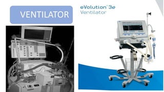
10. A medical ventilator may be defined as machine
designed to mechanically move breatheable air
into and out of the lungs, for a patient who is
physically unable to breathe, or breathing
insufficiently.
Ventilators provide temporary full ventilatory
support or respiratory assistance to patients who
cannot breathe on their own or who require
assistance to maintain adequate ventilation.
Introduction
11. Two types of ventilator
A. Negative-Pressure Ventilators
B. Positive-Pressure Ventilators
Introduction
12. Negative-Pressure Ventilators
Same the natural respiration
In the natural inspiration is a result of negative pressure in
the pleural cavity generated by distention of the diaphragm
Introduction
14. Positive-Pressure Ventilators
During inspiration, the inspiratory flow delivery system creates a positive pressure in the tubes connected to
the patient airway, called patient circuit , and the exhalation control system closes a valve at the outlet of the
tubing to the atmosphere.
When the ventilator switches to exhalation, the inspiratory flow delivery system stops the positive pressure
and the exhalation system opens the valve to allow the patient’s exhaled breath to flow to the atmosphere
positive-pressure ventilators have been very successful in treating a variety of breathing disorders and have
become more popular than negative-pressure ventilators.
Positive-pressure ventilators have been employed to treat patients ranging from neonates to adults.
Introduction
15. A high-frequency ventilator
A high-frequency ventilator uses positive pressure to deliver breaths at frequencies much
higher than the normal breathing rate (e.g., >100 breaths/min).
High-frequency ventilators were developed in an effort to reduce barotrauma and some
of the deleterious hemodynamic effects associated with the high tidal volumes and
positive pressure used with conventional ventilators.
These ventilators are available for patients who cannot tolerate the airway pressures
needed for ventilation at typical volume
Introduction
17. Controls
- Controls are used to select breathing mode and ventilation pattern
parameters (e.g., tidal volume, breathing rate).
- Several parameters can be independently set, such as length of the
inspiratory or expiratory phase, rate of mechanical breaths, (I:E ratio),
waveform shape, tidal volume, minute volume , peak inspiratory flow,
peak pressure, and positive end-expiratory pressure (PEEP).
- The I:E ratio is an indication of the partitioning of a breath into
inspiration and expiration.
Principle of operation
19. Mandatory Mode
When the patients need the ventilator to completely take over the task
of ventilating their lungs.
Spontaneous Mode
Some patients are able to initiate a breath and breathe on their own,
but may need oxygen-enriched air flow or slightly elevated airway
pressure.
Ventilation Modes
20. Mandatory Mode:
A. Volume controlled ventilation: which presently is more popular, refers to delivering a
specified tidal volume to the patient during the inspiratory phase.
B. Pressure controlled ventilation: refers to raising the airway pressure to a level .
-Continuous mandatory ventilation (CMV): the tidal volume and frequency are fixed and
there is no synchronization with the patient’s respiratory efforts.
-Intermediate mandatory ventilation (IMV): was developed from CMV to allow patients to
breath spontaneously in between the mandatory breaths.
Ventilation Modes
21. Spontaneous Mode
1)Continuous Positive Airway Pressure (CPAP) mode:
-In this mode, the ventilator maintains a positive
pressure at the airway as the patient attempts to
inspire.
-The therapist sets the sensitivity level lower than
PEEP. When the patient attempts to breathe, the
pressure drops below the sensitivity level and the
ventilator responds by supplying breathable gases to
raise the pressure back to the PEEP level.
Ventilation Modes
22. 2) Pressure Support in Spontaneous Mode:
(PSSM)
- This mode is similar to the CPAP mode with
the exception that during the inspiration the
ventilator attempts to maintain the patient
airway pressure at a level above PEEP.
- In this mode, when the patient’s airway
pressure drops below the sensitivity line, the
ventilator raises the airway pressure above
PEEP, selected by the therapist. The ventilator
stops the flow of breathable gases when the
patient starts to exhale and controls the
exhalation valve to achieve the set PEEP level.
Ventilation Modes
23. 3) Synchronized intermittent
mandatory ventilation (SIMV)
mode:
- In this mode, delivers controlled
breaths at a set frequency and
also allows the patient to breathe
spontaneously with no assistance
during the periods between the
controlled breaths.
Ventilation Modes
24. 4) Airway-pressure-release ventilation (APRV) mode:
- APRV may be used to treat acute lung injury in patients who require
mechanical support.
5) Neurally Adjusted Ventilator Assist (NAVSA) mode:
- A probe senses the electrical activity of the diaphragm, which triggers
the ventilator
Ventilation Modes