2. Introduction
It is well known that an early diagnosis of canine displacement
and prediction of subsequent impaction is important to decrease
the patient’s need for surgical exposure and prolonged orthodontic
treatment with additional costs and various complications.1–3
Different interceptive treatment alternatives for palatally displaced
canines (PDCs) have been the focus of numerous prospective studies
for the last 15 years,4–9
despite systematic reviews reporting that the
scientific evidence is too sparse to support an interceptive approach.10,11
Since then, several randomized clinical trials have come to the same
conclusion; namely, that extraction of the deciduous canine is an effec-
tive interceptive treatment in patients with PDC.12,13
However, not all
permanent canines erupt, as pointed out in the study by Naoumova
et al.14
, where the authors tried to identify which cases benefit from
extraction of the preceding deciduous canine. A small mesioangular
angle, a long distance from the canine cusp tip to the midline and a
short distance from the canine cusp tip to the maxillary dental arch
plane, measured with cone beam computed tomography (CBCT),
were suggested as predictors of a successful outcome, with the distance
between the canine cusp tip to the midline being the best predictor. In
the literature, there are many other suggestions of predictors of canine
eruption.Ericson and Kurol15
found that a more mesially located crown
or a more horizontally positioned PDC, measured with angles and sec-
tors, reduced the chance of eruption after deciduous canine extraction.
These results were confirmed by Power and Short,16
who found that an
angulation of 31 degrees or more to the midline decreases the chance
of successful eruption. Additional reported predictors are the vertical
distance from the canine tip to the occlusal plane,14,17–19
from the canine
to the first premolar angle, and the distance from the canine cusp tip to
the midline, measured with CBCT.19,20
Comparing sector location21,22
and angulation22
as predictors of
possible impaction of the permanent canine indicates that the sec-
tor location is a better predictor, and that canines overlapping the
adjacent lateral incisor will become impacted in 78–82% of cases.
Predictors based on aetiology for early identification of patients who
may later develop PDCs have also been widely investigated. The aeti-
ology of PDCs appears to be multifactorial with a genetic complex
that controls other concomitant dental anomalies.23
The suggested
associated dental anomalies seen in the literature are agenesis of the
second premolars, small size of or agenesis of the lateral maxillary
incisors, infraocclusion of the primary molars, enamel hypoplasia,
ectopic eruption of the first permanent maxillary molars, distal
angulation of the lower second premolars, morphological devia-
tions of the maxillary incisors and the dentition in general. Since
some of these tooth disturbances may occur before the maxillary
canine becomes palatally displaced, they can be used as early risk
indicators.24–31
Although eruption prediction based on the position of the per-
manent canine has been reported in several previous studies, none of
the above-mentioned studies had a prospective randomized control
design, except for part I of the present study14
using CBCT images.
Accordingly, it is important to identify cut-off points for a successful
outcome of interceptive deciduous canine extraction on the pano-
ramic radiograph (PAN), as this is more extensively used in daily
practice than CBCT. It is also important to examine whether there
are any side effects on the dentition from extracting the deciduous
canine, especially with unilateral extraction, and to assess other den-
tal deviations. None of the clinical studies referred to above,4–9,15,16
evaluating the success of interceptive treatment in patients with PDC,
has reported any side effects, except for one study, which noted that
the maxillary midline was not affected by unilateral extraction.12
Aims
The primary aim of this trial was to:
• Analyse whether possible predictors and cut-off points can be
found on the PAN, when considering whether interceptive ex-
traction of the deciduous canine is beneficial or not during the
mixed dentition in patients with a PDC.
The secondary aims were to:
• Report any side effects on the dentition by unilateral extraction
of the deciduous canine.
• Describe the frequency of other dental deviations than PDC on
the PANs of the present sample.
Hypothesis
The null hypothesis was that a successful outcome (emergence of the
permanent maxillary canine through the gingiva) following intercep-
tive extraction of the deciduous canine is not influenced by the pos-
ition or the angulation of the PDC measured on a PAN. The second
hypothesis was that unilateral extraction of the deciduous canine
does not cause any side effects on the dentition.
Materials and methods
Ethical issue
The research ethics committee of the Sahlgrenska Academy at the
University of Gothenburg, Sweden (Reg. no. 578-08) and the radi-
ation protection committee, Sahlgrenska Academy at the University
of Gothenburg, Sweden approved this study. Before participation,
informed consent was provided by the child and the parent or by an
adult with parental responsibilities and rights in accordance with the
Declaration of Helsinki.
Registration
This trial was registered in the Research and Development database
in ‘FoU i Sverige’, http://www.fou.nu/is/sverige, registration number
211141.
Subjects
Study setting and eligibility criteria
Dental general practitioners (DGPs) from 15 Public Dental Clinics
in Gothenburg, Västra Götaland County Council, Sweden, identi-
fied patients during the period from September 2008 to January
2011, and the consulting orthodontist invited the potential
patients to participate in the study. A more detailed explanation
of the subjects and the study setting can be found in part I of
this trial.13
The inclusion criteria were:
• Children aged 10–13 years with maxillary unilateral or bilateral
PDCs;
• Persisting deciduous canines;
• No previous experience of orthodontic treatment.
Palatal displacement of the maxillary permanent canine was con-
sidered if there was an absence of a labial bulge and/or presence
of a palatal bulge and when the canine crown was diagnosed on
intraoral radiographs as being palatally positioned, using Clark’s
rule.32
Intraoral radiographs were taken by the DGPs.
The exclusion criteria were:
• Crowding in the lateral part of the maxilla exceeding 2 mm;
• On-going orthodontic treatment;
European Journal of Orthodontics, 2018
2
Downloaded from https://academic.oup.com/ejo/advance-article-abstract/doi/10.1093/ejo/cjy002/4859689
by Durham University Library user
on 16 February 2018
3. J. Naoumova and H. Kjellberg 3
• Resorption of the adjacent teeth, grade 3 and 4 according to
Ericson and Kurol,3
either at the start or during the trial and
caused by the displaced canine;
• Craniofacial syndromes;
• Odontomas, cysts;
• Cleft lip and/or palate.
Trial design and randomization
This randomized clinical trial (RCT) was designed as a parallel trial
with an allocation ratio of 1:1. Patients enrolled were randomly allo-
cated to extraction of the deciduous canine (EG) or to a control group
(CG). Patients with bilateral PDCs were randomized to have either
the right or the left deciduous canine extracted. The permuted block
randomization method was used. The allocations were concealed in
sequentially numbered sealed opaque envelopes that were opened by
a dental nurse after written consent was obtained. An intention-to-
treat analysis (ITT) was performed in the study; thus, all participants
were analysed according to the intervention to which they were allo-
cated, regardless of what treatment they actually received and of sub-
sequent withdrawal from treatment or deviation from the protocol.
Dropouts were considered as unsuccessful outcomes.
Treatment protocol and process
All patients underwent a clinical examination, including intra-
and extra-oral photography, at baseline (T0), after 6 (T1) and
after 12 (T2) months, at the Orthodontic Specialist Clinic in
Gothenburg, Sweden. Radiographic examination was performed at
the Department of Oral and Maxillofacial Radiology, the Institute
of Odontology at the Sahlgrenska Academy, Gothenburg, Sweden,
consisting of CBCT (study I, II),13,14
and PANs were taken in con-
nection with the clinical examination. If the canine was clinically
visible at the 6-month or 12-month control, no radiographs were
taken. After 12 months, an individual treatment plan was decided
on for the PDCs that had not erupted. Canines that had improved
their position, judged from measurements made on the radiographs,
were followed until they emerged through the gingiva. In the CG,
the deciduous canine was extracted if there was no mobility of the
tooth. However, if the canine was impaired or had not changed its
position after 12 months, surgical exposure followed by orthodontic
treatment was initiated (Figure 1).
Blinding
Data from the PAN at T0 were compiled as blinded data to the oper-
ator. Blinding was not possible at T2 since the extraction site was
visible. However, all data were blinded to the statistician who made
the analysis. One orthodontist made the measurements on the PAN
and on the clinical images, while an oral radiologist performed the
radiographic assessment of dental deviations.
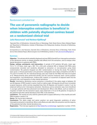
Measures of treatment effect
The initial canine position was assessed on the PAN using the
method first described by Ericson and Kurol.15
The following linear
and angular measurements were measured using the Facad Software
(version 3.0, Ilexis AB, Linköping, Sweden) (Figure 2):
• Alpha angle: the angle formed by the long axis of the canine
and the midline.
• D distance: the distance in mm from the canine cusp tip to the
occlusal plane.
• Sector: mesiodistal crown position in sector 1–5.
The root development of each PDC was assessed with the method
developed by Nolla.33
The side effects were assessed by clinical examination and by
visual assessment on intra-oral and extra-oral images at T0 and at
T2. The following parameters were recorded for patients in the EG:
• Midline change in the upper arch (measured clinically with
a ruler in centric relation between the two central incisors
Figure 1. Flowchart describing the protocol and the patients included in the
study. N, amount of patients of PDCs: palatal displaced canines; SD, standard
deviation; PAN, panoramic radiograph;T0, baseline;T1, 6 months control;T2,
12 months control.
Figure 2. Measurements made on the panoramic radiograph according to
the method first described by Ericson and Kurol15
: a-angle, angle formed by
the long axis of the canine and the midline; d-distance, distance in mm from
the canine cusp tip to the occlusal plane (OP); and sector, mesiodistal crown
position in sector 1–5.
Downloaded from https://academic.oup.com/ejo/advance-article-abstract/doi/10.1093/ejo/cjy002/4859689
by Durham University Library user
on 16 February 2018
4. using a reference line: vertical line through the Glabella and
Subnasale in the natural head position).
• Rotation or movement of the adjacent teeth into the extrac-
tion site.
The following deviations inspired by Sørensen et al.29
were registered
on the PAN and supplemented with CBCT when needed:
• Morphology:
- Invaginations: fillings at the normal locations of invagina-
tions and in teeth with radiographically distinct enamel
notching;
- Narrow-shaped incisor crowns: narrower incisal width
than the width at the column;
- Taurodontic molar roots: mesotaurodontia and hypertau-
rodontia were recorded according to the criteria defined
by Schulze34
;
- Malformed roots of the upper incisors: equal length of
the root or shorter than the height of the crown, slender/
narrow root.
• Agenesis, except the third molars.
• Eruptional deviations beyond canine displacement.
The following outcome measures were assessed:
Primary outcomes
• To compare successful (defined as permanent maxillary ca-
nine emerged through the gingiva) and unsuccessful cases
(PDCs that did not erupt, despite extraction of the de-
ciduous canine), and to identify predictors with possible
cut-off points regarding which cases would benefit from de-
ciduous canine extraction.
Secondary outcomes
• To document whether extraction of the deciduous canine
affects the upper midline and/or causes rotation or move-
ment into the extraction site of the adjacent teeth.
• To describe the frequency of other dental deviations in the
current sample.
Statistics
Sample size calculation
The sample size calculation has previously been described in part
I of this study.13
The statistical analysis was performed using SAS, version 9.3
for Windows (SAS Institute Inc., Cary, North Carolina, USA). P
values less than 0.05 were considered statistically significant. For
numerical variables, arithmetic means and standard deviations
were calculated. Dependent and independent t-tests were used for
comparison of baseline variables between and within the groups.
The bilateral PDC group was tested for independence with Fisher’s
exact test and McNemar’s test.13
Independent t-tests were used to
test whether there were any significant differences in the numerical
values between successful and non-successful outcomes. Fisher’s
exact test was used to calculate differences in categorical data.
To detect possible predictors and to determine cut-off points
for successful and unsuccessful outcomes, respectively, logistic
regression analysis and receiver operating characteristic (ROC)
curve analysis were performed. The accuracy of the clinical test
was measured by the area under the curve (AUC). The following
rough guideline was used for the interpretation of the AUC: 0.50
to 0.75 = fair; 0.75 to 0.92 = good; 0.92 to 0.97 = very good; 0.97
to 1.00 = excellent.
The method of error was calculated using the Dahlberg formula35
on 20 randomly selected subjects, and measured on two separate
occasions with 3 months in between.
Harm
No harm was detected during the study.
Results
The repeatability for angular measurements was 0.5 degree and
for the distance measurements 0.2 mm. The assessment of canine
displacement, root development and dental deviations showed
a reproducibility of 100%. One operator performed all the
measurements (JN).
Participant flow
Three patients declined to participate before the randomization pro-
cedure. Thus, in total, 67 patients were randomly allocated to the EG
or the CG. Forty-five patients had a unilateral PDC (29 girls, mean
age ± SD: 11.2 ± 1.1; 16 boys, mean age ± SD: 11.7 ± 0.8) and 22
patients had bilateral PDCs (11 girls, mean age ± SD: 11.5 ± 0.9; 11
boys, mean age ± SD: 11.6 ± 0.8) (Figure 1).
Baseline findings
The unilateral and bilateral groups did not show any statistically
significant differences regarding right and left extraction side. There
were no significant difference in gender and age between the EG and
CG; however, there were more females than males and more 10- to
11-year-olds than individuals aged 12–13 years old, in total. No sig-
nificant differences were seen either for the radiographic baseline
measurements between the EG and the CG or between the unilateral
and bilateral group (Table 1). All patients had PDCs with root de-
velopment of Nolla stage33
8–10 (8: two thirds of root completed, 9:
root almost completed, 10: root completed).
More detailed information about patient characteristics, success
rate between the groups, mean eruption time, and the number of
patients who had surgical exposure and orthodontic treatment or
root resorption are presented in part I of this trial.
Primary outcomes
Predictors and cut-off points
In the present sample, there were no PDCs located in sector 1 or
5. The PDCs that showed spontaneous eruption in the non-extrac-
tion group had a significantly smaller alpha angle and were posi-
tioned in a ‘lower sector’; i.e. showed less overlapping of the incisors
in comparison with canines that erupted spontaneously after extrac-
tion of the deciduous canine. In the group that showed no eruption,
either with or without extraction of the deciduous canine, the PDCs
were positioned significantly more horizontally (i.e. larger alpha
angle) in a higher sector, and root development was more advanced
(Figure 3, Table 2). The children who showed no spontaneous erup-
tion were also older than the children in the eruption group.
A logistic regression analysis was made on the significant base-
line variables that affected the main outcome (Table 2). The analysis
revealed that extraction of the deciduous canine was the variable
most effecting eruption of the PDC followed by alpha angle, sec-
tor measurement, and the age of the patient (Table 3). ROC curve
European Journal of Orthodontics, 2018
4
Downloaded from https://academic.oup.com/ejo/advance-article-abstract/doi/10.1093/ejo/cjy002/4859689
by Durham University Library user
on 16 February 2018
5. J. Naoumova and H. Kjellberg 5
analyses were therefore made on two radiographic variables in
patients with erupted PDCs; i.e.‘easy cases of PDCs’, and in patients
in the EG where no eruption was seen and who had to be treated
with surgical exposure; i.e. ‘severe cases of PDCs’.
The ROC analysis showed that both the alpha angle and the sec-
tor could be used to determine a cut-off point; thus, we can reject
our null hypothesis.
‘Easy cases of PDCs’; i.e. canines erupted in the CG, had a
cut-off point of 20 degrees of alpha angle with a sensitivity of
0.931 and a specificity of 0.948 (P = 0.000), and an AUC of 0.980
(95% confidence interval [CI] 0.972 to 1.000). For spontaneous
eruption without prior deciduous canine extraction it was essen-
tial that the PDCs were located initially in sector 2 (AUC; 0.932
(95% CI 0.841 to 0.990); sensitivity 0.912; specificity 0.856;
P = 0.000) (Figures 4 and 5).
‘Severe cases of PDCs’; i.e. canines that did not erupt in the CG,
had a cut-off of 30 degrees alpha angle (sensitivity, 0.935; speci-
ficity, 0.898; P = 0.000). The AUC was 0.940 (95% CI 0.924 to
1.000), suggesting a very good discriminatory power. PDCs located
in sector 4 did not erupt despite deciduous canine extraction (AUC;
0.990 (95% CI 0.990 to 1.000); specificity, 0.950; sensitivity, 0.913,
P = 0.000) (Figures 4 and 6).
Secondary outcomes
Side effects
Fifteen out of 35 patients showed minor side effects 1 year after the
extraction of the deciduous canine. Eight of these patients had surgi-
cal exposure of the canine.An increasing midline shift of 0.5–1.5 mm
to the extraction side was noticed in 6 of the 35 patients after 1 year.
In 37% of the patients, rotation (n = 6 premolars) or movement
(n = 4 premolars, n = 4 laterals) into the extraction sites was seen.
These side effects were seen already after 6 months and after that, no
additional rotation/movement occurred (Supplementary Figures 7
and 8). The null hypothesis regarding the side effects can be rejected.
Dental deviations
Only 16% of the patients had no deviations in the dentition, all of
them in the unilateral group (Table 4). All patients with bilateral
PDCs had some malformations in the dentition and females had
more malformations than males. Eruptional deviations of 36 teeth
were found in 39% of the patients (n = 26 patients); most of them
premolars (n = 12) in the maxilla in the unilateral group, while in
the bilateral group, an almost equal number of upper (n = 8) and
lower ectopic premolars (n = 7) was noted. Five teeth were ectopic
canines in the lower arch (n = 3 in the unilateral group and n = 2 in
the bilateral group). In the unilateral group, four teeth were buccally
Table 1.
Baseline
variables
(T0)
for
the
unilateral
and
bilateral
groups
with
mean,
standard
deviations
(SD),
and
p
values.
Unilateral
PDC
(patients,
n = 45)
Bilateral
PDCs
(patients,
n = 22)
Variable
at
T0
EG
(n = 24
PDC),
mean
±
SD,
11.2 ± 0.9
CG
(n = 21
PDC),
mean
±
SD,
11.4 ± 1.1
P-value*
EG
(n = 22
PDC),
mean
±
SD,
11.6 ± 1.0
CG
(n = 22
PDC),
mean
±
SD,
11.6 ± 1.0
P-value*
EG–CG,
P-value*
Alpha
angle
(°)**
26.9 ± 5.2
25.9 ± 4.9
0.806
25.4 ± 5.0
26.6 ± 4.5
0.898
NS
d
distance
(mm)**
15.0 ± 2.6
15.9 ± 2.6
0.535
15.9 ± 2.8
15.4 ± 2.5
0.513
NS
Sector
1,
n
(%)**
0
(0)
0
(0)
1.000
0
(0)
0
(0)
1.000
NS
Sector
2,
n
(%)**
14
(31)
12
(27)
0.245
11
(25)
10
(23)
0.352
NS
Sector
3,
n
(%)**
7
(15.5)
7
(15.5)
1.000
8
(18)
8
(18)
1.000
NS
Sector
4,
n
(%)**
3
(7)
2
(4)
0.358
4
(9)
3
(7)
0.452
NS
Sector
5,
n
(%)**
0
(0)
0
(0)
1.000
0
(0)
0
(0)
1.000
NS
Root
development
of
PDC***
8.6 ± 1.0
8.1 ± 0.6
0.292
8.4 ± 0.8
8.45 ± 0.8
1.000
NS
CG,
control
group;
EG,
extraction
group;
PDC,
palatally
displaced
canines;
SD,
standard
deviation;
NS,
not
significant.
*P
value
<0.05
is
considered
statistically
significant.
**Described
in
Figure 1.
***According
to
Nolla
study.
33
Figure 3. Percentage of spontaneous eruptions of palatal displaced canines
after extraction of the deciduous canine (left drawing) or non-extraction
(right drawing), by sector before extraction.
Downloaded from https://academic.oup.com/ejo/advance-article-abstract/doi/10.1093/ejo/cjy002/4859689
by Durham University Library user
on 16 February 2018
6. displaced in the maxilla. Agenesis of premolars was observed in 15%
of the patients (n = 10 patients), more in the bilateral group (n = 14)
than in the unilateral group (n = 6), and 16 out of 20 cases of agen-
esis referred to premolars in the mandible. Only one patient had
agenesis of laterals.
An equal number of invaginations (n = 58% of the patients,
n = 39 patients), narrow-shaped crown of laterals (n = 43% of the
patients, n = 29 patients), and malformed incisor roots (n = 15% of
the patients, n = 10 patients) was seen in the unilateral and bilateral
group. Sixteen taurodontic molars (n = 3 patients) were found only
in the unilateral group. Suppementary Figure 9 shows two PANs
exemplifying other dental deviations found in the current PDC
patients.
Discussion
Interceptive extraction of the deciduous canines in patients with
PDC during the mixed dentition has been shown to be an effective
measure.11–12
However, since not all canines erupt spontaneously,
efforts have been made to develop guidelines for when intercep-
tive extraction is beneficial,11,15–22
which was also the focus of
the present study. Our findings show that both the alpha angle
and sector measurements made on PAN are good predictors of
whether the canine will erupt spontaneously or not. This is in
accordance with previous studies,15–17
while other studies report
that the pre-treatment alpha angle is not correlated with a suc-
cessful outcome.36,37
The percentage of spontaneous eruption of PDCs decreased,
the more mesially located the canine crown, which is similar to
what Ericson and Kurol15
found, although the numbers differ from
the present study. This can be explained by the authors consider-
ing both canine eruption and improvement of the canine eruption
path as successful outcomes, and by previous studies also includ-
ing PDCs located in sector 1. It could be debated whether or not
canines in sector 1 should be defined as PDCs instead of normally
erupting canines. Looking at the literature there are no consensus
about the precise definition of normally erupting canines or PDCs
as highlighted by Hadler-Olsen et al.38
The wide range of inclusion
of canines with a certain alpha angle or sector measurements6,9,22,38
makes it difficult to compare the studies.
Sector measurements have shown to be the single most impor-
tant prognostic factor,14,16
but according to our study, both the alpha
angle and sectors are similarly good predictors. A cut-off point of
an alpha angle of more than 30 degrees was found to be associated
with a notably decreased chance of successful eruption, which has
also been reported previously by Power and Short.16
The novelty
in the current study is a more detailed description on when inter-
ceptive extraction is beneficial; i.e. in cases with an alpha angle of
20–30 degrees, canines located in sector 2–3, and when the operator
could wait and observe; for instance, for canines with an alpha angle
of less than 20 degrees located in sector 2.
Additional predictors reported in the literature are the vertical
distance from the canine tip to the occlusal plane measured on
PAN17,18
—a finding that is not in accordance with our study or with
other earlier studies.4,15,16
In most of the previous studies,4–9,12,15–20
as well as in the pre-
sent study, the effect of extracting the deciduous canine was assessed
on PAN using the measurement method developed by Ericson and
Kurol.15
However, lately, predictors retrieved from CBCT examina-
tions have also been presented. A small mesioangular angle, a long
distance from the canine cusp tip to the midline and a short distance
Table 2.
Differences
in
baseline
measures
(T0)
between
canines
that
erupted
spontaneously
after
1 year
and
non-erupting
canines.
Erupted
(n = 40
patients,
48
PDC)
Non-erupted
(n = 27
patients,
41
PDC)
EG
CG
EG
CG
Variables
at
T0
Mean
±
SD
a
Mean
±
SD
a
Differences
mean
(95%
CI)
P-value,
EG–CG*
Mean
±
SD
Mean
±
SD
Differences
mean
(95%
CI)
P-value,
EG–CG
P-value,
erupted
versus
non-erupted*
Alpha
angle
(°)**
24.8 ± 3.5
17.7 ± 2.5
8.1
(6.1,
8.1)
0.000
31.9 ± 4.2
31.6 ± 3.7
0.3
(−2.2,
2.9)
0.777
0.000
d
distance
(mm)**
15.7 ± 2.4
15.7 ± 1.9
−0.0
(−1.3,
1.3)
0.998
15.2 ± 2.8
15.7 ± 2.9
−0.5
(−2.4,
1.4)
0.597
0.550
Sector**
3.0 ± 0.4
2.0 ± 0.6
1.0
(0.2,
0.8)
0.000
3.6 ± 0.6
3.8 ± 0.5
−0.2
(0.1,
0,3)
0.890
0.005
Root
development
of
PDC***
8.2 ± 0.7
8.1 ± 0.5
0.1
(−0.1,
0.3)
0.800
8.9 ± 1.0
8.7 ± 0.8
0.2
(0,
0.4)
0.638
0.050
Age,
years
11.0 ± 0.9
10.7 ± 0.99
0.1
(0.6,
0.7)
0.676
12.0 ± 1.0
11.9 ± 1.0
1.0
(2.3,
0.8)
0.112
0.000
SD,
standard
deviation;
CI,
confidence
interval;
EG,
extraction
group;
CG,
control
group. Bold
indicates
the
values
are
significant.
*P
value
<0.05
is
considered
statistically
significant.
**Described
in
Figure 1.
***According
to
Nolla
study.
33
European Journal of Orthodontics, 2018
6
Downloaded from https://academic.oup.com/ejo/advance-article-abstract/doi/10.1093/ejo/cjy002/4859689
by Durham University Library user
on 16 February 2018
7. J. Naoumova and H. Kjellberg 7
from the canine cusp tip to the maxillary dental arch have been sug-
gested as predictors of a successful outcome.14,18
In daily practice, PAN and intra-oral radiographs are more fre-
quently used than CBCT, making cut-off points on 2D images of
greater clinical value. However, since it is difficult to have a stand-
ardized projection with intra-oral radiographs, reliable predictors
cannot be achieved and the use of PAN is therefore an acceptable
substitute for CBCT for predicting the outcome.
The root development of the displaced canine was assessed accord-
ing to the method developed by Nolla.33
Canines that did not erupt,
regardless of whether or not deciduous canine extraction had been per-
formed, had significantly more advanced root development than the
canines that did erupt. Similar results have been shown previously.6,8
Minor midline shifts in the maxilla were observed in 17% of the
patients after one-sided deciduous canine extraction, which is contrast
with the findings in a previous study.12
The differences may depend on
how the midline was measured. In our study, the midline was assessed
clinically, while in the other study it was measured on study casts.
Rotation or movement into the extraction site was noticed in 37%
of the patients after 6 months, with no additional changes during the
Table 3. Logistic regression calculated on the baseline means fromTable 2 that affected successful eruption of the PDC.
Variables at T0 β df OR 95% CI Sig.
Extraction versus non-extraction 2.833 1 288.877 8.846 to >999.999 0.0014
Alpha angle (°) −2.120 1 8.333 1.988 to 34.488 0.0037
D-distance (mm) −0.504 1 1.037 0.863 to 1.109 0.1617
Sector −1.350 1 7.500 1.450 to 6.386 0.0029
Root development of PDC −1.032 1 2.249 0.964 to 2.739 0.0620
Age, years −1.732 1 5.649 1.464 to 21.739 0.0120
β, beta; df, degree of freedom; OR, odds ratio; CI, confidence interval; Sig., statistical significance. Bold indicates the values are significant.
Figure 4. Schematic drawing with cut-off points for sector and alpha angle,
showing when extraction of the deciduous canine in patients with palatal
displaced canine is beneficial.
Figure 5. Eleven-year-old boy with bilateral palatal displaced canines. Sixty-
three was randomized for extraction and at the 12-month control, 23 (baseline
alpha angle: 23.5 degree, sector: 2) was under eruption as well as 13 (baseline
alpha angle: 19.7 degree, sector: 2). Looking at the case retrospectively and
applying the cut-off points, it was a good choice to extract the deciduous
canine on the left side only.
Figure 7. Eleven-year-old girl with 23 palatal displaced canines. Note the
distal movement of the lateral incisor and the mesial movement and the
slight rotation of the first premolar into the extraction site at the 6-month
control (T1). No further movement was seen at the 12-month control (T2).
Patient had surgical exposure of 23.
Figure 6. Twelve-year-old girl with bilateral palatal displaced canines.
Fifty-three was randomized for extraction and at the 12-month control, 13
(baseline alpha angle: 35.2 degree, sector: 4) and 23 (baseline alpha angle:
21.2 degree, sector: 3) had become more ectopically positioned and were
surgically exposed. Looking at the case retrospectively and applying the cut-
off points, it would have been beneficial to extract the deciduous canine on
the left side at baseline and to expose surgically the permanent canine on
the right side.
Downloaded from https://academic.oup.com/ejo/advance-article-abstract/doi/10.1093/ejo/cjy002/4859689
by Durham University Library user
on 16 February 2018
8. rest of the observation period. This is slightly different from what
Bazargani et al.12
found; namely, that the space at the extraction site
continued to decrease during their observation time of 18 months.
Thus, the null hypothesis regarding the side effects can be rejected.
Even though no other dental deviations than PDCs were assessed
in a larger population sample, the findings in the present study were
largely similar to previous studies, including other eruptional devia-
tions such as ectopic premolars or mandibular canines, agenesis of
premolars, small size of lateral maxillary incisors, invaginations
and taurodontia.24–30,39
Almost 80% in the unilateral group and all
patients in the bilateral group had some malformations and these
were more often observed in females than in males, supporting the
theory of the aetiology of PDCs being multifactorial with a genetic
complex controlling other dental anomalies.23
These dental devia-
tions, especially those occurring before the maxillary canines become
palatally displaced, may be used as early clinical predictors. Since
only patients with no space deficiencies were included in the trial, this
could explain why there were no ectopic eruptions of the first per-
manent maxillary molars or other dental anomalies associated with
space deficiency.39
Clinical implications
Our recommendation is to use the cut-off points presented in this
article as guidelines to decide whether an interceptive extraction
should be performed or not. A prospective study assessing the effect-
iveness of using the guidelines would be a clinically relevant future
study. Interceptive extraction is most likely to be beneficial in cases
with an alpha angle of 20–30 degrees located in sector 2–3. Selecting
cases that would benefit from interceptive extraction would be eco-
nomically advantageous to both patients and clinicians.
Unnecessary extractions could most likely be avoided in PDC
cases with alpha less than 20 degrees, located in sector 2, which
would reduce the number of patients being exposed to the potential
pain and discomfort after extraction.40
However, it is the clinician’s
responsibility to follow and observe the permanent canine until it
erupts in order not to miss a feasible change in the eruption path.
A reasonable follow-up period with apical radiographs to monitor
the eruption would be after approximately 10 months instead of
6 months, which has been the recommendation earlier.15
This inter-
val can also be used in cases where the deciduous canine is extracted,
since part I of the present study showed that the majority of the
permanent canines erupted after 12 months with the latest after
22 months.13
In addition, the CBCT showed, both in the extracted
and non-extracted cases, that the degree of resorption of the adjacent
teeth was low (grade 2) at baseline and did not increase significantly
during the 12-month observation period.14
Future studies assessing
the length of the control intervals may reveal whether our recom-
mendations can be further extended.
In severe PDC cases; i.e. an alpha angle of more than 30 degrees,
located in sector 4, where interceptive extraction most likely is not
effective, treatments such as surgical exposure, with simultaneous
extraction of the deciduous canine, followed by the treatment of
aligning the retained canine, could begin earlier. This might decrease
the risk of the canine becoming more impacted but also minimize the
risk of root resorption of the adjacent teeth.
Limitations
Since the criterion for exclusion was crowding in the maxilla
exceeding 2 mm, a conclusion cannot be drawn as to whether
crowded cases would also benefit from deciduous canine extrac-
tion or which side effects might occur in such cases. Other dental
Table 4.
Prevalence
of
dental
deviations
in
the
unilateral
and
bilateral
(palatally
displaced
canine)
PDC
group.
Dental
deviations
PDC
group
Invaginations,
N = 58%
Narrow-shaped
incisor
crowns,
N = 43%
Malformed
roots,
N = 15%
Agenesis,
N = 15%
Taurodontic
molars,
N = 0.5%
Eruptional
deviations,
N = 39%
Other
malformations
Unilateral
PDC
(n = 45
patients)
UJ:
31
incisors
UJ:
21
incisors
UJ:
9
incisors
UJ:
1
premolar;
LJ:
5
premolars
UJ:
10
molars;
LJ:
6
molars
UJ:
12
premolars,
4
BDC;
LJ:
3
BDC
—
Bilateral
PDCs
(n = 22
patients)
UJ:
28
incisors
UJ:
25
incisors
UJ:
7
incisors
UJ:
3
premolars,
2
incisors;
LJ:
11
premolars
UJ:
8
premolars;
LJ:
7
premolars,
2
canines
—
The
percentage
expresses
percentage
of
patients
in
the
total
PDC
group.
PDC,
palatally
displaced
canine;
BDC,
bucally
displaced
canine;
UJ,
upper
jaw;
LJ,
lower
jaw.
European Journal of Orthodontics, 2018
8
Downloaded from https://academic.oup.com/ejo/advance-article-abstract/doi/10.1093/ejo/cjy002/4859689
by Durham University Library user
on 16 February 2018
9. J. Naoumova and H. Kjellberg 9
deviations might also have been noted if crowded cases had been
included. However, including crowded cases would have meant the
inclusion of a confounding factor. In our opinion, crowded cases
with PDC will also need treatment of the crowding, in addition
to extraction of the deciduous canine. Gaining space with rapid
maxillary expansion5,6
or with headgear4,7,41
has shown to have a
positive effect on PDCs. Resorption of adjacent teeth with grades
3 and 4 was also excluded, as these cases require another treat-
ment strategy, such as surgical exposure of the impacted canine
followed by orthodontic traction and, in some cases, extraction of
the resorbed tooth.
Patients were included by their chronological age instead of
their dental developmental stage, which is a limitation as there is
poor correlation between the dental and chorological age. However,
since most clinicians use the chronological in their daily practice
when assessing dental development, we have kept this protocol. The
majority of the permanent canines erupt between the ages of 10 and
13 years. However, in patients with PDCs, the dental development,
according to several studies, is delayed and it is therefore important
to consider the overall stage of dental development of the child when
assessing PDCs.31
Side effects were assessed unblinded, as the extraction site was
visible and could not be blocked. An alternative would have been to
use an assessor who had no knowledge of the study.
Strict cut-off points were used in this trial to maximize the sensi-
tivity and the specificity. The limitation of the ROC curve analysis is
that the cut-off points may differ in different studies, depending on
whether the operator finds it more important to have high sensitiv-
ity rather than high specificity and vice versa. Since no PDCs were
located either in sector 1 or 5, no conclusion can be drawn as to how
the canine would react in these sectors, but it is reasonable to believe
that a PDC in a lower or higher sector would follow the same path
as those in sector 2 or 4.
Generalizability
The results can be generalized on a Caucasian population aged
10–13 years with PDCs located in sector 2–4 and no space defi-
ciency in the maxilla using the determined cut-off points. Even
though one operator performed all the extractions, the treatment
outcomes could be generalized for a larger number of operators, as
several general practitioners and orthodontic specialists performed
the screening of the patients.
Conclusions
• Alpha angles and sectors measured on a PAN are good
predictors of which PDCs may benefit from an interceptive
deciduous canine extraction.
• Minor side effects are seen after deciduous canine extraction.
• The majority of the patients had dental deviations other than
PDC in the dentition.
Supplementary material
Supplementary data are available at European Journal of
Orthodontics online.
Funding
The Local Research and Development Board for Gothenburg and
Södra Bohuslän; the Health & Medical Care Committee of the
Regional Executive Board, Västra Götaland Region.
Acknowledgement
The authors wish to acknowledge Jüri Kürol, professor emeritus, to inspire
us to this study. We would also like to express our gratitude to Reet Palm,
oral maxillofacial radiologist, for her valuable help with the measurements,
the dental general practitioners of the Public Dental Clinics in Gothenburg,
the consulting orthodontists at the Orthodontic Clinic, University Clinics of
Odontology, Gothenburg, Sweden, and all participants for making this study
possible.
Conflict of Interest
None to declare.
References
1. Becker, A. and Chaushu, S. (2003) Success rate and duration of orthodon-
tic treatment for adult patients with palatally impacted maxillary canines.
American Journal of Orthodontics and Dentofacial Orthopedics, 124,
509–514.
Figure 9. Right PAN: bilateral palatal displaced canines (PDCs) with additional
dental deviations, such as agenesis of mandibular second premolars,
narrow-shaped laterals dens invagination on the laterals. Left PAN: unilateral
PDC with additional dental deviations such as ectopic maxillary second
premolars, narrow-shaped laterals, taurodontic first and second maxillary
and mandibular molars.
Figure 8. Twelve-year-old girl with 13 palatal displaced canines. Note the distal movement of the lateral incisor into the extraction site at the 6-month control
(T1). No further movement was seen at the 12-month control (T2). Patient had surgical exposure of 13.
Downloaded from https://academic.oup.com/ejo/advance-article-abstract/doi/10.1093/ejo/cjy002/4859689
by Durham University Library user
on 16 February 2018
10. 2. Stewart, J.A., Heo, G., Glover, K.E., Williamson, P.C., Lam, E.W.
and Major, P.W. (2001) Factors that relate to treatment duration for
patients with palatally impacted maxillary canines. American Journal of
Orthodontics and Dentofacial Orthopedics, 119, 216–225.
3. Ericson, S. and Kurol, J. (2000) Incisor root resorptions due to ectopic
maxillary canines imaged by computerized tomography: a comparative
study in extracted teeth. The Angle Orthodontist, 70, 276–283.
4. Baccetti, T., Leonardi, M. and Armi, P. (2008) A randomized clinical study
of two interceptive approaches to palatally displaced canines. European
Journal of Orthodontics, 30, 381–385.
5. Baccetti, T., Mucedero, M., Leonardi, M. and Cozza, P. (2009) Interceptive
treatment of palatal impaction of maxillary canines with rapid maxillary
expansion: a randomized clinical trial. American Journal of Orthodontics
and Dentofacial Orthopedics, 136, 657–661.
6. Sigler, L.M., Baccetti, T. and McNamara, J.A., Jr. (2011) Effect of rapid
maxillary expansion and transpalatal arch treatment associated with
deciduous canine extraction on the eruption of palatally displaced canines:
a 2-center prospective study. American Journal of Orthodontics and
Dentofacial Orthopedics, 139, e235–e244.
7. Armi, P., Cozza, P. and Baccetti, T. (2011) Effect of RME and headgear
treatment on the eruption of palatally displaced canines: a randomized
clinical study. The Angle Orthodontist, 81, 370–374.
8. Baccetti, T., Sigler, L.M. and McNamara, J.A., Jr. (2011) An RCT on treat-
ment of palatally displaced canines with RME and/or a transpalatal arch.
European Journal of Orthodontics, 33, 601–607.
9. Alessandri Bonetti, G., Zanarini, M., Incerti Parenti, S., Marini, I. and
Gatto, M.R. (2011) Preventive treatment of ectopically erupting maxil-
lary permanent canines by extraction of deciduous canines and first
molars: a randomized clinical trial. American Journal of Orthodontics and
Dentofacial Orthopedics, 139, 316–323.
10. Saeed, A.A., Kaklamanos, E.G. and Athanasiou, A.E. (2017) Effectiveness
of extraction of primary canines for interceptive management of pala-
tally displaced permanent canines: a systematic review and meta-analysis.
European Journal of Orthodontics. doi: 10.1093/ejo/cjx042.
11. Naoumova, J., Kurol, J. and Kjellberg, H. (2011) A systematic review
of the interceptive treatment of palatally displaced maxillary canines.
European Journal of Orthodontics, 33, 143–149.
12. Bazargani, F., Magnuson, A. and Lennartsson, B. (2014) Effect of intercep-
tive extraction of deciduous canine on palatally displaced maxillary canine: a
prospective randomized controlled study. The Angle Orthodontist, 84, 3–10.
13. Naoumova, J., Kurol, J. and Kjellberg, H. (2015) Extraction of the decidu-
ous canine as an interceptive treatment in children with palatal displaced
canines—part I: shall we extract the deciduous canine or not? European
Journal of Orthodontics, 37, 209–218.
14. Naoumova, J., Kürol, J. and Kjellberg, H. (2015) Extraction of the decidu-
ous canine as an interceptive treatment in children with palatally displaced
canines—part II: possible predictors of success and cut-off points for a
spontaneous eruption. European Journal of Orthodontics, 37, 219–229.
15. Ericson, S. and Kurol, J. (1988) Early treatment of palatally erupting max-
illary canines by extraction of the primary canines. European Journal of
Orthodontics, 10, 283–295.
16. Power, S.M. and Short, M.B. (1993) An investigation into the response of
palatally displaced canines to the removal of deciduous canines and an
assessment of factors contributing to favourable eruption. British Journal
of Orthodontics, 20, 215–223.
17. Smailienė, D., Sidlauskas, A., Lopatienė, K., Guzevičienė, V. and
Juodžbalys, G. (2011) Factors affecting self-eruption of displaced perma-
nent maxillary canines. Medicina (Kaunas, Lithuania), 47, 163–169.
18.
Sajnani, A.K. and King, N.M. (2012) Early prediction of maxillary
canine impaction from panoramic radiographs. American Journal of
Orthodontics and Dentofacial Orthopedics, 142, 45–51.
19. Alqerban, A., Jacobs, R., Fieuws, S. and Willems, G. (2015) Radiographic
predictors for maxillary canine impaction. American Journal of
Orthodontics and Dentofacial Orthopedics, 147, 345–354.
20. Alqerban, A., Storms, A.S., Voet, M., Fieuws, S. and Willems, G. (2016)
Early prediction of maxillary canine impaction. Dento Maxillo Facial
Radiology, 45, 20150232. doi: 10.1259/dmfr.20150232
21.
Lindauer, S.J., Rubenstein, L.K., Hang, W.M., Anderson, W.C. and
Isaacson, R.J. (1992) Canine impaction identified early with panoramic
radiographs. Journal of American Dental Association, 123, 91–97.
22. Warford, J.H., Jr, Grandhi, R.K. and Tira, D.E. (2003) Prediction of
maxillary canine impaction using sectors and angular measurement.
American Journal of Orthodontics and Dentofacial Orthopedics, 124,
651–655.
23. Peck, S., Peck, L. and Kataja, M. (1994) The palatally displaced canine
as a dental anomaly of genetic origin. The Angle Orthodontist, 64,
249–256.
24. Bjerklin, K., Kurol, J. and Valentin, J. (1992) Ectopic eruption of maxillary
first permanent molars and association with other tooth and developmen-
tal disturbances. European Journal of Orthodontics, 14, 369–375.
25. Chaushu, S., Sharabi, S. and Becker, A. (2002) Dental morphologic char-
acteristics of normal versus delayed developing dentitions with palatally
displaced canines. American Journal of Orthodontics and Dentofacial
Orthopedics, 121, 339–346.
26.
Paschos, E., Huth, K.C., Fässler, H. and Rudzki-Janson, I. (2005)
Investigation of maxillary tooth sizes in patients with palatal canine
displacement. Journal of orofacial orthopedics = Fortschritte der
Kieferorthopadie: Organ/official journal Deutsche Gesellschaft fur
Kieferorthopadie, 66, 288–298.
27. Baccetti, T. (1998) A controlled study of associated dental anomalies. The
Angle Orthodontist, 68, 267–274.
28. Becktor, K.B., Steiniche, K. and Kjaer, I. (2005) Association between
ectopic eruption of maxillary canines and first molars. European Journal
of Orthodontics, 27, 186–189.
29. Sørensen, H.B.,Artmann, L., Larsen, H.J. and Kjaer, I. (2009) Radiographic
assessment of dental anomalies in patients with ectopic maxillary canines.
International Journal of Paediatric Dentistry, 19, 108–114.
30.
Shalish, M., Chaushu, S. and Wasserstein, A. (2009) Malposition of
unerupted mandibular second premolar in children with palatally dis-
placed canines. The Angle Orthodontist, 79, 796–799.
31. Odeh, R., Townsend, G., Mihailidis, S., Lähdesmäki, R., Hughes, T. and
Brook, A. (2015) Infraocclusion: dental development and associated
dental variations in singletons and twins. Archives of Oral Biology, 60,
1394–1402.
32. Clark, C.A. (1910) A Method of ascertaining the relative position of
unerupted teeth by means of film radiographs. Proceedings of the Royal
Society of Medicine, 3, 87–90.
33. Nolla, C.M. (1960) The development of permanent teeth. Journal of
Dentistry of Children, 27, 254–266.
34. Schulze, C. (1987) Anomalien und Missbildungen der Zahnform und
Zahngrösse. In: Anomalien und Missbildungen der Menschlichen Zähne.
Quintessenz Verlags-GmbH, Berlin, pp. 219–228.
35.
Dahlberg G. (1940) Statistical Methods for Medical and Biological
Students. George Allen and Unwin, Berlin, pp. 122–132.
36. Baccetti, T., Crescini, A., Nieri, M., Rotundo, R. and Pini Prato, G.P.
(2007) Orthodontic treatment of impacted maxillary canines: an appraisal
of prognostic factors. Progress in Orthodontics, 8, 6–15.
37. Zuccati, G., Ghobadlu, J., Nieri, M. and Clauser, C. (2006) Factors associ-
ated with the duration of forced eruption of impacted maxillary canines:
a retrospective study. American Journal of Orthodontics and Dentofacial
Orthopedics, 130, 349–356.
38. Hadler-Olsen, S., Pirttiniemi, P., Kerosuo, H., Bolstad Limchaichana, N.,
Pesonen, P., Kallio-Pulkkinen, S. and Lähdesmäki, R. (2015) Root resorp-
tions related to ectopic and normal eruption of maxillary canine teeth—a
3D study. Acta Odontologica Scandinavica, 73, 609–615.
39. Peck, S. (2009) Dental anomaly patterns (DAP). The Angle Orthodontist,
79, 1015–1016.
40. Naoumova, J., Kjellberg, H., Kurol, J. and Mohlin, B. (2012) Pain, dis-
comfort, and use of analgesics following the extraction of primary canines
in children with palatally displaced canines. International Journal of
Paediatric Dentistry, 22, 17–26.
41. Silvola, A.S., Arvonen, P., Julku, J., Lähdesmäki, R., Kantomaa, T. and
Pirttiniemi, P. (2009) Early headgear effects on the eruption pattern of the
maxillary canines. The Angle Orthodontist, 79, 540–545.
European Journal of Orthodontics, 2018
10
Downloaded from https://academic.oup.com/ejo/advance-article-abstract/doi/10.1093/ejo/cjy002/4859689
by Durham University Library user
on 16 February 2018