SlideShare a Scribd company logo
1 of 106
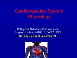
Cardiovascular System
Physiology
Dr.Raghda Alkhateeb, Cardiovascular
Surgeon, Lecturer M.B.Ch.B, CABHS, JBCS
Nursing College of Sulaymaniyah
HEAD AND NECK MAJOR ARTERIES
HOW DOES THE VEIN LOOK LIKE IN
REALITY???
CVS
INTRODUCTION
The major vessels that carry blood to and from
the heart are:
• • inferior vena cava 1 conveys deoxygenated blood
(blood low in oxygen) from the lower extremities of the
body to the heart
• • superior vena cava 1 or 2: coveys deoxygenated
blood from the upper extremities of the body to the
heart
• • aorta 1: conveys oxygenated blood (blood high in
oxygen) away from the heart
• pulmonary trunk or say pulmonary artery 1: from right
ventricle of heart to the lungs for gas exchange.
• Pulmonary veins 4: carry oxygenated blood from the
lungs to the heart.
half-
moon-
shaped
Involuntary
Functional unit of the
cardiomyocyte
Pulmonary and Systemic Circulations
• Blood whose oxygen content has become partially depleted
and carbon dioxide content has increased as a result of
tissue metabolism returns to the right atrium. This blood
then enters the ventricle, which pumps it into the
pulmonary trunk and pulmonary arteries. The pulmonary
arteries branch to transport blood to the lungs, where gas
exchange occurs between the lung capillaries and the
alveoli of the lungs. Oxygen diffuses from the air to the
capillary blood; while carbon dioxide diffuses in the
opposite direction. The blood that returns to the left
atrium by way of the pulmonary veins is therefore enriched
in oxygen and partially depleted of carbon dioxide. The
blood that is ejected from the right ventricle to the lungs
and back to the left atrium completes one circuit: called the
pulmonary circulation.
• Oxygen-rich blood in the left atrium enters the
left ventricle and is pumped into a very large,
elastic artery; the aorta. The aorta ascends for a
short distance, makes a U-turn, and then
descends through the thoracic and abdominal
cavities. Arterial branches from the aorta supply
oxygen-rich blood to all of the organ systems and
are thus part of the systemic circulation. As a
result of cellular respiration, the oxygen
concentration is lower and the carbon dioxide
concentration is higher in the tissues than in the
capillary blood. Blood that drains into the
systemic veins is thus partially depleted of oxygen
and increased in carbon dioxide content.
These veins empty into two large veins; the
superior and inferior venae cavae that return
the oxygen-poor blood to the right atrium.
This completes the systemic circulation; from
the heart (left ventricle), through the organ
systems, and back to the heart (right atrium)
• Blood sequentially flows through the heart in 1
direction through the following structures:
• Deoxygenated blood enters the heart via
the superior vena cava (SVC) and the inferior
vena cava (IVC) →
• Right atrium (RA) → tricuspid valve → right
ventricle (RV) → pulmonary valve →
• Pulmonary trunk →
pulmonary arteries → lungs (blood is oxygenated)
→ pulmonary veins →
• Left atrium (LA) → mitral valve → left ventricle
(LV) → aortic valve → aorta →
• Systemic arteries → capillaries (blood is
deoxygenated) → veins → SVC/IVC → back to the
heart
Cardiac cycle
The cardiac events that occur from the
beginning of one heart beat to the beginning
of the next are called the cardiac cycle. Each
cycle is initiated by spontaneous generation of
an action potential in the sinus node which
travels rapidly through both atria and then
through the A-V bundle into the ventricles.
Because of this special arrangement of the
conducting system from the atria into the
ventricles, there is a delay of more than 0.1
second during passage of the cardiac impulse
from the atria into the ventricles. This allows
the atria to contract, pumping blood into the
ventricles before the strong ventricular
contraction begins. Thus, the atria act as
primer pumps for the ventricles, and the
ventricles in turn provide the major source of
power for moving blood through the body’s
vascular system.
In a normal heart, cardiac activity is repeated
in a regular cycle. At a normal heart rate of
about 72 beats/minute; for the atria, the cycle
lasts for about 0.15 second in systole and 0.65
second in diastole. For the ventricles, the
duration of each cardiac cycle lasts about 0.8
second. If the heart rate increases, the
diastole decreases, which means that the
heart beating very fast may not remain
relaxed long enough to allow complete filling
of the ventricles before the next contraction.
For the ventricles, the two major phases of the
cardiac cycle are:
The diastole; a period of ventricular relaxation
in which the ventricles fill with blood and it
last for about 0.5 second.
The systole; a period of ventricular contraction
and blood ejection, lasting about 0.3 second.
DIASTOLE AND SYSTOLE
The heart derives energy from aerobic metabolism
via many different types of nutrients.
Sixty percent of the energy to power the heart is
derived from fat (free fatty acids and triglycerides),
35% from carbohydrates,
and 5% from amino acids and ketone bodies from
proteins.
These proportions vary widely with available dietary
nutrients. Malnutrition will not result in the death of
heart tissue in the way that oxygen deficiency will,
because the body has glucose reserves that sustain
the vital organs of the body and the ability to recycle
and use lactate aerobically.
Action Potential
• Cardiac muscle fibers contract via
excitation-contraction coupling, using a
mechanism unique to cardiac muscle
called calcium -induced calcium release.
• Excitation-contraction coupling
describes the process of converting an
electrical stimulus ( action potential ) into a
mechanical response (muscle
contraction).
•
Calcium-induced calcium release involves the
conduction of calcium ions into the
cardiomyocyte, triggering further release of
ions into the cytoplasm.
• Calcium prolongs the duration of muscle cell
depolarization before repolarization occurs.
Contraction in cardiac muscle occurs due to
the binding of the myosin head to adenosine
triphosphate ( ATP ), which then pulls the
actin filaments to the center of the
sarcomere, the mechanical force of
contraction.
• excitation contraction coupling (ECC): The
physiological process of converting an
electrical stimulus to a mechanical
response.
• calcium-induced calcium release (CICR):
A process whereby calcium can trigger
release of further calcium from the muscle
sarcoplasmic reticulum.
Action Potential
The function of the heart valves
• The atrioventricular valves (AV valves) are
composed of thin membranous cusps (fibrous
flaps of tissue covered with endothelium),
which hangdown in the ventricular cavities
during diastole. After atrial contraction and
just before ventricular contraction, the AV
valves begin to close and the leaflets (cusps)
come together by mean of backflow of the
blood in the ventricles towards the atria.
The AV valves include:
• The mitral valve; the left AV valve; bicuspid
valve, which consists of two cusps (anterior
and posterior), located between left atrium
and left ventricle.
• The tricuspid valve; the right AV valve, which
consists of three cusps, located between right
atrium and right ventricle.
The function of AV valves is to prevent
backflow (prevent regurgitation; leakage) of
blood into the atria during ventricular
contraction. Normally they allow blood to
flow from the atrium to the ventricle but
prevent backward flow from the ventricle to
the atria. The atrioventricular valves contain
and supported by papillary muscles.
The aortic and pulmonary valves each consist of
three semilunar cusps that resemble pockets
projecting into the lumen of aorta and pulmonary
trunk. They contain no papillary muscle. During
diastole the cusps of these valves become closely
approximated to prevent regurgitation of blood
from aorta and pulmonary arteries into the
ventricles. During systole the cusps are open
towards arterial wall, leaving a wide opening for
ejection of blood from the ventricles. In other
words, the pulmonary and aortic valves allow
blood to flow into the arteries during ventricular
contraction (systole) but prevent blood from
moving in the opposite direction during
ventricular relaxation (diastole).
• *All valves close and open passively. That is,
they close when a backward pressure gradient
pushes blood backward, and they open when
a forward pressure gradient forces blood in
the forward direction.
• *There are no valves at entrance of superior,
inferior vena cava and pulmonary veins into
the atria. What prevents the backflow of
blood from the atria toward the veins is the
compression of these veins by the atrial
contraction.
Tricuspid valve real view
ECHOCARDIOGRAPHY
ECHOCARDIOGRAPHY VIEW
CARDIAC OUTPUT
Cardiac output is the amount of blood pumped out by each side of the
heart in one minute. It is the product of the heart rate and
the stroke volume.
Cardiac output 4.0–8.0 L/minute
Stroke volume. Stroke volume is the volume of blood pumped out by
a ventricle with each heartbeat.
Regulation of stroke volume. According to Starling’s law of the heart,
the critical factor controlling stroke volume is how much the cardiac
muscle cells are stretched just before they contract; the more they
are stretched, the stronger the contraction will be; and anything
that increases the volume or speed of venous return also increases
stroke volume and force of contraction.
Factors modifying basic heart rate. The most important external
influence on heart rate is the activity of the autonomic nervous
system, as well as physical factors (age, gender, exercise, and body
temperature).
CARDIAC OUTPUT DISTRIBUTION
Stroke Volume
Stroke volume refers to the amount of blood ejected from the
heart during a single beat.
It is a measure of the contractility of the heart based on end
diastolic volume (EDV), mathematically described as
SV=EDV-ESV
indexed SV 25-45/m2
EDV is the volume of blood in the ventricles at the end of
diastole,
while ESV is the volume of blood left inside the ventricles at
the end of systole,
another indicator known as the ejection fraction (EF) is used
to evaluate stroke volume and contractility.
Mean Arterial Pressure
Cardiac output is an indicator of mean
arterial blood pressure (MAP), the average
measure of blood pressure within the
body. It is described as MAP = CO×TPR
(total peripheral resistance)
TPR is a measure of resistance in the
blood vessels, which acts as the force by
which blood must overcome to flow
through the arteries determined by the
diameter of the blood vessels.
ELECTROCARDIOGRAM
Both electrical activity and sound accompany the
beating of the heart. The pattern of electrical
activity produced by each heart beat cycle is
called the electrocardiogram (ECG).
The cardiac cycle involves a sequential
contraction of the atria and the ventricles. The
combined electrical activity of the different
myocardial cells produces electrical currents that
spread through the body fluids. These currents
are large enough to be detected by recording
electrodes placed on the skin.
ECG LEADS
ECG LEADS
NORMAL ECG
Components of Normal ECG complex
P wave-
Represents the spread of electrical activity
(wave of negativity) over the atria after the
initial depolarization of the SA node.
QRS complex
Represents the spread of the negativity wave
(depolarization) through the ventricular
musculature. A small amount of atrial
repolarization also occurs at the same time.
PR interval
Time from the beginning of the P wave to
the beginning of the QRS complex,
interval between activation of the SA node
and the beginning of ventricular
depolarization. Any abnormal lengthening
of this interval suggests some interference
with conduction of the impulse through the
atria, atria-ventricular (AV) node, bundle of
His and Purkinje fibres.
T wave
Represents the repolarization of the ventricular
musculature. It is of longer duration and lower
amplitude than the depolarization wave (QRS
complex), which indicates that the ventricular
repolarization process is less synchronized and
slower than the depolarization process.
QT interval
Represents the time from the beginning of the
QRS complex to the end of the T wave that is from
the beginning of ventricular depolarization to the
end of ventricular repolarization. The QT interval
varies with the heart rate, becoming shorter as the
heart rate increases.
PR segment
From the end of the P wave to the beginning of the
QRS complex. During this time the impulse is
travelling through the AV node, AV bundle and the
Purkinje fibres. These structures are within the
heart myocardium; therefore during this time there
is no change in the negativity of the surface of the
heart, and we say that the record is isoelectric (no
change in potential is occurring).
ST segment
From the end of the S wave to the beginning of
the T wave. During this time the heart is
completely depolarized, and therefore the record
is isoelectric. The position and the shape of the ST
segment are important in diagnosis.
Heart Sounds
• When the stethoscope is placed on the chest
wall over the heart, two sounds are normally
heard during each cardiac cycle (1st & 2nd
heart sounds). Heart sounds are associated
with closure of the valves with their
associated vibration of the flaps of the valves
and the surrounding blood under the
influence of the sudden pressure changes
that develop across the valve. That is, heart
sound does not produced by the opening of
the valve because this opening is a slow
developing process that makes no noise.
• 1-The first heart sound (S1): is caused by
closure of the AV valves when ventricles
contract at systole. The vibration is soft, low-
pitched lub.
• 2-The second heart sound (S2): is caused by
closure of the aortic and pulmonary valves
when the ventricles relax at the beginning of
diastole. The vibration is loud, high-pitched
dup. It is rapid sound because these valves
close rapidly and continue for only a short
period i.e., rapid, short and of higher pitch
dup.
• 3-The third heart sound (S3): is caused by
rapid filling of the ventricles, by blood that
flow with a rumbling motion into the almost
filled ventricles; at the middle one third (1/3)
of diastole i.e., it is caused by the vibrations
of the ventricular walls during the period of
rapid ventricular filling that follows the
opening of AV valves. It is a low-pitched
sound and can be heard after the S2. It is
heard in normal heart; in children and in adult
during exercise. It is also heard in anemia,
and AV valve regurgitation.
• 4-The fourth heart sound (S4): it is an
atrial sound when the atria contract (at late
diastole). It is a vibration sound (similar to
that of S3) associated with the flow of
blood into the ventricle. It is not heard in
normal hearts but occurs during ventricular
overload as in severe anemia,
Thyrotoxicosis (hyperthyroidism) or in
reduced ventricular compliance and in
hypertension. If present, it is heard before
S1. (S4, S1, S2, S3).
Pathologies to know!!!
• • myocardial infarction: Necrosis of heart muscle
caused by an interruption to the supply of blood to
the heart, often as a result of prolonged ischemia.
• • ischemia: Oxygen deprivation in tissues due to
mechanical obstruction of the blood supply, such
as by a narrowed or blocked artery or clot.
• • angina: Chest pain that indicates ischemia in
the heart. It may be either transient (unstable) or
stable, and stable anginas typically lead to
infarction.

More Related Content

Similar to Cardiovascular Physiology Lecture notes for Nursing Students.pptx 1-1.pptx

Physiologyofheart 140406090507-phpapp01
Physiologyofheart 140406090507-phpapp01Physiologyofheart 140406090507-phpapp01
Physiologyofheart 140406090507-phpapp01Brijesh Pratap Singh
 
the cardiovascular system and Physiology of heart
the cardiovascular system and Physiology of heartthe cardiovascular system and Physiology of heart
the cardiovascular system and Physiology of heartbhupendra kumar
 
Physiologyofheart 140406090507-phpapp01
Physiologyofheart 140406090507-phpapp01Physiologyofheart 140406090507-phpapp01
Physiologyofheart 140406090507-phpapp01Brijesh Pratap Singh
 
Transport in animals
Transport in animalsTransport in animals
Transport in animalsSian Ferguson
 
The Circulatory System in Humans
The Circulatory System in HumansThe Circulatory System in Humans
The Circulatory System in HumansGift Mokgotho
 
Cardiovascular System, Heart, Blood Vessel, ECG, Hypertension, Arrhythmia
Cardiovascular System, Heart, Blood Vessel, ECG, Hypertension, Arrhythmia Cardiovascular System, Heart, Blood Vessel, ECG, Hypertension, Arrhythmia
Cardiovascular System, Heart, Blood Vessel, ECG, Hypertension, Arrhythmia Audumbar Mali
 
cardiovascular-system-1216560905497953-8 (1).pptx
cardiovascular-system-1216560905497953-8 (1).pptxcardiovascular-system-1216560905497953-8 (1).pptx
cardiovascular-system-1216560905497953-8 (1).pptxsafuraqazi
 
Transport Systems in Mammals
Transport Systems in MammalsTransport Systems in Mammals
Transport Systems in MammalsTrischa Pretorius
 
heart physiology -conduction system nursing
heart physiology -conduction system nursingheart physiology -conduction system nursing
heart physiology -conduction system nursingfarhakhan359998
 
The cardiovascular system.pptx
The cardiovascular system.pptxThe cardiovascular system.pptx
The cardiovascular system.pptxJagruti Marathe
 
Cardiovascular System
Cardiovascular SystemCardiovascular System
Cardiovascular Systemscuffruff
 

Similar to Cardiovascular Physiology Lecture notes for Nursing Students.pptx 1-1.pptx (20)

Physiologyofheart 140406090507-phpapp01
Physiologyofheart 140406090507-phpapp01Physiologyofheart 140406090507-phpapp01
Physiologyofheart 140406090507-phpapp01
 
the cardiovascular system and Physiology of heart
the cardiovascular system and Physiology of heartthe cardiovascular system and Physiology of heart
the cardiovascular system and Physiology of heart
 
Physiologyofheart 140406090507-phpapp01
Physiologyofheart 140406090507-phpapp01Physiologyofheart 140406090507-phpapp01
Physiologyofheart 140406090507-phpapp01
 
Transport in animals
Transport in animalsTransport in animals
Transport in animals
 
Human heart
Human heartHuman heart
Human heart
 
The Circulatory System in Humans
The Circulatory System in HumansThe Circulatory System in Humans
The Circulatory System in Humans
 
circulation
circulationcirculation
circulation
 
Cardiovascular System, Heart, Blood Vessel, ECG, Hypertension, Arrhythmia
Cardiovascular System, Heart, Blood Vessel, ECG, Hypertension, Arrhythmia Cardiovascular System, Heart, Blood Vessel, ECG, Hypertension, Arrhythmia
Cardiovascular System, Heart, Blood Vessel, ECG, Hypertension, Arrhythmia
 
cardiovascular-system-1216560905497953-8 (1).pptx
cardiovascular-system-1216560905497953-8 (1).pptxcardiovascular-system-1216560905497953-8 (1).pptx
cardiovascular-system-1216560905497953-8 (1).pptx
 
Transport Systems in Mammals
Transport Systems in MammalsTransport Systems in Mammals
Transport Systems in Mammals
 
heart physiology -conduction system nursing
heart physiology -conduction system nursingheart physiology -conduction system nursing
heart physiology -conduction system nursing
 
The cardiovascular system.pptx
The cardiovascular system.pptxThe cardiovascular system.pptx
The cardiovascular system.pptx
 
Cardiovascular System
Cardiovascular SystemCardiovascular System
Cardiovascular System
 
Heart and circulation
Heart and circulation Heart and circulation
Heart and circulation
 
Anatomy and Physiology of the Heart
Anatomy and Physiology of the HeartAnatomy and Physiology of the Heart
Anatomy and Physiology of the Heart
 
Circulation
CirculationCirculation
Circulation
 
Cardiovascular System unit VIII - Copy Std.pptx
Cardiovascular System unit VIII - Copy Std.pptxCardiovascular System unit VIII - Copy Std.pptx
Cardiovascular System unit VIII - Copy Std.pptx
 
Cardiovascular System.pptx
Cardiovascular System.pptxCardiovascular System.pptx
Cardiovascular System.pptx
 
Cardiovascular system
Cardiovascular systemCardiovascular system
Cardiovascular system
 
The heart
The heartThe heart
The heart
 

More from Raghda ALKhateeb

CNS AND PNS NURSING .pptx1-1.pptx
CNS AND PNS NURSING .pptx1-1.pptxCNS AND PNS NURSING .pptx1-1.pptx
CNS AND PNS NURSING .pptx1-1.pptxRaghda ALKhateeb
 
Vital Signs Review .pptx1-1.pptx
Vital Signs Review .pptx1-1.pptxVital Signs Review .pptx1-1.pptx
Vital Signs Review .pptx1-1.pptxRaghda ALKhateeb
 
coronarycameralfistula-170307133357.pdf
coronarycameralfistula-170307133357.pdfcoronarycameralfistula-170307133357.pdf
coronarycameralfistula-170307133357.pdfRaghda ALKhateeb
 

More from Raghda ALKhateeb (6)

MCQ CVS Physiology
MCQ CVS PhysiologyMCQ CVS Physiology
MCQ CVS Physiology
 
Integumentary sytem .pdf
Integumentary sytem .pdfIntegumentary sytem .pdf
Integumentary sytem .pdf
 
CNS AND PNS NURSING .pptx1-1.pptx
CNS AND PNS NURSING .pptx1-1.pptxCNS AND PNS NURSING .pptx1-1.pptx
CNS AND PNS NURSING .pptx1-1.pptx
 
Vital Signs Review .pptx1-1.pptx
Vital Signs Review .pptx1-1.pptxVital Signs Review .pptx1-1.pptx
Vital Signs Review .pptx1-1.pptx
 
Blood Physiology.pptx
Blood Physiology.pptxBlood Physiology.pptx
Blood Physiology.pptx
 
coronarycameralfistula-170307133357.pdf
coronarycameralfistula-170307133357.pdfcoronarycameralfistula-170307133357.pdf
coronarycameralfistula-170307133357.pdf
 

Recently uploaded

Russian Call Girl Brookfield - 7001305949 Escorts Service 50% Off with Cash O...
Russian Call Girl Brookfield - 7001305949 Escorts Service 50% Off with Cash O...Russian Call Girl Brookfield - 7001305949 Escorts Service 50% Off with Cash O...
Russian Call Girl Brookfield - 7001305949 Escorts Service 50% Off with Cash O...narwatsonia7
 
Call Girls Colaba Mumbai ❤️ 9920874524 👈 Cash on Delivery
Call Girls Colaba Mumbai ❤️ 9920874524 👈 Cash on DeliveryCall Girls Colaba Mumbai ❤️ 9920874524 👈 Cash on Delivery
Call Girls Colaba Mumbai ❤️ 9920874524 👈 Cash on Deliverynehamumbai
 
Call Girl Chennai Indira 9907093804 Independent Call Girls Service Chennai
Call Girl Chennai Indira 9907093804 Independent Call Girls Service ChennaiCall Girl Chennai Indira 9907093804 Independent Call Girls Service Chennai
Call Girl Chennai Indira 9907093804 Independent Call Girls Service ChennaiNehru place Escorts
 
VIP Call Girls Pune Vrinda 9907093804 Short 1500 Night 6000 Best call girls S...
VIP Call Girls Pune Vrinda 9907093804 Short 1500 Night 6000 Best call girls S...VIP Call Girls Pune Vrinda 9907093804 Short 1500 Night 6000 Best call girls S...
VIP Call Girls Pune Vrinda 9907093804 Short 1500 Night 6000 Best call girls S...Miss joya
 
Call Girls Doddaballapur Road Just Call 7001305949 Top Class Call Girl Servic...
Call Girls Doddaballapur Road Just Call 7001305949 Top Class Call Girl Servic...Call Girls Doddaballapur Road Just Call 7001305949 Top Class Call Girl Servic...
Call Girls Doddaballapur Road Just Call 7001305949 Top Class Call Girl Servic...narwatsonia7
 
Russian Call Girls Chickpet - 7001305949 Booking and charges genuine rate for...
Russian Call Girls Chickpet - 7001305949 Booking and charges genuine rate for...Russian Call Girls Chickpet - 7001305949 Booking and charges genuine rate for...
Russian Call Girls Chickpet - 7001305949 Booking and charges genuine rate for...narwatsonia7
 
Call Girls Chennai Megha 9907093804 Independent Call Girls Service Chennai
Call Girls Chennai Megha 9907093804 Independent Call Girls Service ChennaiCall Girls Chennai Megha 9907093804 Independent Call Girls Service Chennai
Call Girls Chennai Megha 9907093804 Independent Call Girls Service ChennaiNehru place Escorts
 
Call Girls Horamavu WhatsApp Number 7001035870 Meeting With Bangalore Escorts
Call Girls Horamavu WhatsApp Number 7001035870 Meeting With Bangalore EscortsCall Girls Horamavu WhatsApp Number 7001035870 Meeting With Bangalore Escorts
Call Girls Horamavu WhatsApp Number 7001035870 Meeting With Bangalore Escortsvidya singh
 
Bangalore Call Girls Marathahalli 📞 9907093804 High Profile Service 100% Safe
Bangalore Call Girls Marathahalli 📞 9907093804 High Profile Service 100% SafeBangalore Call Girls Marathahalli 📞 9907093804 High Profile Service 100% Safe
Bangalore Call Girls Marathahalli 📞 9907093804 High Profile Service 100% Safenarwatsonia7
 
Low Rate Call Girls Pune Esha 9907093804 Short 1500 Night 6000 Best call girl...
Low Rate Call Girls Pune Esha 9907093804 Short 1500 Night 6000 Best call girl...Low Rate Call Girls Pune Esha 9907093804 Short 1500 Night 6000 Best call girl...
Low Rate Call Girls Pune Esha 9907093804 Short 1500 Night 6000 Best call girl...Miss joya
 
Hi,Fi Call Girl In Mysore Road - 7001305949 | 24x7 Service Available Near Me
Hi,Fi Call Girl In Mysore Road - 7001305949 | 24x7 Service Available Near MeHi,Fi Call Girl In Mysore Road - 7001305949 | 24x7 Service Available Near Me
Hi,Fi Call Girl In Mysore Road - 7001305949 | 24x7 Service Available Near Menarwatsonia7
 
VIP Call Girls Tirunelveli Aaradhya 8250192130 Independent Escort Service Tir...
VIP Call Girls Tirunelveli Aaradhya 8250192130 Independent Escort Service Tir...VIP Call Girls Tirunelveli Aaradhya 8250192130 Independent Escort Service Tir...
VIP Call Girls Tirunelveli Aaradhya 8250192130 Independent Escort Service Tir...narwatsonia7
 
Call Girl Service Bidadi - For 7001305949 Cheap & Best with original Photos
Call Girl Service Bidadi - For 7001305949 Cheap & Best with original PhotosCall Girl Service Bidadi - For 7001305949 Cheap & Best with original Photos
Call Girl Service Bidadi - For 7001305949 Cheap & Best with original Photosnarwatsonia7
 
Low Rate Call Girls Ambattur Anika 8250192130 Independent Escort Service Amba...
Low Rate Call Girls Ambattur Anika 8250192130 Independent Escort Service Amba...Low Rate Call Girls Ambattur Anika 8250192130 Independent Escort Service Amba...
Low Rate Call Girls Ambattur Anika 8250192130 Independent Escort Service Amba...narwatsonia7
 
Call Girls Service Bellary Road Just Call 7001305949 Enjoy College Girls Service
Call Girls Service Bellary Road Just Call 7001305949 Enjoy College Girls ServiceCall Girls Service Bellary Road Just Call 7001305949 Enjoy College Girls Service
Call Girls Service Bellary Road Just Call 7001305949 Enjoy College Girls Servicenarwatsonia7
 
Russian Call Girls in Chennai Pallavi 9907093804 Independent Call Girls Servi...
Russian Call Girls in Chennai Pallavi 9907093804 Independent Call Girls Servi...Russian Call Girls in Chennai Pallavi 9907093804 Independent Call Girls Servi...
Russian Call Girls in Chennai Pallavi 9907093804 Independent Call Girls Servi...Nehru place Escorts
 
Bangalore Call Girls Majestic 📞 9907093804 High Profile Service 100% Safe
Bangalore Call Girls Majestic 📞 9907093804 High Profile Service 100% SafeBangalore Call Girls Majestic 📞 9907093804 High Profile Service 100% Safe
Bangalore Call Girls Majestic 📞 9907093804 High Profile Service 100% Safenarwatsonia7
 
Call Girls Service Surat Samaira ❤️🍑 8250192130 👄 Independent Escort Service ...
Call Girls Service Surat Samaira ❤️🍑 8250192130 👄 Independent Escort Service ...Call Girls Service Surat Samaira ❤️🍑 8250192130 👄 Independent Escort Service ...
Call Girls Service Surat Samaira ❤️🍑 8250192130 👄 Independent Escort Service ...CALL GIRLS
 
Call Girls Service Jaipur Grishma WhatsApp ❤8445551418 VIP Call Girls Jaipur
Call Girls Service Jaipur Grishma WhatsApp ❤8445551418 VIP Call Girls JaipurCall Girls Service Jaipur Grishma WhatsApp ❤8445551418 VIP Call Girls Jaipur
Call Girls Service Jaipur Grishma WhatsApp ❤8445551418 VIP Call Girls Jaipurparulsinha
 
Call Girls Whitefield Just Call 7001305949 Top Class Call Girl Service Available
Call Girls Whitefield Just Call 7001305949 Top Class Call Girl Service AvailableCall Girls Whitefield Just Call 7001305949 Top Class Call Girl Service Available
Call Girls Whitefield Just Call 7001305949 Top Class Call Girl Service Availablenarwatsonia7
 

Recently uploaded (20)

Russian Call Girl Brookfield - 7001305949 Escorts Service 50% Off with Cash O...
Russian Call Girl Brookfield - 7001305949 Escorts Service 50% Off with Cash O...Russian Call Girl Brookfield - 7001305949 Escorts Service 50% Off with Cash O...
Russian Call Girl Brookfield - 7001305949 Escorts Service 50% Off with Cash O...
 
Call Girls Colaba Mumbai ❤️ 9920874524 👈 Cash on Delivery
Call Girls Colaba Mumbai ❤️ 9920874524 👈 Cash on DeliveryCall Girls Colaba Mumbai ❤️ 9920874524 👈 Cash on Delivery
Call Girls Colaba Mumbai ❤️ 9920874524 👈 Cash on Delivery
 
Call Girl Chennai Indira 9907093804 Independent Call Girls Service Chennai
Call Girl Chennai Indira 9907093804 Independent Call Girls Service ChennaiCall Girl Chennai Indira 9907093804 Independent Call Girls Service Chennai
Call Girl Chennai Indira 9907093804 Independent Call Girls Service Chennai
 
VIP Call Girls Pune Vrinda 9907093804 Short 1500 Night 6000 Best call girls S...
VIP Call Girls Pune Vrinda 9907093804 Short 1500 Night 6000 Best call girls S...VIP Call Girls Pune Vrinda 9907093804 Short 1500 Night 6000 Best call girls S...
VIP Call Girls Pune Vrinda 9907093804 Short 1500 Night 6000 Best call girls S...
 
Call Girls Doddaballapur Road Just Call 7001305949 Top Class Call Girl Servic...
Call Girls Doddaballapur Road Just Call 7001305949 Top Class Call Girl Servic...Call Girls Doddaballapur Road Just Call 7001305949 Top Class Call Girl Servic...
Call Girls Doddaballapur Road Just Call 7001305949 Top Class Call Girl Servic...
 
Russian Call Girls Chickpet - 7001305949 Booking and charges genuine rate for...
Russian Call Girls Chickpet - 7001305949 Booking and charges genuine rate for...Russian Call Girls Chickpet - 7001305949 Booking and charges genuine rate for...
Russian Call Girls Chickpet - 7001305949 Booking and charges genuine rate for...
 
Call Girls Chennai Megha 9907093804 Independent Call Girls Service Chennai
Call Girls Chennai Megha 9907093804 Independent Call Girls Service ChennaiCall Girls Chennai Megha 9907093804 Independent Call Girls Service Chennai
Call Girls Chennai Megha 9907093804 Independent Call Girls Service Chennai
 
Call Girls Horamavu WhatsApp Number 7001035870 Meeting With Bangalore Escorts
Call Girls Horamavu WhatsApp Number 7001035870 Meeting With Bangalore EscortsCall Girls Horamavu WhatsApp Number 7001035870 Meeting With Bangalore Escorts
Call Girls Horamavu WhatsApp Number 7001035870 Meeting With Bangalore Escorts
 
Bangalore Call Girls Marathahalli 📞 9907093804 High Profile Service 100% Safe
Bangalore Call Girls Marathahalli 📞 9907093804 High Profile Service 100% SafeBangalore Call Girls Marathahalli 📞 9907093804 High Profile Service 100% Safe
Bangalore Call Girls Marathahalli 📞 9907093804 High Profile Service 100% Safe
 
Low Rate Call Girls Pune Esha 9907093804 Short 1500 Night 6000 Best call girl...
Low Rate Call Girls Pune Esha 9907093804 Short 1500 Night 6000 Best call girl...Low Rate Call Girls Pune Esha 9907093804 Short 1500 Night 6000 Best call girl...
Low Rate Call Girls Pune Esha 9907093804 Short 1500 Night 6000 Best call girl...
 
Hi,Fi Call Girl In Mysore Road - 7001305949 | 24x7 Service Available Near Me
Hi,Fi Call Girl In Mysore Road - 7001305949 | 24x7 Service Available Near MeHi,Fi Call Girl In Mysore Road - 7001305949 | 24x7 Service Available Near Me
Hi,Fi Call Girl In Mysore Road - 7001305949 | 24x7 Service Available Near Me
 
VIP Call Girls Tirunelveli Aaradhya 8250192130 Independent Escort Service Tir...
VIP Call Girls Tirunelveli Aaradhya 8250192130 Independent Escort Service Tir...VIP Call Girls Tirunelveli Aaradhya 8250192130 Independent Escort Service Tir...
VIP Call Girls Tirunelveli Aaradhya 8250192130 Independent Escort Service Tir...
 
Call Girl Service Bidadi - For 7001305949 Cheap & Best with original Photos
Call Girl Service Bidadi - For 7001305949 Cheap & Best with original PhotosCall Girl Service Bidadi - For 7001305949 Cheap & Best with original Photos
Call Girl Service Bidadi - For 7001305949 Cheap & Best with original Photos
 
Low Rate Call Girls Ambattur Anika 8250192130 Independent Escort Service Amba...
Low Rate Call Girls Ambattur Anika 8250192130 Independent Escort Service Amba...Low Rate Call Girls Ambattur Anika 8250192130 Independent Escort Service Amba...
Low Rate Call Girls Ambattur Anika 8250192130 Independent Escort Service Amba...
 
Call Girls Service Bellary Road Just Call 7001305949 Enjoy College Girls Service
Call Girls Service Bellary Road Just Call 7001305949 Enjoy College Girls ServiceCall Girls Service Bellary Road Just Call 7001305949 Enjoy College Girls Service
Call Girls Service Bellary Road Just Call 7001305949 Enjoy College Girls Service
 
Russian Call Girls in Chennai Pallavi 9907093804 Independent Call Girls Servi...
Russian Call Girls in Chennai Pallavi 9907093804 Independent Call Girls Servi...Russian Call Girls in Chennai Pallavi 9907093804 Independent Call Girls Servi...
Russian Call Girls in Chennai Pallavi 9907093804 Independent Call Girls Servi...
 
Bangalore Call Girls Majestic 📞 9907093804 High Profile Service 100% Safe
Bangalore Call Girls Majestic 📞 9907093804 High Profile Service 100% SafeBangalore Call Girls Majestic 📞 9907093804 High Profile Service 100% Safe
Bangalore Call Girls Majestic 📞 9907093804 High Profile Service 100% Safe
 
Call Girls Service Surat Samaira ❤️🍑 8250192130 👄 Independent Escort Service ...
Call Girls Service Surat Samaira ❤️🍑 8250192130 👄 Independent Escort Service ...Call Girls Service Surat Samaira ❤️🍑 8250192130 👄 Independent Escort Service ...
Call Girls Service Surat Samaira ❤️🍑 8250192130 👄 Independent Escort Service ...
 
Call Girls Service Jaipur Grishma WhatsApp ❤8445551418 VIP Call Girls Jaipur
Call Girls Service Jaipur Grishma WhatsApp ❤8445551418 VIP Call Girls JaipurCall Girls Service Jaipur Grishma WhatsApp ❤8445551418 VIP Call Girls Jaipur
Call Girls Service Jaipur Grishma WhatsApp ❤8445551418 VIP Call Girls Jaipur
 
Call Girls Whitefield Just Call 7001305949 Top Class Call Girl Service Available
Call Girls Whitefield Just Call 7001305949 Top Class Call Girl Service AvailableCall Girls Whitefield Just Call 7001305949 Top Class Call Girl Service Available
Call Girls Whitefield Just Call 7001305949 Top Class Call Girl Service Available
 

Cardiovascular Physiology Lecture notes for Nursing Students.pptx 1-1.pptx

  • 1. Cardiovascular System Physiology Dr.Raghda Alkhateeb, Cardiovascular Surgeon, Lecturer M.B.Ch.B, CABHS, JBCS Nursing College of Sulaymaniyah
  • 2.
  • 3.
  • 4.
  • 5. HEAD AND NECK MAJOR ARTERIES
  • 6. HOW DOES THE VEIN LOOK LIKE IN REALITY???
  • 7.
  • 9.
  • 10.
  • 11.
  • 12.
  • 13.
  • 14.
  • 15.
  • 16.
  • 17.
  • 18.
  • 19.
  • 20.
  • 21.
  • 22.
  • 23.
  • 24.
  • 25.
  • 26.
  • 27. The major vessels that carry blood to and from the heart are: • • inferior vena cava 1 conveys deoxygenated blood (blood low in oxygen) from the lower extremities of the body to the heart • • superior vena cava 1 or 2: coveys deoxygenated blood from the upper extremities of the body to the heart • • aorta 1: conveys oxygenated blood (blood high in oxygen) away from the heart • pulmonary trunk or say pulmonary artery 1: from right ventricle of heart to the lungs for gas exchange. • Pulmonary veins 4: carry oxygenated blood from the lungs to the heart.
  • 29.
  • 30.
  • 31.
  • 33.
  • 34.
  • 35. Functional unit of the cardiomyocyte
  • 36.
  • 37. Pulmonary and Systemic Circulations • Blood whose oxygen content has become partially depleted and carbon dioxide content has increased as a result of tissue metabolism returns to the right atrium. This blood then enters the ventricle, which pumps it into the pulmonary trunk and pulmonary arteries. The pulmonary arteries branch to transport blood to the lungs, where gas exchange occurs between the lung capillaries and the alveoli of the lungs. Oxygen diffuses from the air to the capillary blood; while carbon dioxide diffuses in the opposite direction. The blood that returns to the left atrium by way of the pulmonary veins is therefore enriched in oxygen and partially depleted of carbon dioxide. The blood that is ejected from the right ventricle to the lungs and back to the left atrium completes one circuit: called the pulmonary circulation.
  • 38.
  • 39. • Oxygen-rich blood in the left atrium enters the left ventricle and is pumped into a very large, elastic artery; the aorta. The aorta ascends for a short distance, makes a U-turn, and then descends through the thoracic and abdominal cavities. Arterial branches from the aorta supply oxygen-rich blood to all of the organ systems and are thus part of the systemic circulation. As a result of cellular respiration, the oxygen concentration is lower and the carbon dioxide concentration is higher in the tissues than in the capillary blood. Blood that drains into the systemic veins is thus partially depleted of oxygen and increased in carbon dioxide content.
  • 40. These veins empty into two large veins; the superior and inferior venae cavae that return the oxygen-poor blood to the right atrium. This completes the systemic circulation; from the heart (left ventricle), through the organ systems, and back to the heart (right atrium)
  • 41.
  • 42. • Blood sequentially flows through the heart in 1 direction through the following structures: • Deoxygenated blood enters the heart via the superior vena cava (SVC) and the inferior vena cava (IVC) → • Right atrium (RA) → tricuspid valve → right ventricle (RV) → pulmonary valve → • Pulmonary trunk → pulmonary arteries → lungs (blood is oxygenated) → pulmonary veins → • Left atrium (LA) → mitral valve → left ventricle (LV) → aortic valve → aorta → • Systemic arteries → capillaries (blood is deoxygenated) → veins → SVC/IVC → back to the heart
  • 43.
  • 44.
  • 45.
  • 46. Cardiac cycle The cardiac events that occur from the beginning of one heart beat to the beginning of the next are called the cardiac cycle. Each cycle is initiated by spontaneous generation of an action potential in the sinus node which travels rapidly through both atria and then through the A-V bundle into the ventricles.
  • 47. Because of this special arrangement of the conducting system from the atria into the ventricles, there is a delay of more than 0.1 second during passage of the cardiac impulse from the atria into the ventricles. This allows the atria to contract, pumping blood into the ventricles before the strong ventricular contraction begins. Thus, the atria act as primer pumps for the ventricles, and the ventricles in turn provide the major source of power for moving blood through the body’s vascular system.
  • 48. In a normal heart, cardiac activity is repeated in a regular cycle. At a normal heart rate of about 72 beats/minute; for the atria, the cycle lasts for about 0.15 second in systole and 0.65 second in diastole. For the ventricles, the duration of each cardiac cycle lasts about 0.8 second. If the heart rate increases, the diastole decreases, which means that the heart beating very fast may not remain relaxed long enough to allow complete filling of the ventricles before the next contraction.
  • 49. For the ventricles, the two major phases of the cardiac cycle are: The diastole; a period of ventricular relaxation in which the ventricles fill with blood and it last for about 0.5 second. The systole; a period of ventricular contraction and blood ejection, lasting about 0.3 second.
  • 51.
  • 52. The heart derives energy from aerobic metabolism via many different types of nutrients. Sixty percent of the energy to power the heart is derived from fat (free fatty acids and triglycerides), 35% from carbohydrates, and 5% from amino acids and ketone bodies from proteins. These proportions vary widely with available dietary nutrients. Malnutrition will not result in the death of heart tissue in the way that oxygen deficiency will, because the body has glucose reserves that sustain the vital organs of the body and the ability to recycle and use lactate aerobically.
  • 53. Action Potential • Cardiac muscle fibers contract via excitation-contraction coupling, using a mechanism unique to cardiac muscle called calcium -induced calcium release. • Excitation-contraction coupling describes the process of converting an electrical stimulus ( action potential ) into a mechanical response (muscle contraction). •
  • 54. Calcium-induced calcium release involves the conduction of calcium ions into the cardiomyocyte, triggering further release of ions into the cytoplasm. • Calcium prolongs the duration of muscle cell depolarization before repolarization occurs. Contraction in cardiac muscle occurs due to the binding of the myosin head to adenosine triphosphate ( ATP ), which then pulls the actin filaments to the center of the sarcomere, the mechanical force of contraction.
  • 55. • excitation contraction coupling (ECC): The physiological process of converting an electrical stimulus to a mechanical response. • calcium-induced calcium release (CICR): A process whereby calcium can trigger release of further calcium from the muscle sarcoplasmic reticulum.
  • 57.
  • 58.
  • 59.
  • 60.
  • 61.
  • 62.
  • 63.
  • 64.
  • 65.
  • 66.
  • 67. The function of the heart valves • The atrioventricular valves (AV valves) are composed of thin membranous cusps (fibrous flaps of tissue covered with endothelium), which hangdown in the ventricular cavities during diastole. After atrial contraction and just before ventricular contraction, the AV valves begin to close and the leaflets (cusps) come together by mean of backflow of the blood in the ventricles towards the atria.
  • 68. The AV valves include: • The mitral valve; the left AV valve; bicuspid valve, which consists of two cusps (anterior and posterior), located between left atrium and left ventricle. • The tricuspid valve; the right AV valve, which consists of three cusps, located between right atrium and right ventricle.
  • 69. The function of AV valves is to prevent backflow (prevent regurgitation; leakage) of blood into the atria during ventricular contraction. Normally they allow blood to flow from the atrium to the ventricle but prevent backward flow from the ventricle to the atria. The atrioventricular valves contain and supported by papillary muscles.
  • 70. The aortic and pulmonary valves each consist of three semilunar cusps that resemble pockets projecting into the lumen of aorta and pulmonary trunk. They contain no papillary muscle. During diastole the cusps of these valves become closely approximated to prevent regurgitation of blood from aorta and pulmonary arteries into the ventricles. During systole the cusps are open towards arterial wall, leaving a wide opening for ejection of blood from the ventricles. In other words, the pulmonary and aortic valves allow blood to flow into the arteries during ventricular contraction (systole) but prevent blood from moving in the opposite direction during ventricular relaxation (diastole).
  • 71. • *All valves close and open passively. That is, they close when a backward pressure gradient pushes blood backward, and they open when a forward pressure gradient forces blood in the forward direction. • *There are no valves at entrance of superior, inferior vena cava and pulmonary veins into the atria. What prevents the backflow of blood from the atria toward the veins is the compression of these veins by the atrial contraction.
  • 72.
  • 76. CARDIAC OUTPUT Cardiac output is the amount of blood pumped out by each side of the heart in one minute. It is the product of the heart rate and the stroke volume. Cardiac output 4.0–8.0 L/minute Stroke volume. Stroke volume is the volume of blood pumped out by a ventricle with each heartbeat. Regulation of stroke volume. According to Starling’s law of the heart, the critical factor controlling stroke volume is how much the cardiac muscle cells are stretched just before they contract; the more they are stretched, the stronger the contraction will be; and anything that increases the volume or speed of venous return also increases stroke volume and force of contraction. Factors modifying basic heart rate. The most important external influence on heart rate is the activity of the autonomic nervous system, as well as physical factors (age, gender, exercise, and body temperature).
  • 78.
  • 79. Stroke Volume Stroke volume refers to the amount of blood ejected from the heart during a single beat. It is a measure of the contractility of the heart based on end diastolic volume (EDV), mathematically described as SV=EDV-ESV indexed SV 25-45/m2 EDV is the volume of blood in the ventricles at the end of diastole, while ESV is the volume of blood left inside the ventricles at the end of systole, another indicator known as the ejection fraction (EF) is used to evaluate stroke volume and contractility.
  • 80.
  • 81. Mean Arterial Pressure Cardiac output is an indicator of mean arterial blood pressure (MAP), the average measure of blood pressure within the body. It is described as MAP = CO×TPR (total peripheral resistance) TPR is a measure of resistance in the blood vessels, which acts as the force by which blood must overcome to flow through the arteries determined by the diameter of the blood vessels.
  • 82.
  • 83. ELECTROCARDIOGRAM Both electrical activity and sound accompany the beating of the heart. The pattern of electrical activity produced by each heart beat cycle is called the electrocardiogram (ECG). The cardiac cycle involves a sequential contraction of the atria and the ventricles. The combined electrical activity of the different myocardial cells produces electrical currents that spread through the body fluids. These currents are large enough to be detected by recording electrodes placed on the skin.
  • 85.
  • 86.
  • 88.
  • 90. Components of Normal ECG complex P wave- Represents the spread of electrical activity (wave of negativity) over the atria after the initial depolarization of the SA node. QRS complex Represents the spread of the negativity wave (depolarization) through the ventricular musculature. A small amount of atrial repolarization also occurs at the same time.
  • 91. PR interval Time from the beginning of the P wave to the beginning of the QRS complex, interval between activation of the SA node and the beginning of ventricular depolarization. Any abnormal lengthening of this interval suggests some interference with conduction of the impulse through the atria, atria-ventricular (AV) node, bundle of His and Purkinje fibres.
  • 92. T wave Represents the repolarization of the ventricular musculature. It is of longer duration and lower amplitude than the depolarization wave (QRS complex), which indicates that the ventricular repolarization process is less synchronized and slower than the depolarization process. QT interval Represents the time from the beginning of the QRS complex to the end of the T wave that is from the beginning of ventricular depolarization to the end of ventricular repolarization. The QT interval varies with the heart rate, becoming shorter as the heart rate increases.
  • 93. PR segment From the end of the P wave to the beginning of the QRS complex. During this time the impulse is travelling through the AV node, AV bundle and the Purkinje fibres. These structures are within the heart myocardium; therefore during this time there is no change in the negativity of the surface of the heart, and we say that the record is isoelectric (no change in potential is occurring). ST segment From the end of the S wave to the beginning of the T wave. During this time the heart is completely depolarized, and therefore the record is isoelectric. The position and the shape of the ST segment are important in diagnosis.
  • 94.
  • 95.
  • 96.
  • 97.
  • 98.
  • 99.
  • 100. Heart Sounds • When the stethoscope is placed on the chest wall over the heart, two sounds are normally heard during each cardiac cycle (1st & 2nd heart sounds). Heart sounds are associated with closure of the valves with their associated vibration of the flaps of the valves and the surrounding blood under the influence of the sudden pressure changes that develop across the valve. That is, heart sound does not produced by the opening of the valve because this opening is a slow developing process that makes no noise.
  • 101. • 1-The first heart sound (S1): is caused by closure of the AV valves when ventricles contract at systole. The vibration is soft, low- pitched lub. • 2-The second heart sound (S2): is caused by closure of the aortic and pulmonary valves when the ventricles relax at the beginning of diastole. The vibration is loud, high-pitched dup. It is rapid sound because these valves close rapidly and continue for only a short period i.e., rapid, short and of higher pitch dup.
  • 102. • 3-The third heart sound (S3): is caused by rapid filling of the ventricles, by blood that flow with a rumbling motion into the almost filled ventricles; at the middle one third (1/3) of diastole i.e., it is caused by the vibrations of the ventricular walls during the period of rapid ventricular filling that follows the opening of AV valves. It is a low-pitched sound and can be heard after the S2. It is heard in normal heart; in children and in adult during exercise. It is also heard in anemia, and AV valve regurgitation.
  • 103. • 4-The fourth heart sound (S4): it is an atrial sound when the atria contract (at late diastole). It is a vibration sound (similar to that of S3) associated with the flow of blood into the ventricle. It is not heard in normal hearts but occurs during ventricular overload as in severe anemia, Thyrotoxicosis (hyperthyroidism) or in reduced ventricular compliance and in hypertension. If present, it is heard before S1. (S4, S1, S2, S3).
  • 104.
  • 105.
  • 106. Pathologies to know!!! • • myocardial infarction: Necrosis of heart muscle caused by an interruption to the supply of blood to the heart, often as a result of prolonged ischemia. • • ischemia: Oxygen deprivation in tissues due to mechanical obstruction of the blood supply, such as by a narrowed or blocked artery or clot. • • angina: Chest pain that indicates ischemia in the heart. It may be either transient (unstable) or stable, and stable anginas typically lead to infarction.