Download as PDF, PPTX

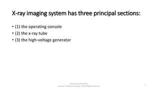
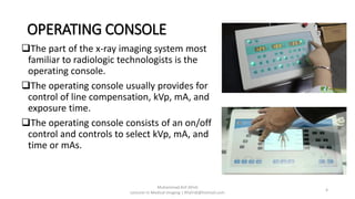
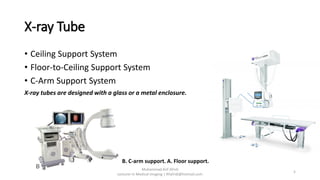
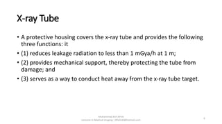
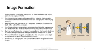


The document discusses the components and operation of an X-ray imaging system. The system has three principal sections: the operating console, X-ray tube, and high-voltage generator. The operating console controls parameters like kVp, mA, and exposure time. X-ray tubes can be attached to ceiling, floor-to-ceiling, or C-arm support systems. The tube is enclosed in a protective housing to reduce radiation leakage and provide mechanical support. The high-voltage generator supplies power to the X-ray tube for image formation when X-rays pass through a patient and expose imaging plates or screens.


![PERI-PROSTHETIC FRACTURE NAIL-PLATE CONSTRUCT [NPC].pptx](https://cdn.slidesharecdn.com/ss_thumbnails/drarunkumardrmohamedashrafperiprostheticfrasturenail-plateconstructnpc-260209164459-7e9d15a1-thumbnail.jpg?width=640&height=640&fit=bounds)


