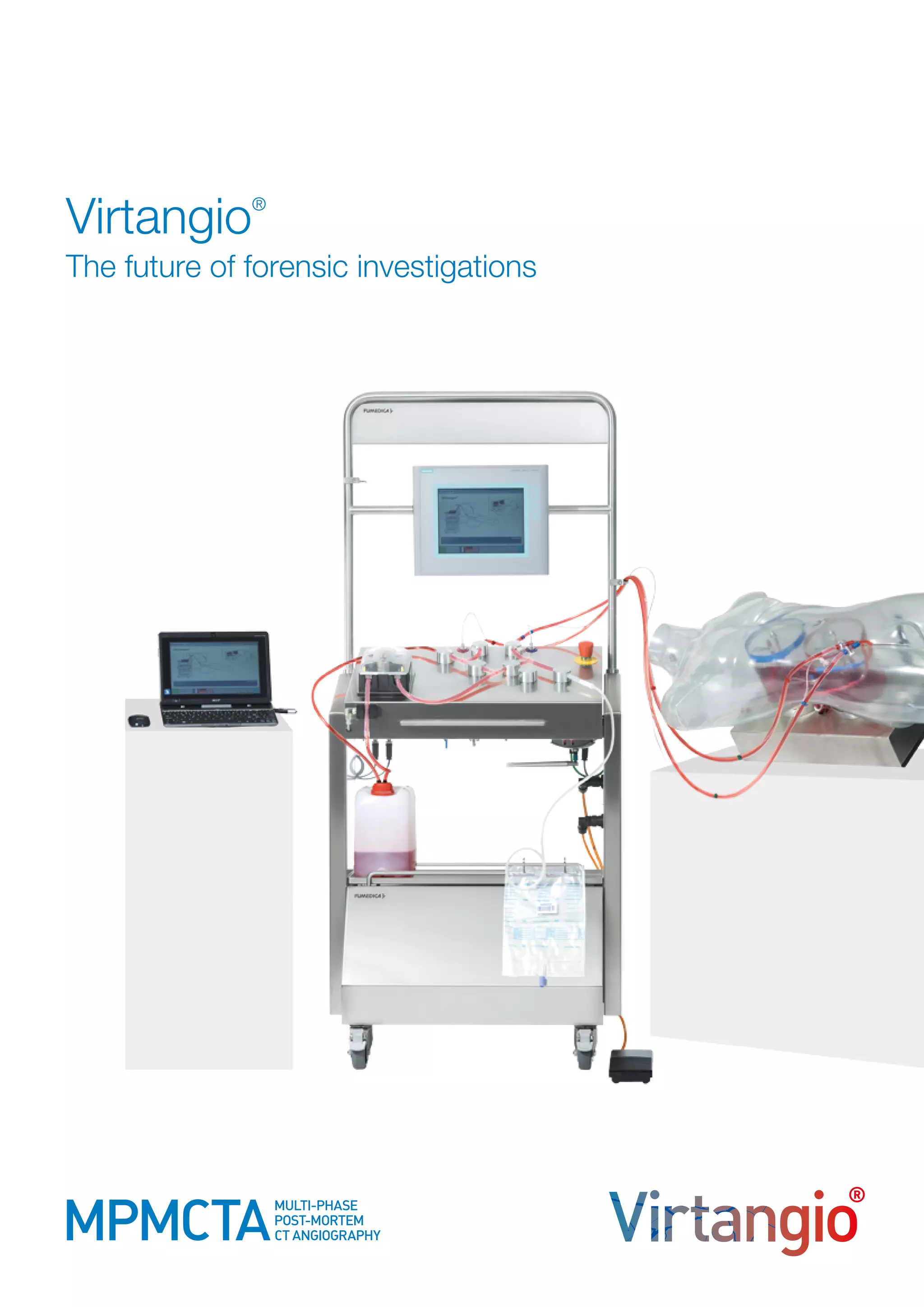
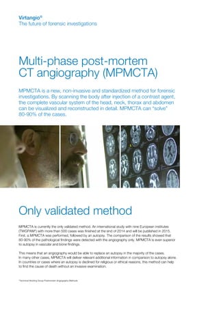
Virtangio®
The future of forensic investigations
Multi-phase post-mortem
CT angiography (MPMCTA)
Virtangio®
The future of forensic investigations
*Technical Working Group Postmortem Angiography Methods
MPMCTA is a new, non-invasive and standardized method for forensic
investigations. By scanning the body after injection of a contrast agent,
the complete vascular system of the head, neck, thorax and abdomen
can be visualized and reconstructed in detail. MPMCTA can “solve”
80-90% of the cases.
Only validated method
MPMCTA is currently the only validated method. An international study with nine European institutes
(TWGPAM*) with more than 500 cases was finished at the end of 2014 and will be published in 2015.
First, a MPMCTA was performed, followed by an autopsy. The comparison of the results showed that
80-90% of the pathological findings were detected with the angiography only. MPMCTA is even superior
to autopsy in vascular and bone findings.
This means that an angiography would be able to replace an autopsy in the majority of the cases.
In many other cases, MPMCTA will deliver relevant additional information in comparison to autopsy alone.
In countries or cases where an autopsy is declined for religious or ethical reasons, this method can help
to find the cause of death without an invasive examination.
Virtangio®
The equipment
•	 the Virtangio®
machine (injection machine)
•	 the single use tubing/cannula set
•	 the contrast agent Angiofil®
The equipment was developed by the company Fumedica AG in Muri/Switzerland in collaboration
with the Legal Medicine Department of the University Lausanne/Switzerland (PD Dr Silke Grabherr).
To perform MPMCTA, a CT scanner is needed.
Virtangio®
The future of forensic investigations
Virtangio®
The use
The Virtangio®
machine is placed beside the CT scanner.
It works independently from the scanner and can be operated
by remote control in the CT command room.
Before starting the angiography, an unenhanced CT scan
of the body is performed. The body preparation for angiography
takes about 15 minutes:
•	small incision in the inguinal region on one side
•	insertion of two cannulas into the femoral artery and vein
•	connect the Virtangio®
machine with the two cannulas fixed
	 in the body with the tubing set
•	entry of case information into the system
The angiography can start and the system guides the
user systematically through the process.
To avoid vascular filling defects, the angiography
consists of three different phases:
•	arterial phase
•	venous phase
•	dynamic phase
During each phase, the machine automatically fills the respective
blood vessels with the contrast agent mixture (Angiofil®
with pa-
raffin oil). While the CT scan in the arterial and venous phase is
performed after the respective injection is complete, the scan for
the dynamic phase is performed during the ongoing injection to
simulate live perfusion. Depending on the CT scanner, performing
the three phases takes between 10 – 30 minutes.
The Virtangio®
machine
provides the following advantages:
•	 injection can be performed volume- or pressure-controlled
•	 standard or individual protocols available
•	 machine stops in case of abnormal pressure loss or increase
•	 injection data are stored automatically
Virtangio®
The future of forensic investigations
MPMCTA can even be performed on putrefied bodies, as long as the vascular system is intact.
The radiological interpretation of the images is done by a forensic
radiologist together with the forensic pathologist. The image qua-
lity is comparable to or even better than corresponding images
of living patients. By comparing the images of the three different
phases and the native CT scan, artefacts and filling defects can
be recognized and pathological findings verified. Trainings and
guidelines for radiological interpretation are available.
If necessary, a conventional autopsy can be performed after the
angiography. With the exception of pulmonary fat embolism, there
is no negative impact of the technique and the contrast agent on
histology or toxicology; nevertheless it is recommended to take
sensitive biopsy samples before the angiography if possible.
Performing MPMCTA with the Virtangio®
equipment is a fast, standardized and minimally invasive
procedure that can be easily implemented into the daily routine. The oily contrast agent does not
mix with remaining blood and avoids an oedemisation of tissues (as frequently observed with most
other contrast agents) due to a micro-embolization of the capillary vessels. The contrast agent can
remain in the body after the examination. The single use tubing/cannula set avoids any contamina-
tion due to its closed system design.
Virtangio®
The future of forensic investigations
Indications
•	trauma cases like traffic accidents,
	 sharp trauma, gunshot, blunt trauma
•	suspicion of cardio-vascular disease
•	suspicion of medical malpractice
•	any kind of unexpected and sudden death
Advantages
•	standardized, validated and minimally
	 invasive procedure
•	controlled vascular filling in three phases
	 (arterial, venous, dynamic)
•	closed system, no contamination
•	oily contrast agent with high radio opacity
•	no mixture with remaining blood
•	no oedemisation, no extravasation
•	training and guidelines for radiological
	interpretation
Virtangio®
The future of forensic investigations
Literature
Application of contrast media in post-mortem imaging (CT and MRI)
Grabherr S et al; Radiol Med 2015 Apr 5. [Epub ahead of print]; PMID: 25841652
 
Postmortem computed tomography angiography, contrast medium administration and toxicological analyses in urine
Palmiere C et al; Leg Med (Tokyo). 2014 Dec 12. pii: S1344-6223(14)00176-X. doi: 10.1016/
j.legalmed.2014.12.005 [Epub ahead of print]; PMID: 25537625
 
Postmortem angiography using femoral cannulation and postmortem microbiology
Palmiere C et al; Int J Legal Med. 2014 Nov 8. [Epub ahead of print]; PMID: 25381195
 
The usefulness of post-mortem CT angiography in injuries caused by falling from considerable heights: Three fatal cases
Mokrane FZ et al; Diagn Interv Imaging. 2014 Oct 23;95(11):1085-1090. doi: 10.1016/j.diii.2013.08.010. [Epub ahead of print]; PMID: 25443333
   
Virtual autopsy with multiphase postmortem computed tomographic angiography versus traditional medical autopsy to investigate unexpected
deaths of hospitalized patients: A cohort study
Wichmann D et al; Ann Intern Med. 2014 Apr 15;160(8):534-41. doi: 10.7326/M13-2211; PMID: 24733194
 
Advances in post-mortem CT angiography
Grabherr S et al; Br J Radiol. 2014 Apr;87(1036):20130488. doi: 10.1259/bjr.20130488; PMID: 24234582
 
Postmortem imaging of sudden cardiac death
Michaud K et al; Int J Legal Med. 2014 Jan;128(1):127-37. doi: 10.1007/s00414-013-0819-6. Epub 2013 Jan 16; PMID: 23322013
 
Postmortem computed tomography angiography vs. conventional autopsy: advantages and inconveniences of each method
Christine C et al; Int J Legal Med. 2013 Sep;127(5):981-9. doi: 10.1007/s00414-012-0814-3. Epub 2013 Jan 6; PMID: 23292183
  
Multi-phase postmortem CT angiography: recognizing technique-related artefacts and pitfalls
Bruguier C et al; Int J Legal Med. 2013 May;127(3):639-52. doi: 10.1007/s00414-013-0840-9. Epub 2013 Mar 21; PMID: 23515679
 
Surgical interventions with fatal outcome: utility of multi-phase postmortem CT angiography
Zerlauth JB et al; Forensic Sci Int. 2013 Feb 10;225(1-3):32-41. doi: 10.1016/j.forsciint.2012.05.013. Epub 2012 Jun 19; PMID: 22721937
For more information or literature, visit www.postmortem-angio.ch
Contact
Production, Sales & Marketing	 TWGPAM
Fumedica AG	 PD Dr Silke Grabherr
Division Postmortem-Angio	 Department of Legal Medicine
Luzernerstrasse 91	 University of Lausanne
CH-5630 Muri AG	 Chemin de la Vulliette 4
info@postmortem-angio.ch	 CH-1000 Lausanne 25
www.postmortem-angio.ch	silke.grabherr@chuv.ch

Virtangio_Folder

  • 1.
    Virtangio® The future offorensic investigations
  • 2.
    Multi-phase post-mortem CT angiography(MPMCTA) Virtangio® The future of forensic investigations *Technical Working Group Postmortem Angiography Methods MPMCTA is a new, non-invasive and standardized method for forensic investigations. By scanning the body after injection of a contrast agent, the complete vascular system of the head, neck, thorax and abdomen can be visualized and reconstructed in detail. MPMCTA can “solve” 80-90% of the cases. Only validated method MPMCTA is currently the only validated method. An international study with nine European institutes (TWGPAM*) with more than 500 cases was finished at the end of 2014 and will be published in 2015. First, a MPMCTA was performed, followed by an autopsy. The comparison of the results showed that 80-90% of the pathological findings were detected with the angiography only. MPMCTA is even superior to autopsy in vascular and bone findings. This means that an angiography would be able to replace an autopsy in the majority of the cases. In many other cases, MPMCTA will deliver relevant additional information in comparison to autopsy alone. In countries or cases where an autopsy is declined for religious or ethical reasons, this method can help to find the cause of death without an invasive examination.
  • 3.
    Virtangio® The equipment • theVirtangio® machine (injection machine) • the single use tubing/cannula set • the contrast agent Angiofil® The equipment was developed by the company Fumedica AG in Muri/Switzerland in collaboration with the Legal Medicine Department of the University Lausanne/Switzerland (PD Dr Silke Grabherr). To perform MPMCTA, a CT scanner is needed. Virtangio® The future of forensic investigations
  • 4.
    Virtangio® The use The Virtangio® machineis placed beside the CT scanner. It works independently from the scanner and can be operated by remote control in the CT command room. Before starting the angiography, an unenhanced CT scan of the body is performed. The body preparation for angiography takes about 15 minutes: • small incision in the inguinal region on one side • insertion of two cannulas into the femoral artery and vein • connect the Virtangio® machine with the two cannulas fixed in the body with the tubing set • entry of case information into the system The angiography can start and the system guides the user systematically through the process. To avoid vascular filling defects, the angiography consists of three different phases: • arterial phase • venous phase • dynamic phase During each phase, the machine automatically fills the respective blood vessels with the contrast agent mixture (Angiofil® with pa- raffin oil). While the CT scan in the arterial and venous phase is performed after the respective injection is complete, the scan for the dynamic phase is performed during the ongoing injection to simulate live perfusion. Depending on the CT scanner, performing the three phases takes between 10 – 30 minutes. The Virtangio® machine provides the following advantages: • injection can be performed volume- or pressure-controlled • standard or individual protocols available • machine stops in case of abnormal pressure loss or increase • injection data are stored automatically Virtangio® The future of forensic investigations MPMCTA can even be performed on putrefied bodies, as long as the vascular system is intact.
  • 5.
    The radiological interpretationof the images is done by a forensic radiologist together with the forensic pathologist. The image qua- lity is comparable to or even better than corresponding images of living patients. By comparing the images of the three different phases and the native CT scan, artefacts and filling defects can be recognized and pathological findings verified. Trainings and guidelines for radiological interpretation are available. If necessary, a conventional autopsy can be performed after the angiography. With the exception of pulmonary fat embolism, there is no negative impact of the technique and the contrast agent on histology or toxicology; nevertheless it is recommended to take sensitive biopsy samples before the angiography if possible. Performing MPMCTA with the Virtangio® equipment is a fast, standardized and minimally invasive procedure that can be easily implemented into the daily routine. The oily contrast agent does not mix with remaining blood and avoids an oedemisation of tissues (as frequently observed with most other contrast agents) due to a micro-embolization of the capillary vessels. The contrast agent can remain in the body after the examination. The single use tubing/cannula set avoids any contamina- tion due to its closed system design. Virtangio® The future of forensic investigations Indications • trauma cases like traffic accidents, sharp trauma, gunshot, blunt trauma • suspicion of cardio-vascular disease • suspicion of medical malpractice • any kind of unexpected and sudden death Advantages • standardized, validated and minimally invasive procedure • controlled vascular filling in three phases (arterial, venous, dynamic) • closed system, no contamination • oily contrast agent with high radio opacity • no mixture with remaining blood • no oedemisation, no extravasation • training and guidelines for radiological interpretation
  • 6.
    Virtangio® The future offorensic investigations Literature Application of contrast media in post-mortem imaging (CT and MRI) Grabherr S et al; Radiol Med 2015 Apr 5. [Epub ahead of print]; PMID: 25841652   Postmortem computed tomography angiography, contrast medium administration and toxicological analyses in urine Palmiere C et al; Leg Med (Tokyo). 2014 Dec 12. pii: S1344-6223(14)00176-X. doi: 10.1016/ j.legalmed.2014.12.005 [Epub ahead of print]; PMID: 25537625   Postmortem angiography using femoral cannulation and postmortem microbiology Palmiere C et al; Int J Legal Med. 2014 Nov 8. [Epub ahead of print]; PMID: 25381195   The usefulness of post-mortem CT angiography in injuries caused by falling from considerable heights: Three fatal cases Mokrane FZ et al; Diagn Interv Imaging. 2014 Oct 23;95(11):1085-1090. doi: 10.1016/j.diii.2013.08.010. [Epub ahead of print]; PMID: 25443333     Virtual autopsy with multiphase postmortem computed tomographic angiography versus traditional medical autopsy to investigate unexpected deaths of hospitalized patients: A cohort study Wichmann D et al; Ann Intern Med. 2014 Apr 15;160(8):534-41. doi: 10.7326/M13-2211; PMID: 24733194   Advances in post-mortem CT angiography Grabherr S et al; Br J Radiol. 2014 Apr;87(1036):20130488. doi: 10.1259/bjr.20130488; PMID: 24234582   Postmortem imaging of sudden cardiac death Michaud K et al; Int J Legal Med. 2014 Jan;128(1):127-37. doi: 10.1007/s00414-013-0819-6. Epub 2013 Jan 16; PMID: 23322013   Postmortem computed tomography angiography vs. conventional autopsy: advantages and inconveniences of each method Christine C et al; Int J Legal Med. 2013 Sep;127(5):981-9. doi: 10.1007/s00414-012-0814-3. Epub 2013 Jan 6; PMID: 23292183    Multi-phase postmortem CT angiography: recognizing technique-related artefacts and pitfalls Bruguier C et al; Int J Legal Med. 2013 May;127(3):639-52. doi: 10.1007/s00414-013-0840-9. Epub 2013 Mar 21; PMID: 23515679   Surgical interventions with fatal outcome: utility of multi-phase postmortem CT angiography Zerlauth JB et al; Forensic Sci Int. 2013 Feb 10;225(1-3):32-41. doi: 10.1016/j.forsciint.2012.05.013. Epub 2012 Jun 19; PMID: 22721937 For more information or literature, visit www.postmortem-angio.ch Contact Production, Sales & Marketing TWGPAM Fumedica AG PD Dr Silke Grabherr Division Postmortem-Angio Department of Legal Medicine Luzernerstrasse 91 University of Lausanne CH-5630 Muri AG Chemin de la Vulliette 4 info@postmortem-angio.ch CH-1000 Lausanne 25 www.postmortem-angio.ch silke.grabherr@chuv.ch