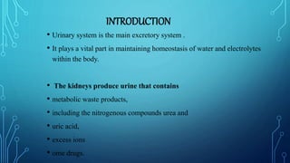
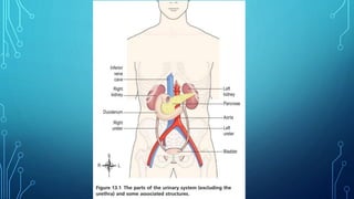
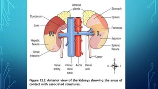
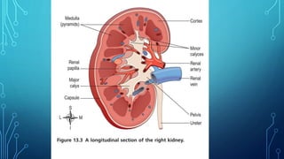
THEHUMANURINARYSYSTEM
By :-
MANOJ VERMA
B. Pharmacy 2nd Sem
UNIVERSITY INSTITUTE OF
PHARMACY,
ORIENTAL UNIVERSITY INDORE.
Guided By :-
Mr. Rahul Sisodiya Sir
Assistant Professor
UNIVERSITY INSTITUTE OF
PHARMACY
ORIENTAL UNIVERSITY
INDORE
INTRODUCTION
• Urinary system is the main excretory system .
• It plays a vital part in maintaining homeostasis of water and electrolytes
within the body.
• The kidneys produce urine that contains
• metabolic waste products,
• including the nitrogenous compounds urea and
• uric acid,
• excess ions
• ome drugs.
PARTS OF URINARY SYSTEM
• 2 kidneys, which secrete urine
• 2 ureters that convey the urine from the kidneys to the
• urinary bladder the urinary bladder, which collects and stores
urine
• The urethra through which urine leaves the body
IMPORTANT TERMS
• Urine is stored in the bladder and excreted by the process of micturition.
KIDNEY
• ANATOMY OF KIDNEY
• LOCATION
• The kidneys lie on the posterior abdominal wall,
• one on each side of the vertebral column,
• behind the peritoneum and below the diaphragm.
• They extend from the level of the 12th thoracic vertebra to the 3rd
lumbar vertebra,
• receiving some protection from the lower rib cage.
• The right kidney is usually slightly lower than the left,
• probably because of the considerable space occupied by the liver.
• Kidneys are bean-shaped organs,
• about 11 cm long, 6 cm wide, 3 cm thick and weigh 150 g.
• They are embedded in, and held in position by, a mass of fat.
• A sheath of fibrous connective tissue, the renal fascia, encloses the
kidney and the renal fat.
ORGANS ASSOCIATED WITH THE KIDNEYS
•Right kidney
• Superiorly – the right adrenal gland
• Anteriorly – the right lobe of the liver, the duodenum and the hepatic flexure
of the colon
• Posteriorly – the diaphragm, and muscles of the
• posterior abdominal wall.
• Left kidney
• Superiorly – the left adrenal gland
• Anteriorly – the spleen, stomach, pancreas, jejunum and
splenic flexure of the colon
• Posteriorly – the diaphragm and muscles of the posterior
abdominal wall.
PHYSIOLOGY OF KIDNEYS
• formation of urine,
• maintaining water, electrolyte and acid–base balance
• excretion of waste products
• production and secretion of erythropoietin, the hormone that stimulates formation of
red blood cells
• production and secretion of renin, an important enzyme in the control of blood
pressure
STRUCTURE OF KIDNEY
• an outer fibrous capsule, surrounding the kidney the cortex,
• a reddish-brown layer of tissue immediately below the capsule and outside
the renal pyramids .
• the medulla, the innermost layer, consisting of pale conical-shaped striations,
the renal pyramids.
• The hilum is the concave medial border of the kidney where the renal blood
and lymph vessels, the ureter and nerves enter
• Urine formed within the kidney passes through a renal papilla at the apex of a
pyramid into a minor calyx
• Several minor calyces merge into a major calyx and two or three major
calyces combine forming the renal pelvis,
MICROSCOPICSTRUCTURE OF THE KIDNEY
THE NEPHRON
• The kidney contains about 1–2 million functional units, the nephrons,
• a tubule closed at one end that joins a collecting duct at the other end. The closed
or blind end is indented to form the cup-shaped glomerular capsule(Bowman’s
capsule),
• which almost completely encloses a network of tiny arterial capillaries, the
glomerulus.
• the proximal convoluted tubule
• the medullary loop (loop of Henle)
• the distal convoluted tubule, leading into a collecting duct.
PROCESS OF URINE FORMATION
• There are three processes involved in the formation of urine
1. filtration
2. selective reabsorption
3. secretion.
FILTRATION
• This takes place through the semipermeable walls of the glomerulus and
glomerular capsule.
• Water and other small molecules readily pass through, although some are
reabsorbed later.
• Blood cells, plasma proteins and other large molecules are too large to filter
through and therefore remain in the capillaries .
• the efferent arteriole is narrower than the afferent arteriole, a capillary
hydrostatic pressure
SELECTIVE REABSORPTION
• Most reabsorption from the filtrate back into
the blood
• takes place in the proximal convoluted
tubule, whose
• walls are lined with microvilli to increase
surface area for absorption.
• Many substances are reabsorbed here,
including some water, electrolytes and
organic nutrients such as glucose.
• Some reabsorption is passive, but some
substances, e.g. glucose, are actively
transported.
• Some ions, e.g. sodium and chloride, can be
absorbed by both active and passive
mechanisms
TUBULAR SECRETION
• Filtration occurs as blood flows through the glomerulus.
• Substances not required and foreign materials,
• e.g. drugs including penicillin and aspirin, may not be entirely
• filtered out of the blood because of the short time it remains in the
glomerulus.
• Such substances are cleared by secretion from the peritubular capillaries into
the filtrate within the convoluted tubules.
• Tubular secretion of hydrogen ions (H+) is important in maintaining normal
blood pH.
COMPOSITION OF URINE
• The specific gravity is between 1020 and 1030, and
• the pH is around 6 (normal range 4.5–8).
• A healthy adult passes from 1000 to 1500 mL per day.
URETERS
• The ureters carry urine from the kidneys to the urinary bladder .
• They are about 25–30 cm long with a diameter of approximately 3 mm.
• The ureter is continuous with the funnel-shaped renal pelvis. It passes
downwards through the abdominal cavity,
• behind the peritoneum in front of the psoas muscle into the pelvic
• cavity, and
• passes obliquely through the posterior wall of the bladder
STRUCTURE OF URETERS
• The walls of the ureters consist of three
layers of tissue which are shown in cross-
section in
• an outer covering of fibrous tissue,
continuous with the fibrous capsule of the
kidney
• • a middle muscular layer consisting of
interlacing smooth muscle fibres that form
a functional unit round the ureter and an
additional outer longitudinal layer in the
lower third
• an inner layer, the mucosa, composed of
transitional epithelium.
FUNCTIONS OF URETERS
• Peristalsis is an intrinsic property of the smooth muscle
• layer that propels urine along the ureter. Peristaltic waves
• occur several times per minute, increasing in frequency
• with the volume of urine produced, sending little spurts
• of urine along the ureter towards the bladder.
URINARY BLADDER
• The urinary bladder is a reservoir for urine.
• It lies in the pelvic cavity and I
• ts size and position vary, depending on the volume of urine it contains.
• When distended, the bladder rises into the abdominal cavity
STRUCTURE OF URINARY BLADDER
• The bladder is roughly pear shaped, but becomes more balloon shaped as it
fills with urine.
• The posterior surface is the base.
• The bladder opens into the urethra at its lowest point, the neck.
• The peritoneum covers only the superior surface before it turns upwards as
the parietal peritoneum, lining the anterior abdominal wall.
• Posteriorly it surrounds the uterus in the female and the rectum in the male
• The bladder wall is composed of three layers:
• the outer layer of loose connective tissue, containing blood and lymphatic
vessels and nerves, covered on the upper surface by the peritoneum
• the middle layer, consisting of interlacing smooth muscle fibres and elastic
tissue loosely arranged in three layers. This is called the detrusor muscle and
when it contracts, it empties the bladder
• the inner mucosa, composed of transitional epithelium that readily permits
distension of the bladder as it fills.
URETHRA
• The urethra is a canal extending from the neck of the bladder to the exterior, at the
external urethral orifice.
• It is longer in the male than in the female
• When bladder control is established, the micturition
• reflex is still stimulated but sensory impulses also pass
• upwards to the brain and there is awareness of the need
• to pass urine as the bladder fills (around 300–400 mL in
• adults). By learned and conscious effort, contraction of
• the external urethral sphincter and muscles of the pelvic
• floor can inhibit micturition until it is convenient to pass urine.
REFERENCE
• Waugh Anne, Grant Allison "Ross and Wilson ANATOMY AND
PHYSIOLOGY in Health and Illness” 13th Edition 2018
• Page No. 369- 391.
*** THANK YOU ***
Urinary system

Urinary system

  • 1.
    THEHUMANURINARYSYSTEM By :- MANOJ VERMA B.Pharmacy 2nd Sem UNIVERSITY INSTITUTE OF PHARMACY, ORIENTAL UNIVERSITY INDORE. Guided By :- Mr. Rahul Sisodiya Sir Assistant Professor UNIVERSITY INSTITUTE OF PHARMACY ORIENTAL UNIVERSITY INDORE
  • 2.
    INTRODUCTION • Urinary systemis the main excretory system . • It plays a vital part in maintaining homeostasis of water and electrolytes within the body. • The kidneys produce urine that contains • metabolic waste products, • including the nitrogenous compounds urea and • uric acid, • excess ions • ome drugs.
  • 3.
    PARTS OF URINARYSYSTEM • 2 kidneys, which secrete urine • 2 ureters that convey the urine from the kidneys to the • urinary bladder the urinary bladder, which collects and stores urine • The urethra through which urine leaves the body
  • 6.
    IMPORTANT TERMS • Urineis stored in the bladder and excreted by the process of micturition.
  • 7.
    KIDNEY • ANATOMY OFKIDNEY • LOCATION • The kidneys lie on the posterior abdominal wall, • one on each side of the vertebral column, • behind the peritoneum and below the diaphragm. • They extend from the level of the 12th thoracic vertebra to the 3rd lumbar vertebra, • receiving some protection from the lower rib cage. • The right kidney is usually slightly lower than the left, • probably because of the considerable space occupied by the liver.
  • 8.
    • Kidneys arebean-shaped organs, • about 11 cm long, 6 cm wide, 3 cm thick and weigh 150 g. • They are embedded in, and held in position by, a mass of fat. • A sheath of fibrous connective tissue, the renal fascia, encloses the kidney and the renal fat.
  • 10.
    ORGANS ASSOCIATED WITHTHE KIDNEYS •Right kidney • Superiorly – the right adrenal gland • Anteriorly – the right lobe of the liver, the duodenum and the hepatic flexure of the colon • Posteriorly – the diaphragm, and muscles of the • posterior abdominal wall.
  • 11.
    • Left kidney •Superiorly – the left adrenal gland • Anteriorly – the spleen, stomach, pancreas, jejunum and splenic flexure of the colon • Posteriorly – the diaphragm and muscles of the posterior abdominal wall.
  • 12.
    PHYSIOLOGY OF KIDNEYS •formation of urine, • maintaining water, electrolyte and acid–base balance • excretion of waste products • production and secretion of erythropoietin, the hormone that stimulates formation of red blood cells • production and secretion of renin, an important enzyme in the control of blood pressure
  • 13.
    STRUCTURE OF KIDNEY •an outer fibrous capsule, surrounding the kidney the cortex, • a reddish-brown layer of tissue immediately below the capsule and outside the renal pyramids . • the medulla, the innermost layer, consisting of pale conical-shaped striations, the renal pyramids. • The hilum is the concave medial border of the kidney where the renal blood and lymph vessels, the ureter and nerves enter • Urine formed within the kidney passes through a renal papilla at the apex of a pyramid into a minor calyx • Several minor calyces merge into a major calyx and two or three major calyces combine forming the renal pelvis,
  • 14.
    MICROSCOPICSTRUCTURE OF THEKIDNEY THE NEPHRON • The kidney contains about 1–2 million functional units, the nephrons, • a tubule closed at one end that joins a collecting duct at the other end. The closed or blind end is indented to form the cup-shaped glomerular capsule(Bowman’s capsule), • which almost completely encloses a network of tiny arterial capillaries, the glomerulus. • the proximal convoluted tubule • the medullary loop (loop of Henle) • the distal convoluted tubule, leading into a collecting duct.
  • 18.
    PROCESS OF URINEFORMATION • There are three processes involved in the formation of urine 1. filtration 2. selective reabsorption 3. secretion.
  • 19.
    FILTRATION • This takesplace through the semipermeable walls of the glomerulus and glomerular capsule. • Water and other small molecules readily pass through, although some are reabsorbed later. • Blood cells, plasma proteins and other large molecules are too large to filter through and therefore remain in the capillaries . • the efferent arteriole is narrower than the afferent arteriole, a capillary hydrostatic pressure
  • 20.
    SELECTIVE REABSORPTION • Mostreabsorption from the filtrate back into the blood • takes place in the proximal convoluted tubule, whose • walls are lined with microvilli to increase surface area for absorption. • Many substances are reabsorbed here, including some water, electrolytes and organic nutrients such as glucose. • Some reabsorption is passive, but some substances, e.g. glucose, are actively transported. • Some ions, e.g. sodium and chloride, can be absorbed by both active and passive mechanisms
  • 21.
    TUBULAR SECRETION • Filtrationoccurs as blood flows through the glomerulus. • Substances not required and foreign materials, • e.g. drugs including penicillin and aspirin, may not be entirely • filtered out of the blood because of the short time it remains in the glomerulus. • Such substances are cleared by secretion from the peritubular capillaries into the filtrate within the convoluted tubules. • Tubular secretion of hydrogen ions (H+) is important in maintaining normal blood pH.
  • 22.
    COMPOSITION OF URINE •The specific gravity is between 1020 and 1030, and • the pH is around 6 (normal range 4.5–8). • A healthy adult passes from 1000 to 1500 mL per day.
  • 23.
    URETERS • The ureterscarry urine from the kidneys to the urinary bladder . • They are about 25–30 cm long with a diameter of approximately 3 mm. • The ureter is continuous with the funnel-shaped renal pelvis. It passes downwards through the abdominal cavity, • behind the peritoneum in front of the psoas muscle into the pelvic • cavity, and • passes obliquely through the posterior wall of the bladder
  • 24.
    STRUCTURE OF URETERS •The walls of the ureters consist of three layers of tissue which are shown in cross- section in • an outer covering of fibrous tissue, continuous with the fibrous capsule of the kidney • • a middle muscular layer consisting of interlacing smooth muscle fibres that form a functional unit round the ureter and an additional outer longitudinal layer in the lower third • an inner layer, the mucosa, composed of transitional epithelium.
  • 25.
    FUNCTIONS OF URETERS •Peristalsis is an intrinsic property of the smooth muscle • layer that propels urine along the ureter. Peristaltic waves • occur several times per minute, increasing in frequency • with the volume of urine produced, sending little spurts • of urine along the ureter towards the bladder.
  • 26.
    URINARY BLADDER • Theurinary bladder is a reservoir for urine. • It lies in the pelvic cavity and I • ts size and position vary, depending on the volume of urine it contains. • When distended, the bladder rises into the abdominal cavity
  • 27.
    STRUCTURE OF URINARYBLADDER • The bladder is roughly pear shaped, but becomes more balloon shaped as it fills with urine. • The posterior surface is the base. • The bladder opens into the urethra at its lowest point, the neck. • The peritoneum covers only the superior surface before it turns upwards as the parietal peritoneum, lining the anterior abdominal wall. • Posteriorly it surrounds the uterus in the female and the rectum in the male
  • 28.
    • The bladderwall is composed of three layers: • the outer layer of loose connective tissue, containing blood and lymphatic vessels and nerves, covered on the upper surface by the peritoneum • the middle layer, consisting of interlacing smooth muscle fibres and elastic tissue loosely arranged in three layers. This is called the detrusor muscle and when it contracts, it empties the bladder • the inner mucosa, composed of transitional epithelium that readily permits distension of the bladder as it fills.
  • 29.
    URETHRA • The urethrais a canal extending from the neck of the bladder to the exterior, at the external urethral orifice. • It is longer in the male than in the female • When bladder control is established, the micturition • reflex is still stimulated but sensory impulses also pass • upwards to the brain and there is awareness of the need • to pass urine as the bladder fills (around 300–400 mL in • adults). By learned and conscious effort, contraction of • the external urethral sphincter and muscles of the pelvic • floor can inhibit micturition until it is convenient to pass urine.
  • 30.
    REFERENCE • Waugh Anne,Grant Allison "Ross and Wilson ANATOMY AND PHYSIOLOGY in Health and Illness” 13th Edition 2018 • Page No. 369- 391.
  • 31.