
Recommended
More Related Content
Similar to ULTRASONICS vs HAND INSTRUMENTATION.pptx
Similar to ULTRASONICS vs HAND INSTRUMENTATION.pptx (20)
Recently uploaded
Recently uploaded (20)
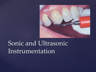
ULTRASONICS vs HAND INSTRUMENTATION.pptx
- 2. Hand instruments like curettes have been used since ages for scaling and root planing. Technically demanding Time consuming Tiring Introduction to electronically powered instrumentation
- 3. Initially introduced in the late 1950’s : Bulky and limited to removal of supragingival calculus Late 1980’s : Slim diameter instrument tips developed 1990’s: New approach to instrumentation and cementu, conservation TODAY : Modern electronically powered slim diameter instrument tips as effecetive as hand instruments HISTORY OF ELECTRONICALLY POWERED DEVICES
- 4. Electronically powered instruments use rapid energy vibrations of a powered instrument tip to fracture calculus from the tooth surface Physical factors playing a role in the mechanism of power scalers include FREQUENCY STROKE WATER FLOW Mechanism of Action
- 5. FREQUENCY : Number of time the insert tip moves back and forth during one cycle . STROKE: How far the instrument tip moves during one cycle. Another term is amplitude WATER FLOW: Water flow can be controlled by a water knob that controls the volume of water being delivered to the insert tip.
- 6. 2 Basic types of electronically powered devices SONIC DEVICE ULTRASONIC DEVICE
- 7. in 1955 Zinner reported the first application of ultrasound for the removal of calculus from tooth surfaces. In the 1960s, recognized as an effective tool in accomplishing the healing of diseased periodontal tissues vibrations with frequencies in the range of 25,000–42,000 Hz Free standing units with electric generator ULTRASONIC SCALERS
- 8. Ultrasonic unit 4 components Electric generator Foot control Hand piece assembly Interchangeable inserts Two types Manual and Automatic Manual – Amplitude, Frequency And water adjustment Automatic- Only frequency And water adjustment
- 9. MAGNETOSTRICTIVE DEVICES ULTRASONIC DEVICES 2 Basic types
- 10. Consists of an electronic generator, a handpiece and Interchangeable instrument inserts. Transducer consists of metal stacks that change dimension when electric energy is applied leading to vibration of the tip Tips move in elliptical or orbital stroke pattern 4 active working surfaces Magnetostrictrive
- 11. Transducer - Stack of flat metal strips (nickel iron alloy), (Cavitron 25kHz, DENTSPLY, Des Plaines, IL) - Rod of ferromagnetic material (Odontoson 42kHz)
- 12. Point Ferromagnetic stack Water Outlet O Ring Grip Tip PARTS OF A MAGNETOSTRICTIVE INSERT
- 13. Length of active tip area energy output and frequency Frequency 18- 45 kHz All surfaces active flexibility in adaptation Frequency Active tip 25kHz Terminal 4.3mm 30kHz 4.2mm 50kHz 2.3mm
- 14. Transducer completely into the hand piece Ceramic discs located in the hand piece : change in dimension as electrical energy is applied to it Tips move in a linear pattern that give it 2 active surfaces Piezon Master – 27-30 kHz (EMS, Nyon) ENAC – 30 kHz (Osada Electronics) Solphy – 27-29 kHz Piezoelectric
- 15. Comparison of Magnetostrictive and Piezoelectric units Magnetostrictive Piezoelectric Mechanism Metal stack or ferrite rod Aligned ceramic discs Tip movement Elliptical Linear Active surfaces 4 (back, face and lateral) 2 (lateral) Positioning of tips Flexible Must be lateral to surface Effective calculus removal Yes Yes
- 16. Directly attach to the dental unit’s high speed handpiece tubing. Air driven scalers Work at a frequency of 2000-6500 Stroke pattern is elliptical to orbital in shape Larger in diameter and universal in design Introduced into the dental market in the 1970s as less expensive alternatives to ultrasonic scalers SONIC SCALERS
- 17. Hand piece hollow rod, a rotor, rubber O- rings Rotor 6mm wide thin metal ring encircles hollow rod Compressed air is forced through hollow rod Air escapes through the 10 holes and causes rotor to vibrate triggers rod to vibrate All surfaces of the tip - active Coolant not necessary
- 18. Sonic scalers used in conjunction with hand instruments were more effective for the removal of subgingival calculus than either method used alone (Gellin et al, 1986). Sonic scalers produce no difference in clinical response compared with ultrasonic scalers and that sonic scalers provide an effetive alternative to hand instruments (Loos et al, 1987 ) Several studies with conlicting results created a controversy
- 20. Instrument Tips
- 21. The type of calculus deposits The location of calculus deposits Instrument tip selection criteria STANDARD DIAMETER SLIM DIAMTERER
- 23. MODIFIED POWER DRIVEN SCALERS
- 24. Developed for use in deeper pockets.5-12mm pockets Straight slim diameter inserts effective in anterior teeth Recommended for use on root surfaces located 4mm or less below the CEJ. Slim periodontal probe type inserts
- 25. Dragoo et al. 1992 examined a manual scaler (universal curette) and ultrasonic scalers (Cavi-tron ) with modified (contra-angled tips EW-P10which resemble a periodontal probe. and an unmodified (P- 10 type) inserts in debridement of hopeless single or multi-rooted teeth. They evaluated the pocket depth, instrument limit, and instrument efficiency. The modified inserts showed added benefits
- 26. Designed to facilitate adaptation to the curved root surfaces of posterior teeth Back of the working end adapts best to the curved root surface The Curved Slim tips
- 27. Yukna et al (1997) - diamond coated ultrasonic tip which resembled the traditional Cavitron scaler tip. - more efficient - roughest surface Diamond Coated ultrasonic tips
- 28. Burnett Power tip Unique slim diameter tip Can withstand higher Power settings and more lateral pressure than typical slim diameter tip
- 29. Furcation Slim Tips More effective than Hand instruments in treating class 2 or 3 furcations ( Hou, G.L et al, 1994) Gracey curettes too wide to enter furcation areas in over 50% of All maxillary and mandibular molars First introduced by Osha and Ishikawa In the year 1987.
- 30. Beuchat et al (2001) - Periosonic - modified version of endodontic system - 2 types of files sonic handpiece - Periosonic 1 reamer , 16mm working tip ( heavy calculus) - Periosonic 2 flexible, 21mm working tip Split mouth study - equally effective - better clinical attachment gain - less recession Periosonic
- 31. Tip motion Scanning laser vibrotomy studies- -variability in tip vibration Three dimensional scanning laser vibrotomy studies- All tips oscillate in an elliptical manner (with varying degrees of ellipticity)
- 32. Tip motion- - Power settings of generator - Shape of the probe Lea SC et al (2009) – probes under load show a reduction in the lateral component of oscillation. Both classes exhibit elliptical motion
- 34. EFFECTIVENESS OF ELECTRONICALLY POWERED INSTRUMENTATION 1)Plaque and calculus removal 2)Bacterial reduction endotoxin removal 3) Root surface conservation 4) Pocket penetration 5) Required time and clinical outcome 7) Irrigation ( lavage) 8) Bactericidal effect
- 35. Powered instrumentation have been shown to be as effective as hand instrumentation They are especially effective in deplaquing Plaque and calculus removal
- 36. Cavitational activity of the ultrasonic scaler is considered effective for removal of plaque and endotoxin. ( Walmsley et al, 1990). Studies suggested that ultrasonic scalers are capable of removing endotoxin located on the root surface without excessive removal of cementum or dentin. ( Checchi et al, 1988) ( Chiew et al, 1991) Bacterial reduction and endotoxin and cementum removal
- 37. Root Surface Removal Rosenberg (1979), Von Vokinberg et al (1976) - Manual instruments remove more root surface Pattison et al (1996): - Ultrasonics better Ritz et al (1991): Sonic 1.72 µm Ultrasonic 4.3 – 7.8 µm Diamond bur 7.9 – 15.5 µm Fine curette 5 – 22 µm
- 38. Required time and clinical outcome Badersten et al (1984): Compared clinical effects no difference Manual instrumentation longer time Copulos et al (1993): ultrasonic and curette equally effective in all clinical parameters measured time spent 3.6 min (US), 5.8 min (manual)
- 39. Busslinger et al (2001) time needed , calculus removal and root roughness in -vitro model curette 126.1 ± 38.2 s piezoelectric 74.1 ± 27.6 s (> efficient but rougher surface) magnetostrictive 104.9 ± 25.4 s It may be concluded that manual scalers require more time in scaling and root planing than power-driven scalers.
- 40. Slim diameter tips penetrate deeper into periodontal pockets Reach the base of the pocket better than hand instruments. Water lavage reaches a depth that is equal to the depth reached by a powered instrument tip Pocket penetration
- 41. Constant stream of water exits near the point of the instrument tip Water stream within the periodontal pocket is termed as the fluid lavage. Washes toxic products and free floating bacteria from the pocket Better vision during instrumentation Irrigation ( lavage)
- 43. Bactericidal effect Cavitation and Acoustic streaming Distruption of cell walls and biofilms even beyond reach Destroy from a distance Just the act of holding an activated ultrasonic tip in the periodontal pocket is destructive
- 44. Recent developments of power driven scalers
- 45. Limitations of electronically powered instrumentation
- 46. AEROSOL PRODUCTION CARDIAC PACEMAKERS REDUCED TACTILE SENSATION INFECTION CONTROL
- 47. Ultrasonic instruments Advantages- Less time Stain removal Flushing of debris, blood, necrotic tissue Less force Antimicrobial effect Less fatigue Patient comfort Disadvantages- Less tactile sensitivity Hampers visibility by constant water spray Hazard by contaminated aerosol Damage to hearing in many pts Magnetostrictive not used in pacemakers
- 48. Scaling and root planing play a pivotal role in the elimination of causative factors of periodontal disease. In the past, it had been generally agreed that excessive root surface removal by hand instruments was necessary to remove the tenacious calculus deposits. However, research over the past years has shown that definitive root surface detoxification can be achieved without excessive cementum removal or aggressive instrumentation. Complete cementum removal is no longer a requisite. Many studies have demonstrated that hand and power-driven instruments are equally effective in reducing the probing depth, attaining attachment level gains and reducing inflammation by removal of plaque bacteria, calculus, and endotoxin. Conclusion
- 49. Power-driven instruments have many advantages over the manual scalers; however, further studies are needed to improve the performance of currently available instruments. These studies would help to provide treatment based on exact information regarding the instrument and technology
- 50. Electrocautery
- 51. The term electrosurgery or radiosurgery is currently used to identify surgical techniques performed on soft tissue using controlled high-frequency electrical currents in the range of 1.5 to 7.5 million cycles per second or megahertz
- 52. 3 classes of active electrodes Incising or excising planing tissue coagulation procedures Single - wire electrodes Loop electrodes Heavy electrodes
- 53. 4 basic types of electrosurgical techniques Electrosection Electrocoagulation Electrofulguration Electrodessication
- 54. Electrosection is used for incisions, excisions and tissue planing They are performed with single-wire active electrodes that can be bent or adapted to accomplish any type of cutting procedure Electrocoagulation provides a wide range of coagulation or haemorrhage control obtained by using the electrocoagulation current Electrofulguration and Electrodessication are not in general use in dentistry
- 55. The most important basic rule of electrosurgery is always keep the tip moving Electrosurgery is not intended to destroy tissue It is a controllable means of sculpturing or modifying oral soft tissue with little discomfort and hemorrhage for the patient
- 56. ADVANTAGES: Electrosurgery permits adequate contouring of the tissue and controls hemorrhage DISADVANTAGES: It cannot be used in patients who have a noncompatible or a poorly shielded cardiac pacemaker The treatment causes an unpleasant odour If the tip contacts the bone, irreparable damage can be done
- 57. HEALING AFTER ELECTROSURGERY When used for deep resections close to the bone, electrosurgery can produce gingival recession bone necrosis Sequestration loss of bone height furcation exposure tooth mobility
Editor's Notes
- Developed with the goal of making calculus removal easy and faster with less patient discomfort and clinician fatigue
- Pg 543 from gehrig
- Sonic sclaers are air driven scalers and attach directly to the dental unit ‘s compressed air line. Ultrasonic devices have an electric generator and do not need to be attached to the dental unit