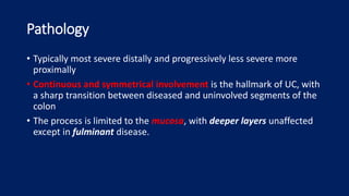
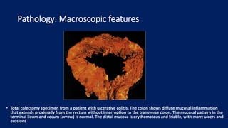
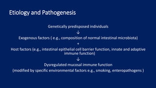
Ulcerative colitis is a chronic inflammatory bowel disease that affects the large intestine. It has a complex interaction of genetic susceptibility, immunity, and environmental factors. The highest rates are seen in Northern Europe and North America. Clinically, it presents with diarrhea, rectal bleeding, abdominal pain, and can range from limited to the rectum to involving the entire colon. The pathology shows continuous superficial inflammation and regenerative changes. Treatment focuses on reducing inflammation and immunosuppression.

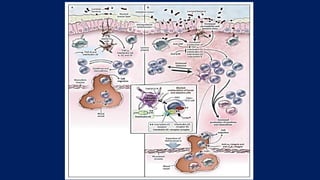
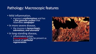
![Intestinal Immune System
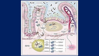
• In the healthy state, the goblet cells secrete a layer of mucus that limits exposure of the intestinal
epithelial cells to bacteria.
• Both the secretion of antimicrobial peptides (e.g., α-defensins) by Paneth cells and the production
of immunoglobulin A (IgA) provide additional protection from luminal microbiota.
• Innate microbial sensing by epithelial cells, dendritic cells, and macrophages is mediated through
pattern-recognition receptors such as toll-like receptors and nucleotide oligomerization domain
(NOD) proteins.
• Dendritic cells present antigens to naive CD4+ T cells in secondary lymphoid organs (Peyer’s
patches and mesenteric lymph nodes), where factors such as the phenotype of the antigen-
presenting cells and the cytokine milieu (transforming growth factor β [TGF-β] and interleukin-10)
modulate differentiation of CD4+ T-cell subgroups with characteristic cytokine profiles (regulatory
T cells [e.g., Treg] and helper T cells [e.g., Th1, Th2, and Th17]), and enterotropic molecules (e.g.,
α4β7) are induced that provide for gut homing of lymphocytes from the systemic circulation.
• These activated CD4+ T cells then circulate to the intestinal lamina propria, where they carry out
effector functions.](https://image.slidesharecdn.com/ulcerativecollitis-190401002623/85/Ulcerative-collitis-10-320.jpg)

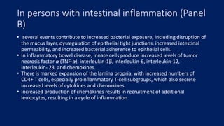
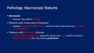
![In healthy persons (Panel A)
• the lamina propria normally contains a diverse array of immune cells
and secreted cytokines.
• These include antiinflammatory mediators (transforming growth
factor β [TGF-β] and interleukin- 10) that down-regulate immune
responses, as well as proinflammatory mediators from both innate
and adaptive immune cells that limit excessive entry of intestinal
microbiota and defend against pathogens.
• Noninflammatory defenses, such as phagocytosis by macrophages,
probably assist in defending against bacteria entering the lamina
propria while minimizing tissue injury.
• A homeostatic balance is maintained between regulatory T cells (e.g.,
Treg) and effector T cells (Th1, Th2, and Th17).](https://image.slidesharecdn.com/ulcerativecollitis-190401002623/85/Ulcerative-collitis-12-320.jpg)