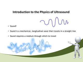
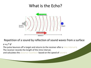
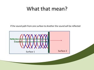
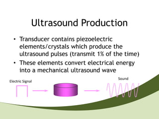
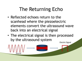
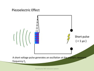
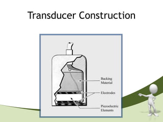
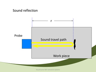
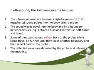
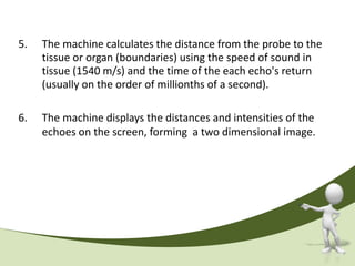
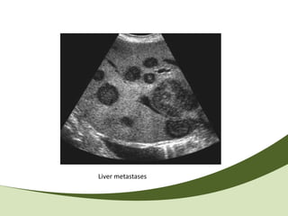
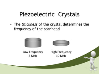
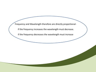
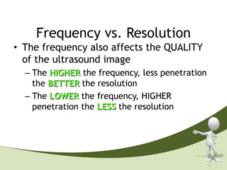
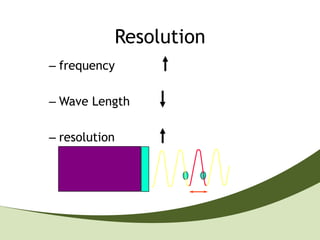
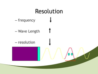
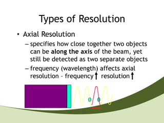
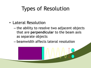
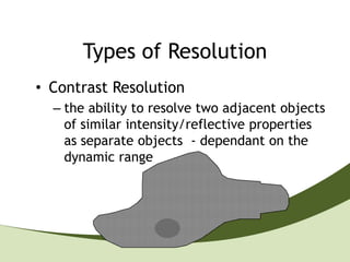
Introduction of Ultrasound
              Physics'


ENGR AHMED A A HASSAN
Introduction to the Physics of Ultrasound
                                                       Amplitude


• Sound?

• Sound is a mechanical, longitudinal wave that travels in a straight line

• Sound requires a medium through which to travel
Cycle
• 1 Cycle = 1 repetitive periodic oscillation


                           Cycle
frequency
1 cycle in 1 second = 1Hz

                    1 second


                               = 1 Hertz
Wavelength
• The length of one complete cycle
• A measurable distance
Wavelength


  Wavelength
Amplitude
• The degree of variance from the norm


                                     Amplitude
Spectrum of sound

Frequency range Hz      Description              Example


      0 - 20           Infrasound            Earth quake


   20 - 20.000       Audible sound          Speech, music


    > 20.000           Ultrasound        , Quartz crystal
Atomic structures
          gas                         liquid                     solid


• low density              • medium density              • high density
• weak bonding forces      • medium bonding              • strong bonding forces
                             forces                      • crystallographic
                                                           structure


                    Krautkramer NDT Ultrasonic Systems
Basic formula


Air            330 m/s
Water                    1480 m/s
Steel, long                                    5920 m/s
Steel, trans                        3250 m/s
Example Sound Speeds
          Medium              sound speed (m/s)
          air (20 C)                343
          water                     1497
          gold                      3240
          brick                     3650
          wood                   3800–4600
          glass                     5100
          steel                     5790
          aluminum                  6420


Spring 2006                                       11
What is the Echo?


Repetition of a sound by reflection of sound waves from a surface
r = c * t2
The pulse bounces off a target and returns to the receiver after a time interval t.
The receiver records the length of this time interval,
and calculates the distance travelled r based on the speed of sound c
What that mean?

If the sound path from one surface to Another the sound will be reflected


                Surface 1                        Surface 2
What is Ultrasound?

• Ultrasound is a mechanical, longitudinal
  wave with a frequency exceeding the
  upper limit of human hearing, which is
  20,000 Hz or 20 kHz.

Medical Ultrasound 2MHz to 16MHz
How the Ultrasound System work?
Piezoelectric material
• AC applied to a piezoelectric crystal causes it
  to expand and contract – generating ultrasound,
  and vice versa

• Naturally occurring - quartz

• Synthetic - Lead zirconate titanate (PZT)
Human Hair
                                    Single
                                    Crystal


Microscopic view of scanhead
Ultrasound Production
  • Transducer contains piezoelectric
    elements/crystals which produce the
    ultrasound pulses (transmit 1% of the time)
  • These elements convert electrical energy
    into a mechanical ultrasound wave
                                       Sound
Electric Signal
The Returning Echo
• Reflected echoes return to the
  scanhead where the piezoelectric
  elements convert the ultrasound wave
  back into an electrical signal
• The electrical signal is then processed
  by the ultrasound system
                                Electric Signal


     Sound
Piezoelectric Effect

 Sound wave
     with
 frequency f


                                                               U(f)


An alternating voltage generates crystal oscillations at the frequency f


                      Krautkramer NDT Ultrasonic Systems
Piezoelectric Effect


                                                            Short pulse
                                                            ( < 1 µs )


A short voltage pulse generates an oscillation at the crystal‘s resonant
frequency f0

                      Krautkramer NDT Ultrasonic Systems
Transducer Construction
Sound reflection

                                  r


 Probe
                    Sound travel path


                                      Work piece


              Krautkramer NDT Ultrasonic Systems
Immersion testing

                            1                            2
 surface =                                                                    water delay
sound entry

  backwall                                                                     Mass


           IP                       1                 IP                      2
                        IE                                           IE

                                    BE                                        BE
                                                                          F

       0        2   4   6       8    10             0        2   4   6    8    10

                        Krautkramer NDT Ultrasonic Systems
In ultrasound, the following events happen:

1. The ultrasound machine transmits high-frequency (1 to 20
   megahertz) sound pulses into the body using a probe.
2. The sound waves travel into the body and hit a boundary
   between tissues (e.g. between fluid and soft tissue, soft tissue
   and bone).
3. Some of the sound waves reflect back to the probe, while
   some travel on further until they reach another boundary and
   then reflect back to the probe .
4. The reflected waves are detected by the probe and relayed to
   the machine.
5.   The machine calculates the distance from the probe to the
     tissue or organ (boundaries) using the speed of sound in
     tissue (1540 m/s) and the time of the each echo's return
     (usually on the order of millionths of a second).

6.   The machine displays the distances and intensities of the
     echoes on the screen, forming a two dimensional image.
Liver metastases
Piezoelectric Crystals
• The thickness of the crystal determines the
  frequency of the scanhead


       Low Frequency   High Frequency
           3 MHz           10 MHz
Frequency and Wavelength therefore are directly proportional-

  if the frequency increases the wavelength must decrease.

  if the frequency decreases the wavelength must increase
Frequency vs. Resolution
• The frequency also affects the QUALITY
  of the ultrasound image
  – The HIGHER the frequency, less penetration
    the BETTER the resolution
  – The LOWER the frequency, HIGHER
    penetration the LESS the resolution
Resolution
– frequency

– Wave Length

– resolution
Resolution
– frequency

– Wave Length

– resolution
Types of Resolution
• Axial Resolution
  – specifies how close together two objects
    can be along the axis of the beam, yet
    still be detected as two separate objects
  – frequency (wavelength) affects axial
    resolution – frequency resolution
Types of Resolution

• Lateral Resolution
  – the ability to resolve two adjacent objects
    that are perpendicular to the beam axis
    as separate objects
  – beamwidth affects lateral resolution
Types of Resolution
• Spatial Resolution
  – also called Detail Resolution

  – the combination of AXIAL and LATERAL
    resolution - how closely two reflectors
    can be to one another while they can be
    identified as different reflectors
Types of Resolution
• Temporal Resolution
  – the ability to accurately locate the
    position of moving structures at particular
    instants in time
  – also known as frame rate
Types of Resolution
• Contrast Resolution
  – the ability to resolve two adjacent objects
    of similar intensity/reflective properties
    as separate objects - dependant on the
    dynamic range
History
1st who is the 1st sono grapher ?

• Bats use a variety of ultrasonic ranging
(echolocation) techniques to detect their prey.
They able to fly without their vision.
They can detect frequencies beyond 100 kHz,
possibly up to 200 kHz.

This discovered in 1793 by Italian Scientist called Spallanzani.
Sir Francis Galton (1800)
• Recognized that by moving the
  plunger (located inside the whistle)
  the size of the cavity could be
  changed to alter the pitch (frequency)
  of sound
• Determined the normal limit of
  human hearing is around 18 kHz
1st Contact B-Scanner (1956)


The first contact B scanner was designed and built by Tom Brown on
the frame of a hospital bed-table. It is seen here with its first picture;
this shows echoes from the skin, at the top of the picture, and from
the bowel.
Automatic Scanner (1959)


Tom Brown developed the world’s first and only fully
automatic scanner in order to give a consistent scanning
pattern. Much of the early research was carried out with this
machine
Fetal Cephalometry (1961)

• Dr. James Willocks
  (seated, scanning)
  developed a technique for
  fetal cephalometry using
  A-Scan equipment and
  electronics.
1st Commercially Produced Medical Scanners
                   (1962)
The Mid ’70s and the Change to Real-time


                                  System 185
  EMI 4200   System 85            Diagnostic Sonar
  Static     Real-time scanners   Ltd.
  Scanner
The Beginnings of 3D (1976)


•   develop the world’s first 3-D
    ultrasound scanner.
•   Sonicaid Multiplanar Scanner
Now a Days:
U ltrasound physiscs

U ltrasound physiscs

  • 1.
    Introduction of Ultrasound Physics' ENGR AHMED A A HASSAN
  • 2.
    Introduction to thePhysics of Ultrasound Amplitude • Sound? • Sound is a mechanical, longitudinal wave that travels in a straight line • Sound requires a medium through which to travel
  • 3.
    Cycle • 1 Cycle= 1 repetitive periodic oscillation Cycle
  • 4.
    frequency 1 cycle in1 second = 1Hz 1 second = 1 Hertz
  • 5.
    Wavelength • The lengthof one complete cycle • A measurable distance
  • 6.
  • 7.
    Amplitude • The degreeof variance from the norm Amplitude
  • 8.
    Spectrum of sound Frequencyrange Hz Description Example 0 - 20 Infrasound Earth quake 20 - 20.000 Audible sound Speech, music > 20.000 Ultrasound , Quartz crystal
  • 9.
    Atomic structures gas liquid solid • low density • medium density • high density • weak bonding forces • medium bonding • strong bonding forces forces • crystallographic structure Krautkramer NDT Ultrasonic Systems
  • 10.
    Basic formula Air 330 m/s Water 1480 m/s Steel, long 5920 m/s Steel, trans 3250 m/s
  • 11.
    Example Sound Speeds Medium sound speed (m/s) air (20 C) 343 water 1497 gold 3240 brick 3650 wood 3800–4600 glass 5100 steel 5790 aluminum 6420 Spring 2006 11
  • 12.
    What is theEcho? Repetition of a sound by reflection of sound waves from a surface r = c * t2 The pulse bounces off a target and returns to the receiver after a time interval t. The receiver records the length of this time interval, and calculates the distance travelled r based on the speed of sound c
  • 13.
    What that mean? Ifthe sound path from one surface to Another the sound will be reflected Surface 1 Surface 2
  • 14.
    What is Ultrasound? •Ultrasound is a mechanical, longitudinal wave with a frequency exceeding the upper limit of human hearing, which is 20,000 Hz or 20 kHz. Medical Ultrasound 2MHz to 16MHz
  • 15.
    How the UltrasoundSystem work?
  • 16.
    Piezoelectric material • ACapplied to a piezoelectric crystal causes it to expand and contract – generating ultrasound, and vice versa • Naturally occurring - quartz • Synthetic - Lead zirconate titanate (PZT)
  • 17.
    Human Hair Single Crystal Microscopic view of scanhead
  • 18.
    Ultrasound Production • Transducer contains piezoelectric elements/crystals which produce the ultrasound pulses (transmit 1% of the time) • These elements convert electrical energy into a mechanical ultrasound wave Sound Electric Signal
  • 19.
    The Returning Echo •Reflected echoes return to the scanhead where the piezoelectric elements convert the ultrasound wave back into an electrical signal • The electrical signal is then processed by the ultrasound system Electric Signal Sound
  • 20.
    Piezoelectric Effect Soundwave with frequency f U(f) An alternating voltage generates crystal oscillations at the frequency f Krautkramer NDT Ultrasonic Systems
  • 21.
    Piezoelectric Effect Short pulse ( < 1 µs ) A short voltage pulse generates an oscillation at the crystal‘s resonant frequency f0 Krautkramer NDT Ultrasonic Systems
  • 22.
  • 23.
    Sound reflection r Probe Sound travel path Work piece Krautkramer NDT Ultrasonic Systems
  • 24.
    Immersion testing 1 2 surface = water delay sound entry backwall Mass IP 1 IP 2 IE IE BE BE F 0 2 4 6 8 10 0 2 4 6 8 10 Krautkramer NDT Ultrasonic Systems
  • 25.
    In ultrasound, thefollowing events happen: 1. The ultrasound machine transmits high-frequency (1 to 20 megahertz) sound pulses into the body using a probe. 2. The sound waves travel into the body and hit a boundary between tissues (e.g. between fluid and soft tissue, soft tissue and bone). 3. Some of the sound waves reflect back to the probe, while some travel on further until they reach another boundary and then reflect back to the probe . 4. The reflected waves are detected by the probe and relayed to the machine.
  • 26.
    5. The machine calculates the distance from the probe to the tissue or organ (boundaries) using the speed of sound in tissue (1540 m/s) and the time of the each echo's return (usually on the order of millionths of a second). 6. The machine displays the distances and intensities of the echoes on the screen, forming a two dimensional image.
  • 27.
  • 28.
    Piezoelectric Crystals • Thethickness of the crystal determines the frequency of the scanhead Low Frequency High Frequency 3 MHz 10 MHz
  • 29.
    Frequency and Wavelengththerefore are directly proportional- if the frequency increases the wavelength must decrease. if the frequency decreases the wavelength must increase
  • 30.
    Frequency vs. Resolution •The frequency also affects the QUALITY of the ultrasound image – The HIGHER the frequency, less penetration the BETTER the resolution – The LOWER the frequency, HIGHER penetration the LESS the resolution
  • 31.
    Resolution – frequency – WaveLength – resolution
  • 32.
    Resolution – frequency – WaveLength – resolution
  • 33.
    Types of Resolution •Axial Resolution – specifies how close together two objects can be along the axis of the beam, yet still be detected as two separate objects – frequency (wavelength) affects axial resolution – frequency resolution
  • 34.
    Types of Resolution •Lateral Resolution – the ability to resolve two adjacent objects that are perpendicular to the beam axis as separate objects – beamwidth affects lateral resolution
  • 35.
    Types of Resolution •Spatial Resolution – also called Detail Resolution – the combination of AXIAL and LATERAL resolution - how closely two reflectors can be to one another while they can be identified as different reflectors
  • 36.
    Types of Resolution •Temporal Resolution – the ability to accurately locate the position of moving structures at particular instants in time – also known as frame rate
  • 37.
    Types of Resolution •Contrast Resolution – the ability to resolve two adjacent objects of similar intensity/reflective properties as separate objects - dependant on the dynamic range
  • 38.
  • 39.
    1st who isthe 1st sono grapher ? • Bats use a variety of ultrasonic ranging (echolocation) techniques to detect their prey. They able to fly without their vision. They can detect frequencies beyond 100 kHz, possibly up to 200 kHz. This discovered in 1793 by Italian Scientist called Spallanzani.
  • 40.
    Sir Francis Galton(1800) • Recognized that by moving the plunger (located inside the whistle) the size of the cavity could be changed to alter the pitch (frequency) of sound • Determined the normal limit of human hearing is around 18 kHz
  • 41.
    1st Contact B-Scanner(1956) The first contact B scanner was designed and built by Tom Brown on the frame of a hospital bed-table. It is seen here with its first picture; this shows echoes from the skin, at the top of the picture, and from the bowel.
  • 42.
    Automatic Scanner (1959) TomBrown developed the world’s first and only fully automatic scanner in order to give a consistent scanning pattern. Much of the early research was carried out with this machine
  • 43.
    Fetal Cephalometry (1961) •Dr. James Willocks (seated, scanning) developed a technique for fetal cephalometry using A-Scan equipment and electronics.
  • 44.
    1st Commercially ProducedMedical Scanners (1962)
  • 45.
    The Mid ’70sand the Change to Real-time System 185 EMI 4200 System 85 Diagnostic Sonar Static Real-time scanners Ltd. Scanner
  • 46.
    The Beginnings of3D (1976) • develop the world’s first 3-D ultrasound scanner. • Sonicaid Multiplanar Scanner
  • 47.