
Recommended
More Related Content
Similar to TM-1.pptx
Similar to TM-1.pptx (20)
Recently uploaded
Recently uploaded (20)
TM-1.pptx
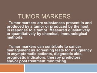
- 1. TUMOR MARKERS • Tumor markers are substances present in and produced by a tumor or produced by the host in response to a tumor. Measured qualitatively or quantitatively by chemical, immunological methods. • Tumor markers can contribute to cancer management as screening tests for malignancy in asymptomatic patients, diagnostic aids, prognostic indicators, therapy predictors, and/or post treatment monitoring.
- 2. Most Frequently Used Tumor Markers ◻ Cancer antigen 125 (CA125) ◻ Alpha-fetoprotein (AFP) ◻ Human chorionic gonadotropin (hCG) ◻ Cancer antigen 19-9 (CA19-9) ◻ Carcinoembryonic antigen (CEA) ◻ Prostate-specific antigen (PSA)
- 3. Cancer antigen 125 (CA125) ◻ Screening for Ovarian Cancer The American College of Obstetricians and Gynecologists recommends that risk-reducing salpingo- oophorectomy (ie, total removal of the ovaries and fallopian tubes) should be offered by age 40 years for BRCA1 or BRCA2 mutation carriers. ◻ For women who decline this operation, the National Comprehensive Cancer Network (NCCN) view is that while there may be circumstances where offering screening with transvaginal ultrasound and/or CA125 may be helpful.
- 4. Cancer antigen 125 (CA125) ◻ Diagnosis CA125 measurement in isolation is not recommended for diagnosis due to its low sensitivity and specificity for ovarian cancer. ◻ CA125 concentrations greater than 95 U/L in postmenopausal women can discriminate such masses with a positive predictive value of 95% ◻ If CA125 is 35 U/L or higher, an ultrasound scan of the abdomen should be arranged to enable calculation of the RMI (U × M × [CA125] cutoff RMI value of 200 )
- 5. Cancer antigen 125 (CA125) ◻ Algorithm can be embedded in specialist software Risk of malignancy algorithm (ROMA) scoring system that includes CA125, HE4, and menopausal ◻ Copenhagen Index (CPH-I), which includes serum HE4, serum CA125, and age without menopausal status or ultrasound ◻ Clearly there are significant drawbacks to the first-line use of CA125 in the early detection of ovarian cancer, but it is the best serum marker available.
- 6. Cancer antigen 125 (CA125) ◻ Prognosis Five-year survival rates in patients with a preoperative CA125 concentration > 65 U/L have been found to be significantly lower than those for patients with CA125 < 65 U/L. ◻ Following primary treatment with surgery and/or chemotherapy, a nadir CA125 concentration (eg, to ≤5 U/L) and a more rapid rate of decrease (eg, ≥50% during the initial two cycles of platinum-based chemotherapy) are generally associated with more favorable prognosis. ◻ Generally, a prolonged CA125 half-life and/or failure to fall to within reference limits is predictive of poor response to treatment and a higher rate of relapse.
- 7. Cancer antigen 125 (CA125) ◻ Monitoring Current ESGO recommendations are that CA125 follow-up should be continued in patients after complete response on primary treatment for ovarian cancer ◻ The UK-based Medical Research Council/European Organization for Research and Treatment of Cancer (MRC-EORTC) conducted a randomized trial to evaluate the utility for such monitoring of CA125 (concluded routine CA125 monitoring is not necessary )
- 8. Cancer antigen 125 (CA125) ◻ SUMMARY: CA125 is the most widely used serum tumor marker for all epithelial ovarian cancers but is most sensitive for serous adenocarcinomas. ◻ Because many benign conditions are associated with increased CA125, careful interpretation of results in the clinical context is paramount. ◻ The serum markers of choice for ovarian germ cell tumors are AFP, hCG, and LDH while for granulosa cell tumors, inhibin is the
- 9. Alpha-fetoprotein (AFP) ◻ AFP is an oncofetal antigen that is appropriately increased in pregnant women and most frequently inappropriately increased in patients with germ cell tumors and HCC . ◻ Screening for HCC high-risk patients include men and women with established cirrhosis due to HBV or HCV infection or genetic hemochromatosis, men with alcohol-related cirrhosis( AFP and ultrasound)
- 10. Alpha-fetoprotein (AFP) ◻ Diagnosis For liver lesions more than 2 cm in size where USS results are typical of HCC and AFP is greater than 200 ng/ml a diagnosis of HCC can be made without proceeding to biopsy. ◻ The AASLD guidelines no longer recommend AFP testing as part of diagnostic evaluation because AFP concentrations may be raised in nonmalignant conditions and may be within normal limits in a substantial number of HCC patients, and because imaging is more definitive, as is also recognized by the NCCN.
- 11. Alpha-fetoprotein (AFP) ◻ AFP in Prognosis. In combination with other factors, AFP concentrations may provide prognostic information in untreated HCC patients and in those being considered for liver resection or transplantation(<1000ng/ml), with high concentrations generally associated with poor prognosis.
- 12. Alpha-fetoprotein (AFP) ◻ AFP in Post treatment Monitoring. Measurement of AFP at follow-up visits is recommended by the NACB, NCCN, and others to monitor disease status after liver resection or liver transplantation, for detection of recurrence or after ablative therapies, and/or during palliative treatment.
- 13. Alpha-fetoprotein (AFP) ◻ Measurements of hCG, AFP, and LDH are integral to the management of patients with GCTs(Germ Cell Tumors) . ◻ Plasma concentrations of AFP and hCG should be measured in any male with a suspicious lump in the testes and in any patient with malignancy of unknown origin.
- 14. Alpha-fetoprotein (AFP) ◻ Serum or plasma concentrations of AFP and/or hCG are increased in 80% to 85% of men with NSGCT, while less than 25% of those with seminomas have raised hCG and none have raised AFP. ◻ Increased AFP precludes a diagnosis of seminoma. Increased AFP in a patient diagnosed with seminoma suggests that the AFP may not be related to the tumor (eg, may be of liver origin) or that yolk sac elements were overlooked.
- 15. Alpha-fetoprotein (AFP) ◻ criteria developed by the International Germ Cell Cancer Collaborative Group (IGCCCG) for Prognostic Categories for Nonseminomas ◻ Good- marker concentrations— all of AFP <1000 ng/ml and hCG <5000 U/L ◻ Intermediate- marker concentrations— any of AFP ≥1000 ng/ml and hCG >5000 U/L ◻ Poor -marker concentrations—any of AFP >10,000 ng/ml and hCG >50,000 U/L
- 16. Alpha-fetoprotein (AFP) ◻ Monitoring. Tumor markers should be measured before and after surgical excision of GCTs because calculation of the tumor marker half-life provides helpful information about the success of treatment. ◻ Provided that disease is confined to the testis or ovary, serum AFP and/or hCG should decline to normal with apparent half-lives of 5 to 6 days for AFP and 1 to 2 days for hCG. ◻ Further treatment with chemotherapy or radiation is required if AFP or hCG remain increased following surgery or if imaging identifies residual or metastatic disease.
- 17. The importance of careful monitoring of tumor markers is illustrated in Fig.
- 18. Human chorionic gonadotropin (hCG) ◻ Screening of women at high risk of developing GTD (ie, women who have had a partial or complete hydatidiform mole in a previous pregnancy) is conveniently performed by measuring hCG in urine or blood. ◻ Estimation of hCG serum concentrations may be helpful in diagnosing molar pregnancies because values greater than two multiples of the median for the gestational date are suggestive of GTD
- 19. Human chorionic gonadotropin (hCG) ◻ Prognosis is assessed according to the International Federation of Gynaecology and Oncology staging system for GTN,142 which includes pretreatment hCG concentration as one of the eight prognostic factors used for scoring. ◻ Women with scores of 7 or higher are at high risk of recurrence and are treated with intravenous multiagent chemotherapy until the hCG concentration has returned to normal and then for a further 6 consecutive weeks
- 20. Human chorionic gonadotropin (hCG) ◻ Following a diagnosis of GTD and surgical evacuation, if hCG has reverted to normal within 56 days, then follow-up will be for 6 months from the date of uterine evacuation. ◻ If hCG has not reverted to normal within 56 days, then follow-up will be for 6 months from normalization of the hCG concentration.
- 21. Cancer antigen 19-9 (CA19-9) ◻ Screening and Diagnosis. The low positive predictive value of CA19-9 means it cannot be used in screening, but in symptomatic patients, it contributes to diagnosis with sensitivity of 80% . ◻ Because CA19-9 may be increased in biliary infection (cholangitis), inflammation, or biliary obstruction (of any etiology), the NCCN recommends that it is best measured after biliary decompression is complete and bilirubin is normal.
- 22. Cancer antigen 19-9 (CA19-9) ◻ Prognosis. Preoperative CA19-9 concentrations correlate with both American Joint Cancer Committee (AJCC) staging and disease burden, so they can complement when assessing whether a pancreatic tumor is resectable. ◻ with concentrations greater than 130 kU/L found in one center to be strongly associated with the identification of subradiographic unresectable disease.
- 23. Cancer antigen 19-9 (CA19-9) ◻ Low postoperative CA19-9 concentrations at 3 months and before adjuvant chemotherapy have also been shown to be independent prognostic factor. ◻ normalization of CA19-9 following neoadjuvant therapy associated with longer median survival among both nonresected and resected groups of patients.
- 24. Cancer antigen 19-9 (CA19-9) ◻ Therapy Prediction. Postoperative CA19-9 concentrations may help to predict the benefit of adjuvant therapy (usually gemcitabine- based). ◻ In one study, patients with CA19-9 concentrations of 90 kU/L or less who received gemcitabine-based adjuvant therapy had a longer disease-free survival than those who did not (26 months vs. 16.7 months),
- 25. Cancer antigen 19-9 (CA19-9) ◻ Monitoring. Rising CA19-9 concentrations suggest progression and can be used, together with radiographic and clinical data, to influence decisions to initiate palliative treatment in a patient whose disease progresses after surgery or to change (or discontinue) chemotherapy in patients progressing during treatment.
- 26. Carcinoembryonic antigen (CEA) ◻ Diagnosis. Depending on the cutoff point chosen, serum CEA will be increased in only 50% of patients at the time of diagnosis, and it cannot be used in isolation to diagnose even advanced disease of colorectal cancer . ◻ Markedly increased concentrations are suggestive of metastatic disease (generally to the liver) but do not necessarily indicate a colorectal primary source.
- 27. Carcinoembryonic antigen (CEA) ◻ Prognosis. A number of studies have shown that increased preoperative CEA concentrations at the time of initial presentation of patients with colorectal cancer are associated with adverse outcome. ◻ It is therefore generally recommended that CEA be measured preoperatively in newly diagnosed colorectal cancer patients to provide both prognostic information and a baseline value for interpreting subsequent concentrations.
- 28. Carcinoembryonic antigen (CEA) ◻ Monitoring. Metaanalyses of some randomized controlled trials concluded that overall survival was significantly improved only when CEA testing was included in the intensive follow-up arm. ◻ Now generally agreed that CEA testing should be performed every 3 to 6 months for 5 years following curative surgery for colorectal cancer. ◻ An increase of 20% compared with the previous CEA (provided CEA was >2.5 μg/L) was considered to be significant.
- 29. Carcinoembryonic antigen (CEA) ◻ Measurement of CEA is also recommended during chemotherapy for metastatic disease, although care in interpretation is required because CEA concentrations may be affected by factors other than tumor progression (eg, liver damage). ◻ CEA-defined responses agree well with radiologic responses (the “gold standard”) in over 90% of cases, enabling the conclusion that use of CEA is as accurate as CT imaging for assessing the response of colorectal cancer liver metastases to chemotherapy.
- 30. Prostate-specific antigen (PSA) ◻ PSA in Screening for Prostate Cancer. Screening asymptomatic men for prostate cancer with PSA is highly controversial ◻ inability of PSA to differentiate aggressive prostate cancers that require treatment from indolent cancers that will not progress ◻ there are significant risks of overdiagnosis and overtreatment of some men.
- 31. Prostate-specific antigen (PSA) ◻ PSA as an Aid to Diagnosis of Prostate Cancer. Men with symptoms suggestive of prostate cancer should be offered a PSA test ◻ Results above the upper limit of the reference interval but less than 10 μg/L are considered to be within a “gray zone” where additional testing (eg, percent free PSA) may be implemented in some centers before proceeding to biopsy.
- 32. Prostate-specific antigen (PSA) PSA in Prognosis and Therapy Prediction of Prostate Cancer The pretreatment serum PSA value correlates with the risk of extraprostatic extension, seminal vesicle invasion, and lymph node involvement and is an independent predictor of response to all forms of treatment. Bone scans are generally not necessary in patients with newly diagnosed prostate cancer who have a PSA of less than 20.0 μg/L
- 33. Prostate-specific antigen (PSA) ◻ Following radical prostatectomy, PSA should decrease to and remain at undetectable concentrations, with measurable concentrations indicative of residual disease or possibly the presence of benign glands. ◻ The rate of fall is likely to be slower in patients who have received radiotherapy, and PSA rarely falls to less than 0.2 μg/L because not all prostate tissue is ablated.
- 34. Prostate-specific antigen (PSA) ◻ In patients receiving hormonal therapy, a low nadir after treatment has prognostic significance and can be quantitatively linked to survival. ◻ Those for whom the PSA nadir is greater than 0.2 μg/L within 8 months of endocrine therapy have a 20-fold greater risk of cancer-specific mortality than those with a PSA nadir of less than 0.2 μg/L
- 35. CONCLUSION here are tumor markers and tumor markers, Their numbers increasing year after year. But are they sensitive and, more importantly, specific? We should get our answers loud and clear.
- 36. THANK YOU