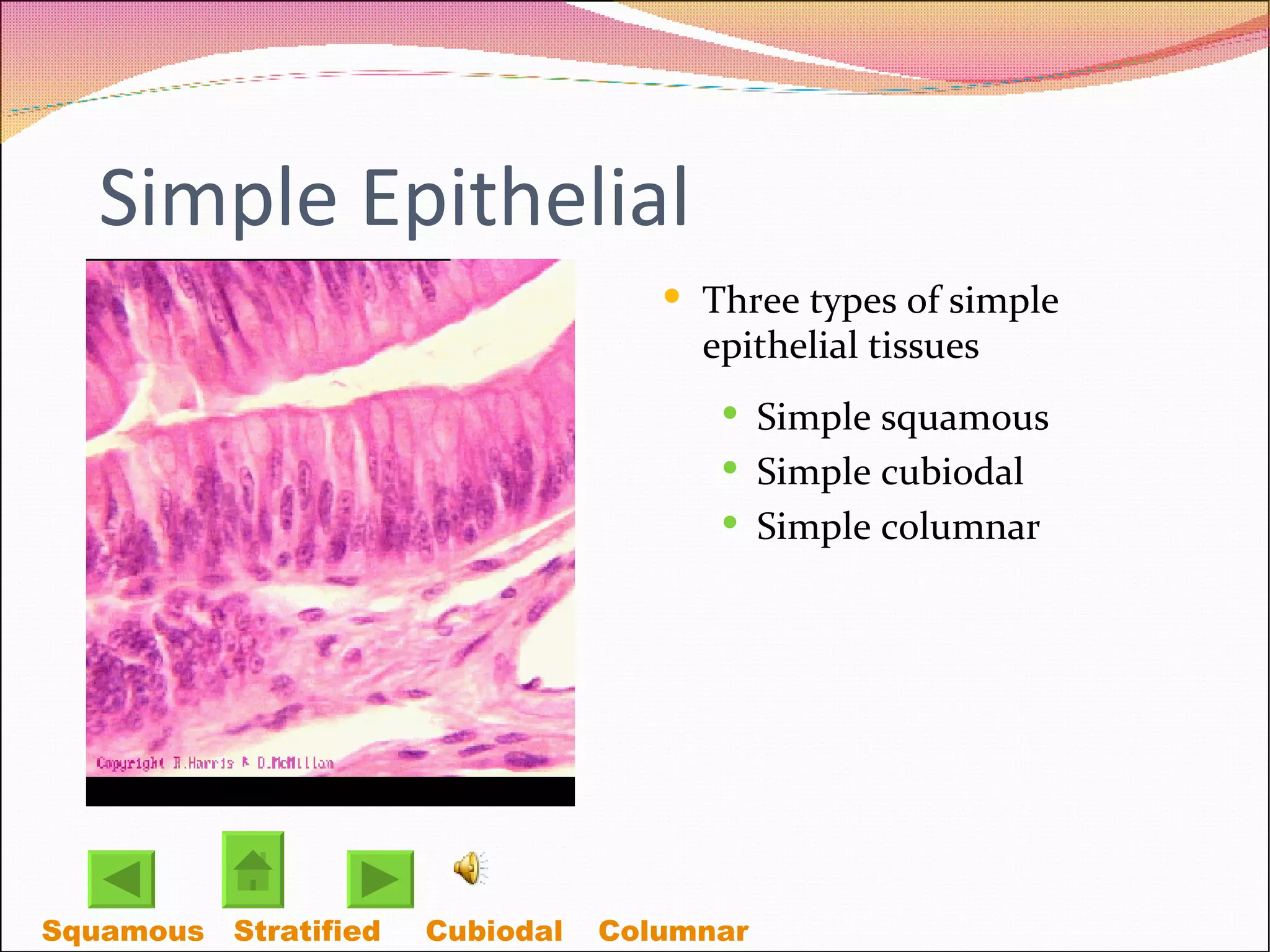
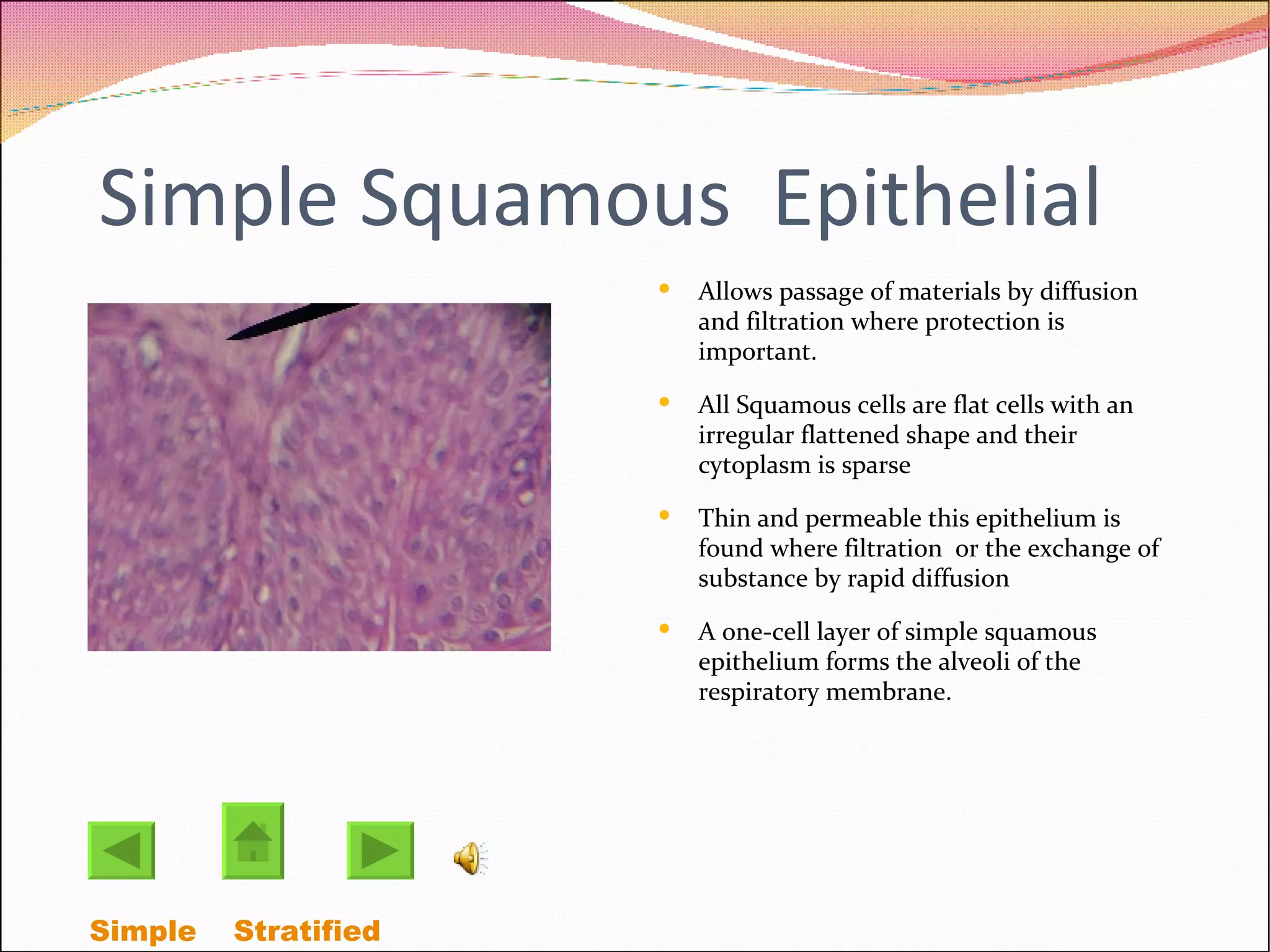
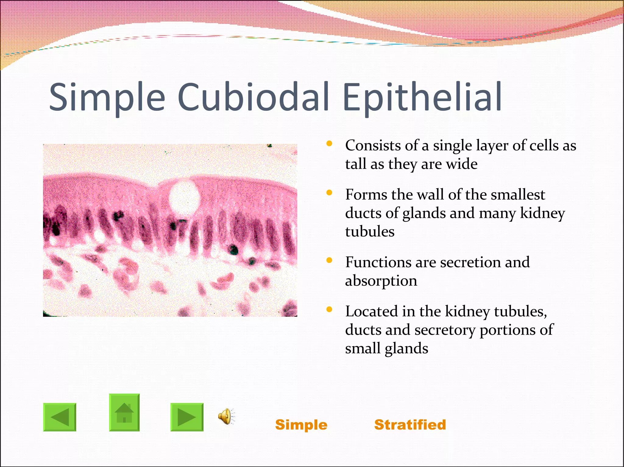
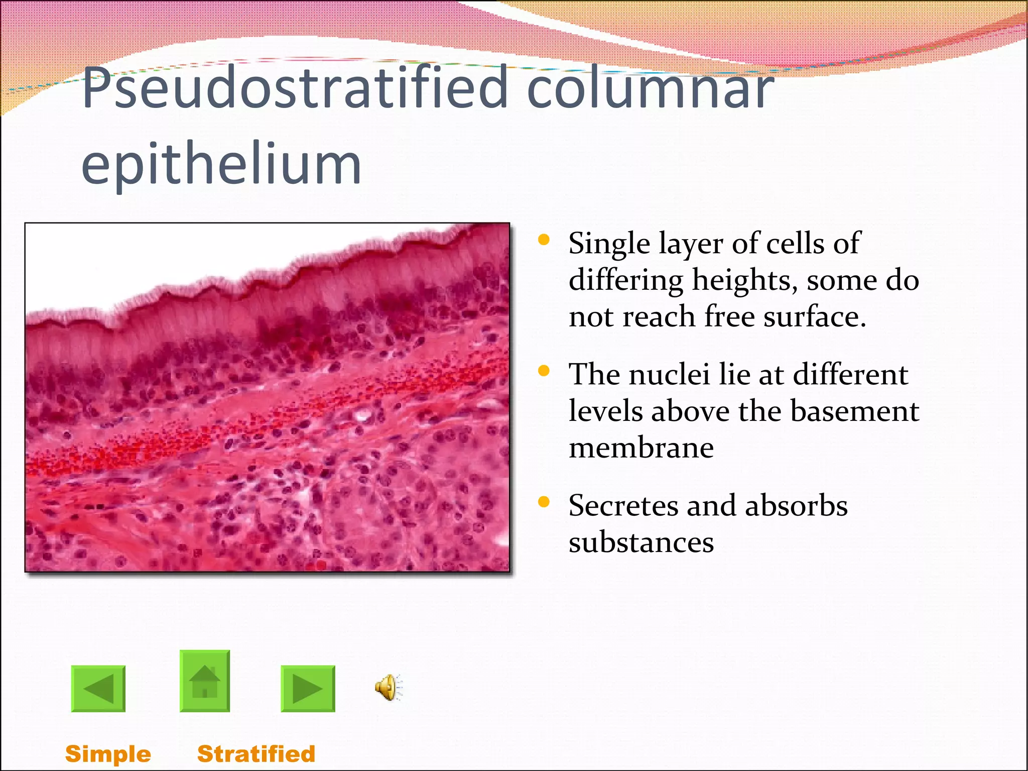
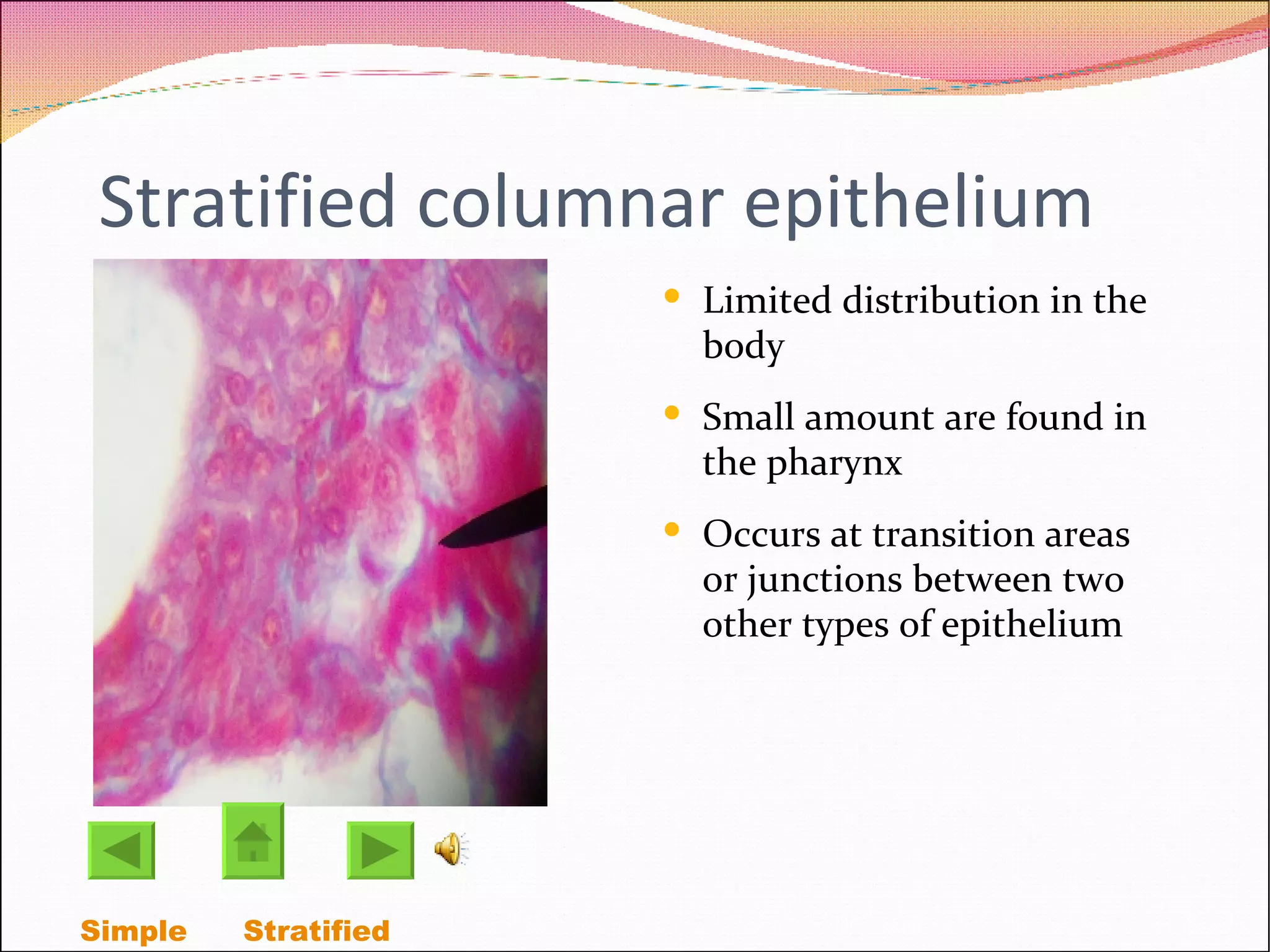
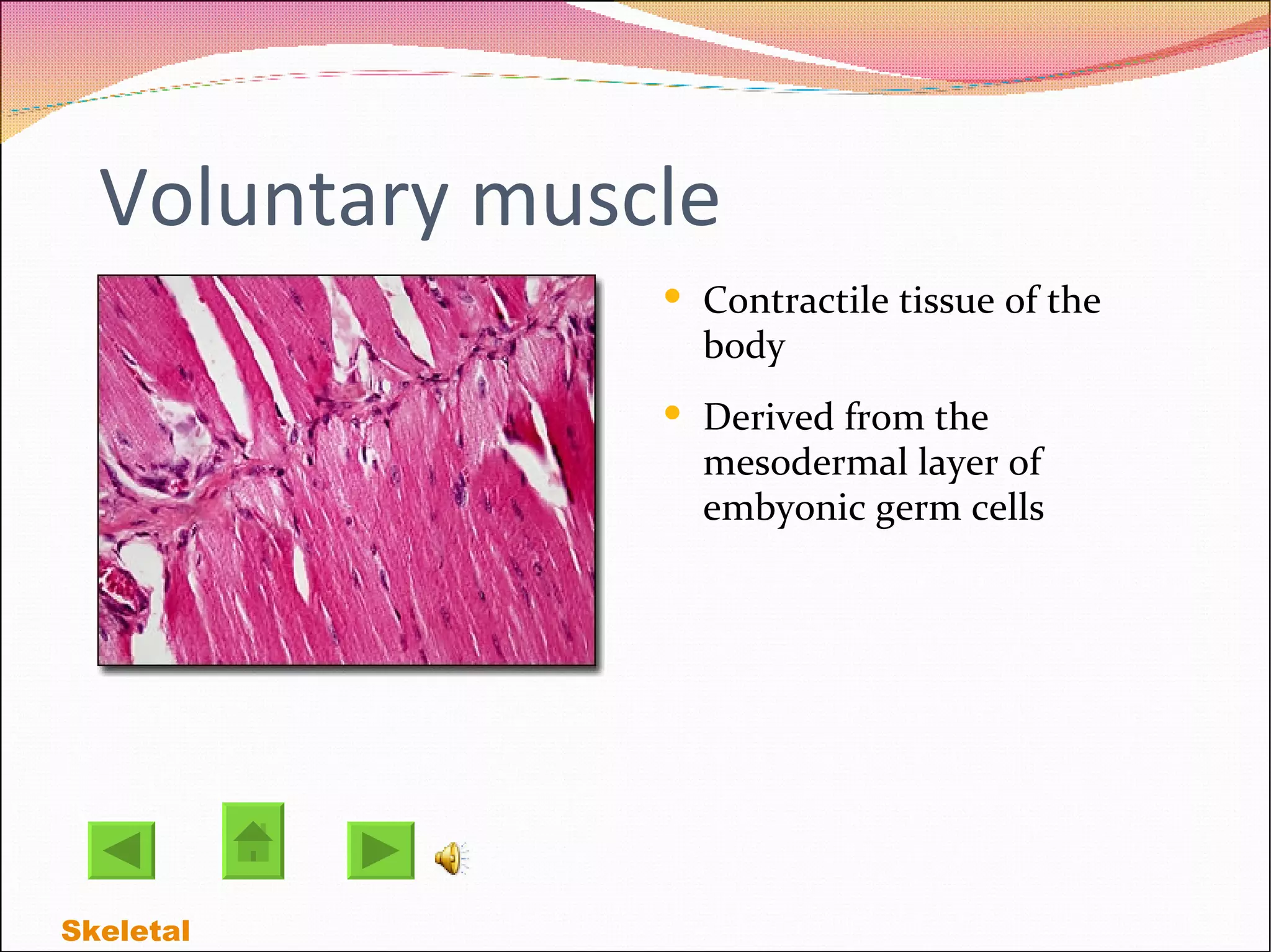
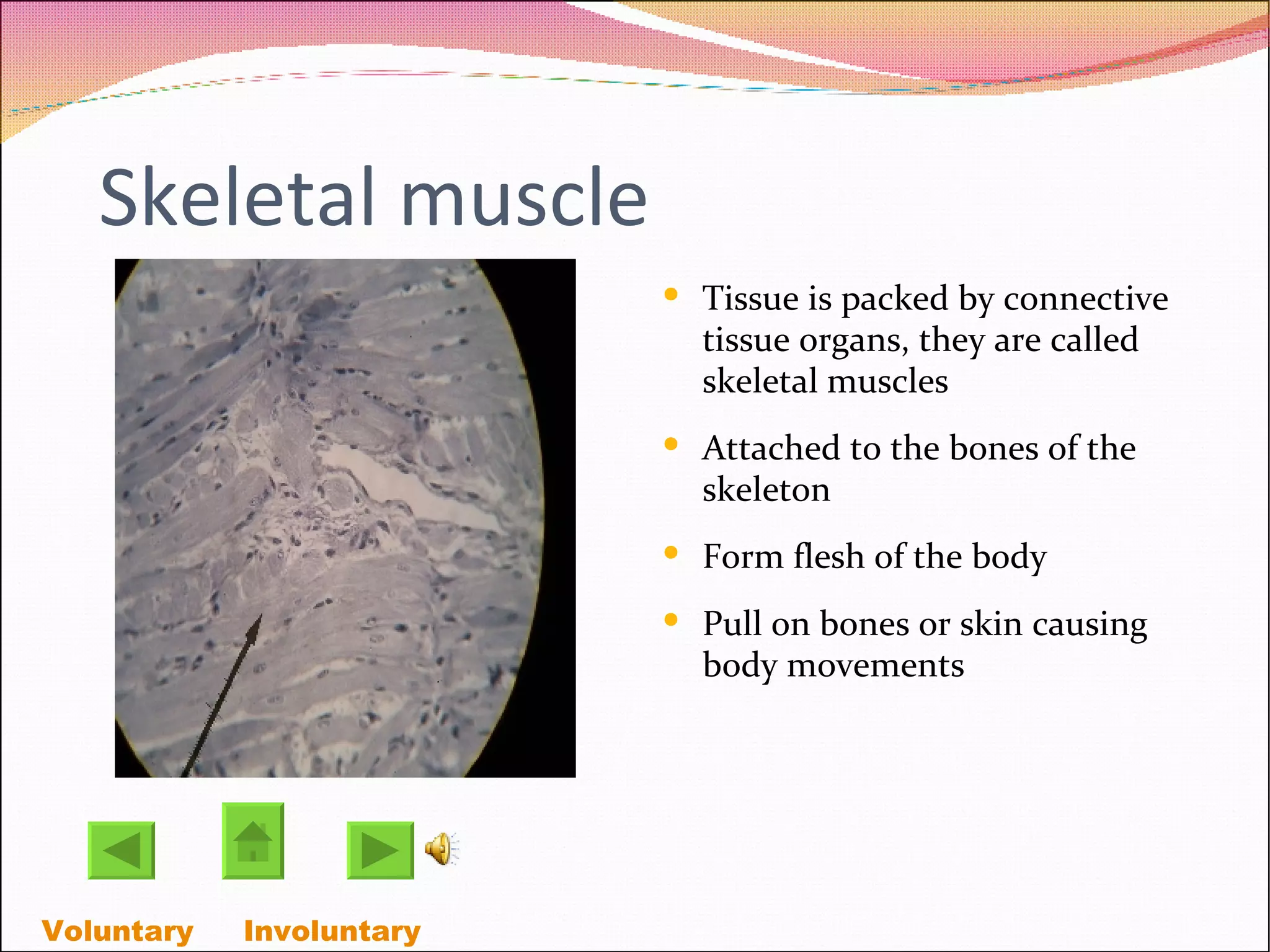
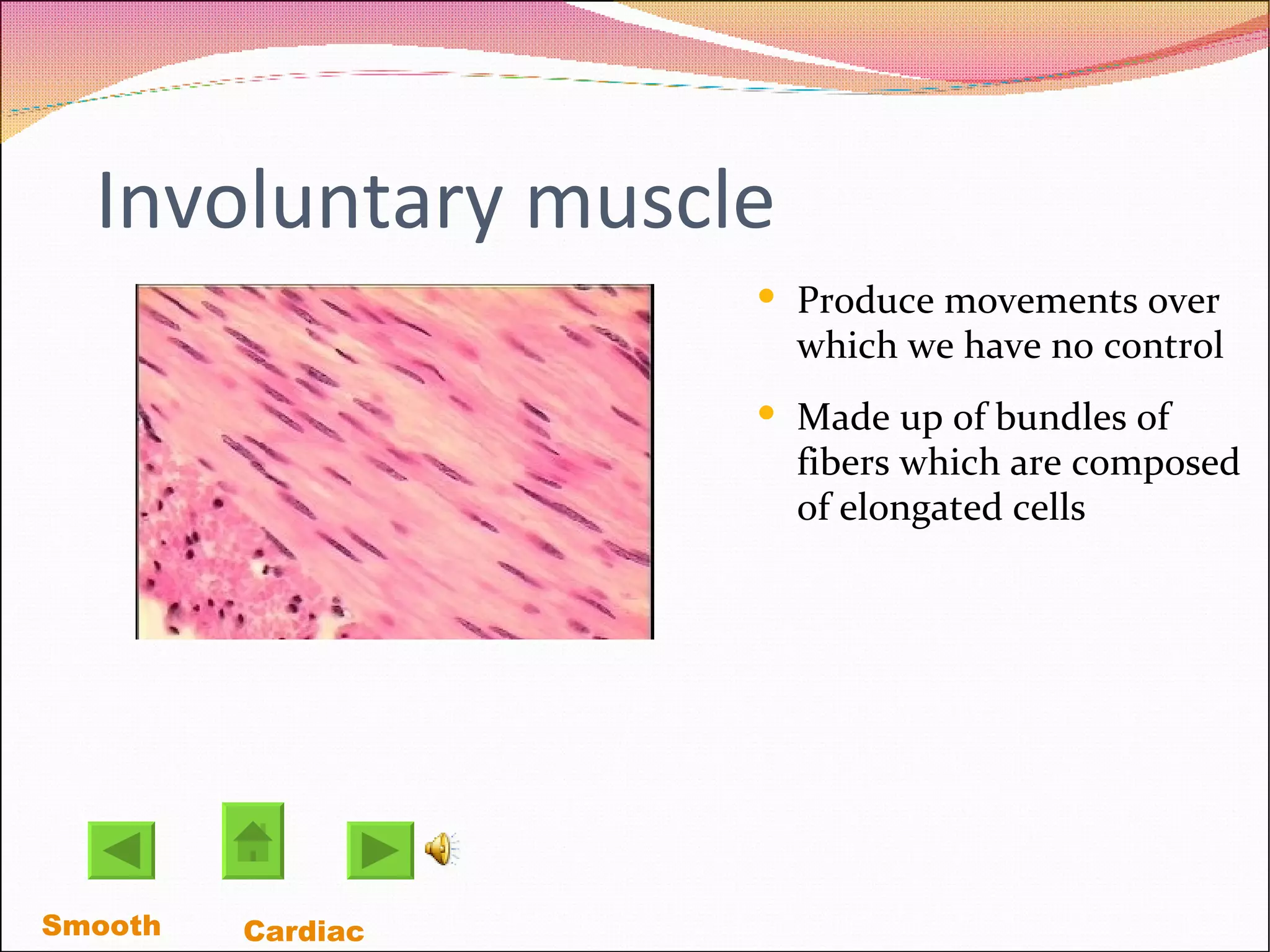
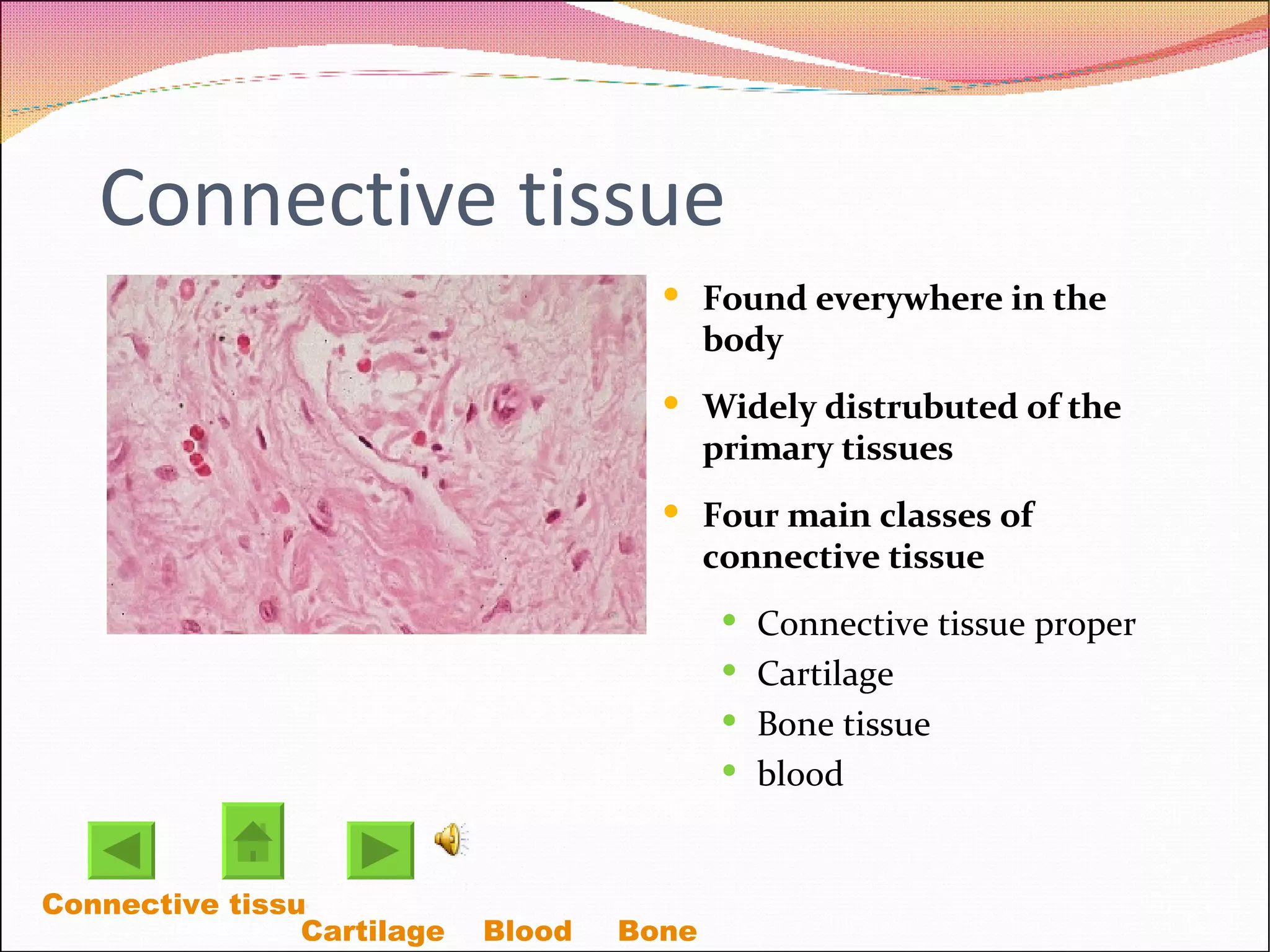
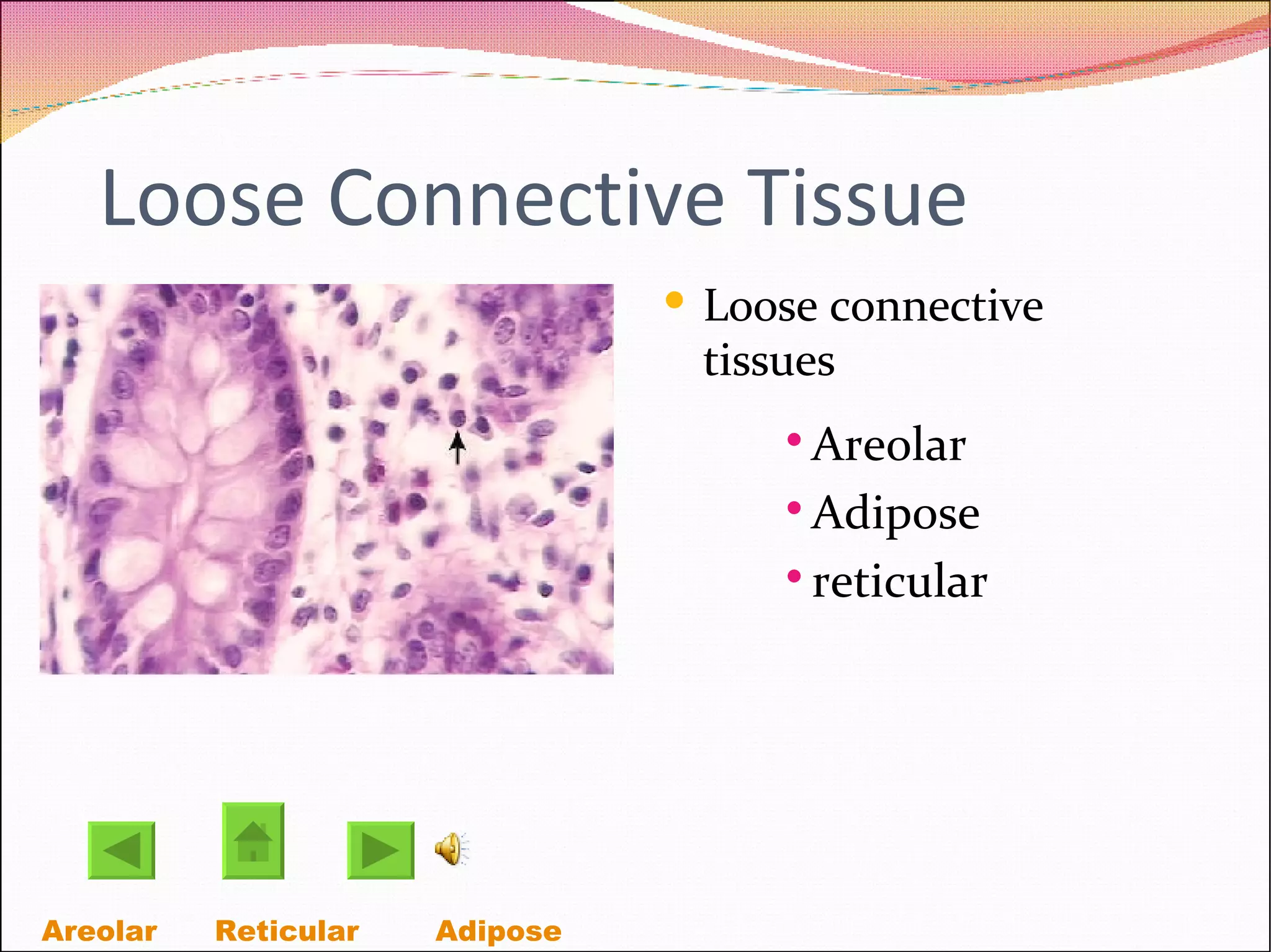
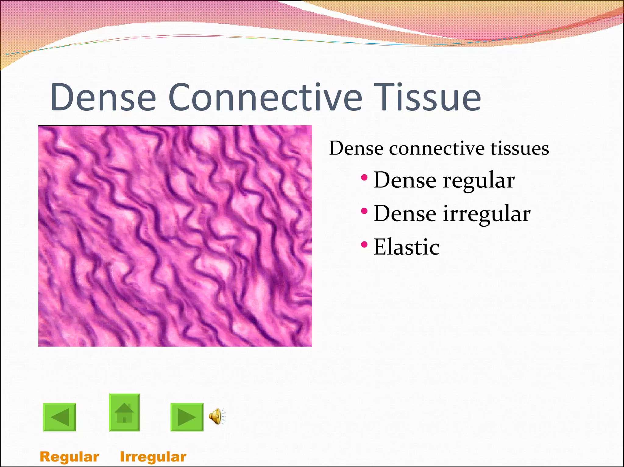
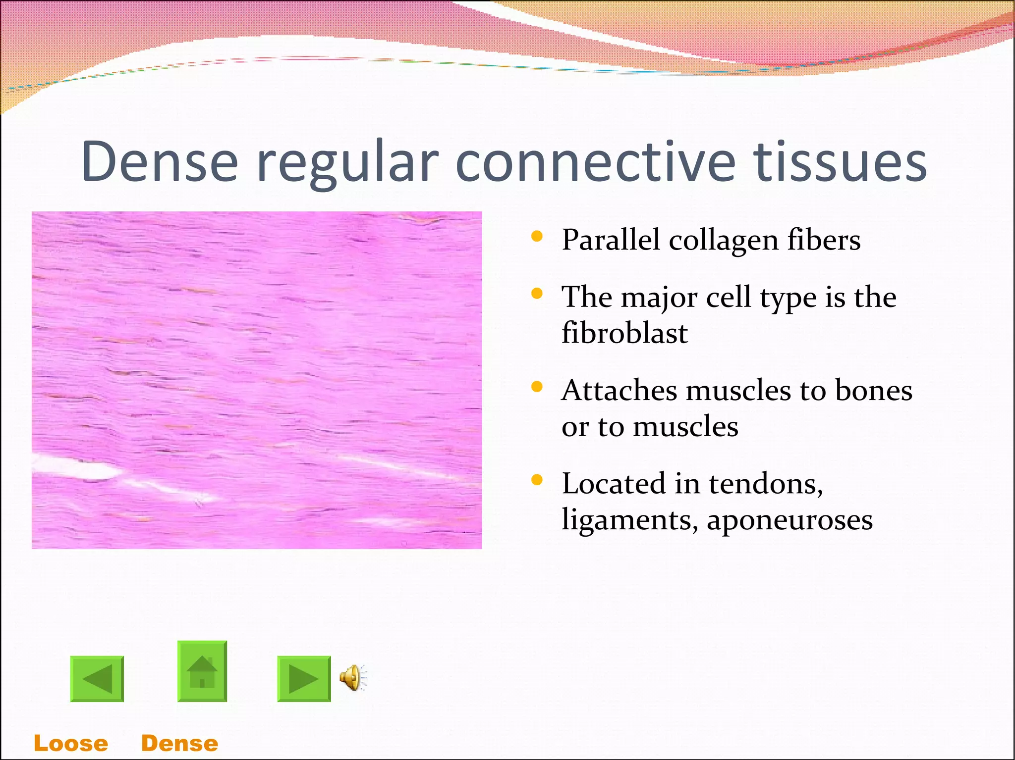
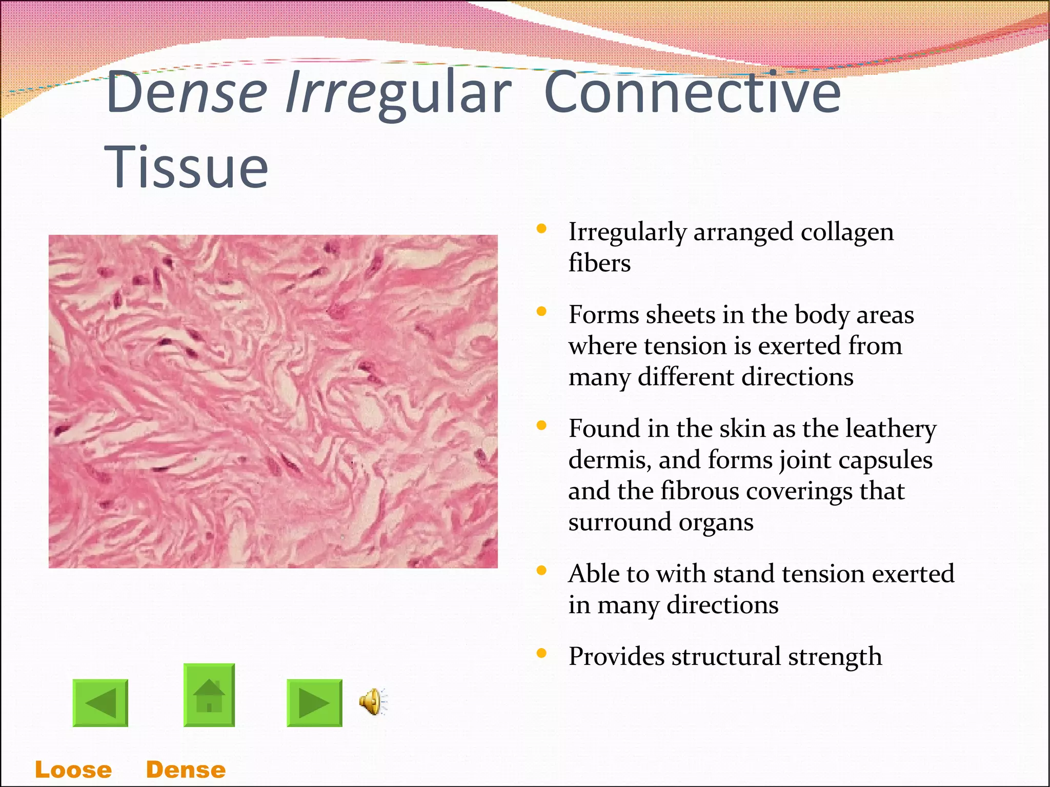
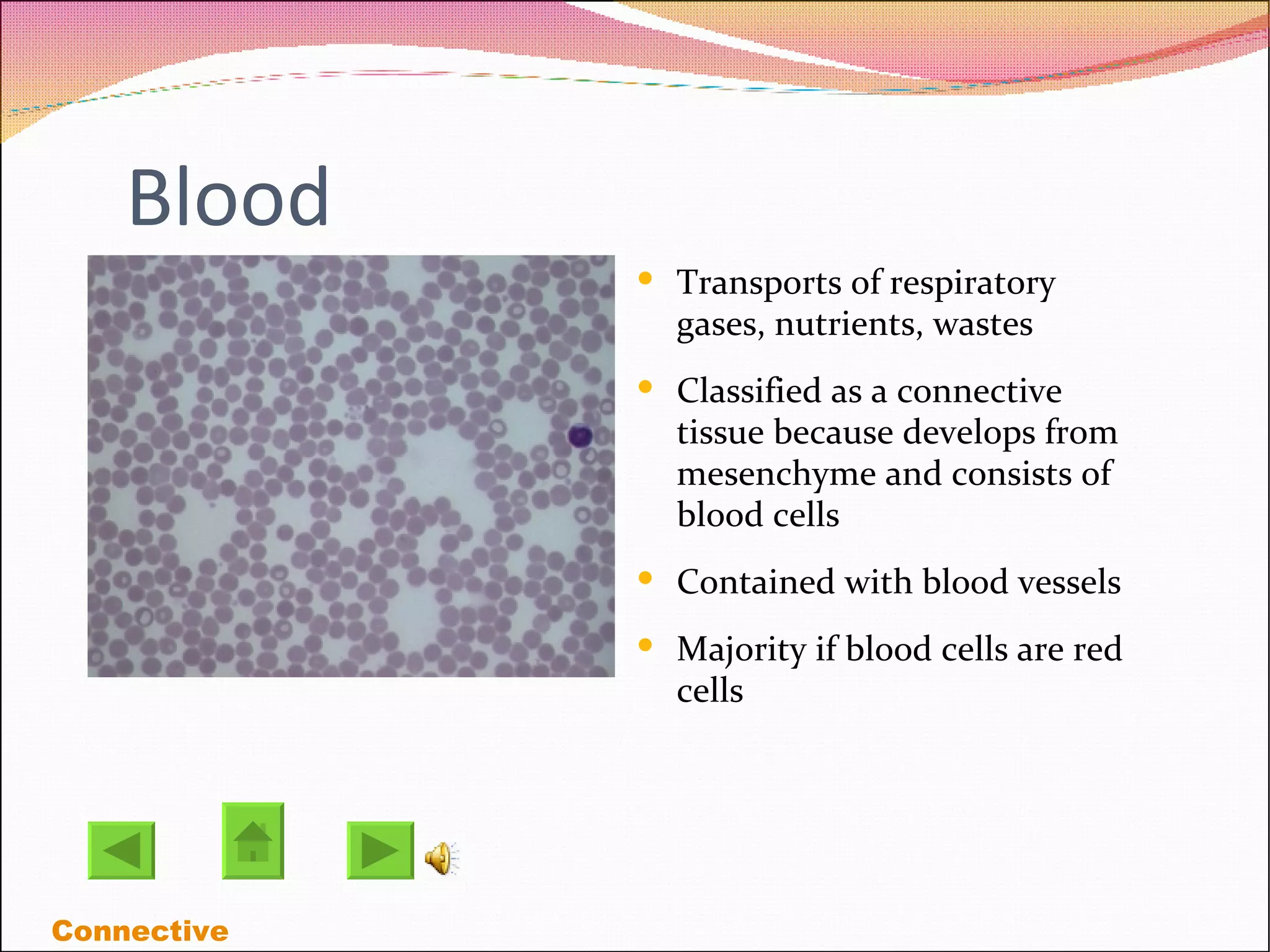
Tissues! Four types of tissues Connective Muscular Epithelial  Nervous
Epithelial Tissues A sheet of cells that covers a body's surface Lines a body's cavity The major function of  epithelial tissue includes protection, secretion, absorption, and filtration. Cells can be thin, flat to cubic to elongated Simple  Stratified
Simple Epithelial Three types of simple epithelial tissues Simple squamous  Simple cubiodal Simple columnar Squamous Cubiodal Stratified  Columnar
Simple Squamous  Epithelial Allows passage of materials by diffusion and filtration where protection is important. All Squamous cells are flat cells with an irregular flattened shape and their cytoplasm is sparse Thin and permeable this epithelium is found where filtration  or the exchange of substance by rapid diffusion A one-cell layer of simple squamous epithelium forms the alveoli of the respiratory membrane. Simple   Stratified
Simple Cubiodal Epithelial Consists of a single layer of cells as tall as they are wide Forms the wall of the smallest ducts of glands and many kidney tubules Functions are secretion and absorption  Located in the kidney tubules, ducts and secretory portions of small glands  Simple   Stratified
Simple columnar epithelial  Columnar epithelial cells occur in one or more layers Functions are absorption, secretion of mucus enzymes  Lines the digestive tract from the stomach through the rectum Displays cilia on their free surfaces, which moves substance or cells through an internal passageway. Simple   Stratified
Stratified Epithelial Contains two or more cell layers The basal cells divide and push apically to replace the older surface cells  More durable than simple epithelial  protects Transitional Squamous Cubiodal  columnar Pseudostratified
Stratified Squamous Epithelial  Thick membrane composed if several layers  Protects underlying tissues in areas subjected to abrasion Found in the areas subjected to wear and tear, and its surface cells are being rubbed away and replaced by division of its basal cells Simple  Stratified
Pseudostratified columnar epithelium Single layer of cells of differing heights, some do not reach free surface.  The nuclei lie at different levels above the basement membrane  Secretes and absorbs substances Simple  Stratified
Stratified cuboidal epithelial Rare Found in the ducts of some of the larger glands Has two layers of cubiodal cells The cells function in secretion and protection Simple  Stratified
Stratified columnar epithelium Limited distribution in the body Small amount are found in the pharynx Occurs at transition areas or junctions between two other types of epithelium Simple  Stratified
Transitional Epithelium Forms the lining of hollow urniary organs The cells of the basal layer are cubiodal or columnar Stretches readily and permits distension of urniary organ by contained urine Resembles both stratified squamous and stratified cubiodal  Simple  Stratified
Muscular Tissues Highly cellular Three types of muscle tissue Skeletal Cardiac smooth Voluntary   Involuntary
Voluntary muscle Contractile tissue of the body Derived from the mesodermal layer of embyonic germ cells Skeletal
Skeletal muscle Tissue is packed by connective tissue organs, they are called skeletal muscles Attached to the bones of the skeleton Form flesh of the body Pull on bones or skin causing body movements Voluntary   Involuntary
Involuntary muscle Produce movements over which we have no control Made up of bundles of fibers which are composed of elongated cells  Smooth   Cardiac
Cardiac Muscle Found in the wall of the heart Its contractions help propel blood through the blood vessels  Located in the walls of the heart Smooth Involuntary
Smooth muscle Its cells have no visible striations Spindle shaped cells with central nuclei Cells arranged closely to form sheets Located in the walls of hollow organs Cardiac Involuntary
Connective tissue Found everywhere in the body Widely distrubuted of the primary tissues Four main classes of connective tissue  Connective tissue proper Cartilage Bone tissue blood Connective tissue Proper Cartilage  Blood Bone
Connective tissue proper Wraps and cushions organs  Holds and conveys issue fluid Located under the epithelia of body Two subclasses Loose connective tissues Areolar Adipose reticular Dense connective tissues Dense regular Dense irregular Elastic Loose Dense
Loose Connective Tissue Loose connective tissues Areolar Adipose reticular Areolar   Reticular Adipose
Dense Connective Tissue Dense connective tissues Dense regular Dense irregular Elastic Regular  Irregular
Areolar connective Tissue Supports and binds other tissues Holds body fluids Defends against infection Stores nutrients as fat Loose    Dense
Reticular Connective Tissue A network of reticular fibers in a typical ground substance  Located in Lymphoid organs  Fibers form a soft internal skeleton that supports other cell types including white blood cells,mast cells and macrophages Loose    Dense
Adipose connective tissue Provides reserve food fuel Insulates against heart loss Supports and protects organs Located under the skin and around kidneys Has high metabolic activity Loose    Dense
Dense regular connective tissues Parallel collagen fibers  The major cell type is the fibroblast Attaches muscles to bones or to muscles Located in tendons, ligaments, aponeuroses Loose    Dense
De nse Irre gular  Connective Tissue Irregularly arranged collagen fibers Forms sheets in the body areas where tension is exerted from many different directions Found in the skin as the leathery dermis, and forms joint capsules and the fibrous coverings that surround organs Able to with stand tension exerted in many directions  Provides structural strength Loose    Dense
Cartilage  Stands up to both tension and compression  Lacks nerve fibers and its avascular Receives nutrients by diffusion Three varieties of cartilage  Hyaline cartilage Elastic cartilage fibrocartilage Hyaline Elastic Fibro cartilage
Hyaline Cartilage Supports and reinforces and resists compressive stress Forms most of the embryonic skeleton,  Covers the ends of long bones in joint cavities Cartilage
Elastic Cartilage Identical to hyaline cartilage Found where strength and stretchability is needed Maintains the shape of a structure while allowing great flexibility Supports the external ear Cartilage
Fibro cartilage Has tensile strength with the ability to absorb compressive shock Has tensile strength with the ability to absorb compressive shock Located in interverebral discs, pubic symphysis, and disc of knee joint Cartilage
Bone (osseous tissue)‏ Hard calcified matrix containing many collagen fibers, lie in lacunae Bone supports and protects  Provides levers for the muscles to act on Stores calcium on other minerals and fat Connective
Blood Transports of respiratory gases, nutrients, wastes  Classified as a connective tissue because develops from mesenchyme and consists of blood cells Contained with blood vessels Majority if blood cells are red cells  Connective
Nervous Tissue Highly specialized nerve cells that generate and conduct nerve impulses Neurons are branching cells Transmits electrical signals from sensory receptors and to effectors with control activity Located in brain, spinal cord, and nerves
Credits http://www.germes-online.com/direct/dbimage/50165020/Facial_Tissue_Box.jpg http://www.buildsize.com/images/bicep.jpg http://cytochemistry.net/Cell-biology/Medical/25092.JPG http://micro.magnet.fsu.edu/primer/anatomy/brightfieldgallery/images/pseudostratifiedcilialarge.jpg http://images.google.com/imgres?imgurl=http://biology.clc.uc.edu/fankhauser/Labs/Anatomy_%26_Physiology/A%26P201/Epithelium/esophagus_400x_PA041991.JPG&imgrefurl=http://biology.clc.uc.edu/ http://biology.ucf.edu/~logiudice/zoo3713/Files/image015.gif http://images.google.com/imgres?imgurl=http://biodidac.bio . http://www.mhhe.com/biosci/ap/histology_mh/smmusc.jpg http://micro.magnet.fsu.edu/primer/anatomy/brightfieldgallery/images/mammalcardiacmusclesmall.jpg http://images.google.com/imgres?imgurl=http://www.pathguy.com/histo/052.jpg&imgrefurl=http:// http://www.udel.edu/biology/Wags/histopage/colorpage/cct/cctrct.GIF http://www.cytochemistry.net/microanatomy/connective_tissue/00004525.jpg http://washington.uwc.edu/about/faculty/schaefer_w/TISSUES/hyaline_cartilage1.jpg http://virtual.yosemite.cc.ca.us/randerson/Lynn's%20Bioslides/119.jpg http://www.nlm.nih.gov/medlineplus/ency/images/ency/fullsize/1679.jpg http://content.answers.com/main/content/wp/en-commons/thumb/3/34/350px-Blood_smear.jpg

Tissues3

  • 1.
    Tissues! Four typesof tissues Connective Muscular Epithelial Nervous
  • 2.
    Epithelial Tissues Asheet of cells that covers a body's surface Lines a body's cavity The major function of epithelial tissue includes protection, secretion, absorption, and filtration. Cells can be thin, flat to cubic to elongated Simple Stratified
  • 3.
    Simple Epithelial Threetypes of simple epithelial tissues Simple squamous Simple cubiodal Simple columnar Squamous Cubiodal Stratified Columnar
  • 4.
    Simple Squamous Epithelial Allows passage of materials by diffusion and filtration where protection is important. All Squamous cells are flat cells with an irregular flattened shape and their cytoplasm is sparse Thin and permeable this epithelium is found where filtration or the exchange of substance by rapid diffusion A one-cell layer of simple squamous epithelium forms the alveoli of the respiratory membrane. Simple Stratified
  • 5.
    Simple Cubiodal EpithelialConsists of a single layer of cells as tall as they are wide Forms the wall of the smallest ducts of glands and many kidney tubules Functions are secretion and absorption Located in the kidney tubules, ducts and secretory portions of small glands Simple Stratified
  • 6.
    Simple columnar epithelial Columnar epithelial cells occur in one or more layers Functions are absorption, secretion of mucus enzymes Lines the digestive tract from the stomach through the rectum Displays cilia on their free surfaces, which moves substance or cells through an internal passageway. Simple Stratified
  • 7.
    Stratified Epithelial Containstwo or more cell layers The basal cells divide and push apically to replace the older surface cells More durable than simple epithelial protects Transitional Squamous Cubiodal columnar Pseudostratified
  • 8.
    Stratified Squamous Epithelial Thick membrane composed if several layers Protects underlying tissues in areas subjected to abrasion Found in the areas subjected to wear and tear, and its surface cells are being rubbed away and replaced by division of its basal cells Simple Stratified
  • 9.
    Pseudostratified columnar epitheliumSingle layer of cells of differing heights, some do not reach free surface. The nuclei lie at different levels above the basement membrane Secretes and absorbs substances Simple Stratified
  • 10.
    Stratified cuboidal epithelialRare Found in the ducts of some of the larger glands Has two layers of cubiodal cells The cells function in secretion and protection Simple Stratified
  • 11.
    Stratified columnar epitheliumLimited distribution in the body Small amount are found in the pharynx Occurs at transition areas or junctions between two other types of epithelium Simple Stratified
  • 12.
    Transitional Epithelium Formsthe lining of hollow urniary organs The cells of the basal layer are cubiodal or columnar Stretches readily and permits distension of urniary organ by contained urine Resembles both stratified squamous and stratified cubiodal Simple Stratified
  • 13.
    Muscular Tissues Highlycellular Three types of muscle tissue Skeletal Cardiac smooth Voluntary Involuntary
  • 14.
    Voluntary muscle Contractiletissue of the body Derived from the mesodermal layer of embyonic germ cells Skeletal
  • 15.
    Skeletal muscle Tissueis packed by connective tissue organs, they are called skeletal muscles Attached to the bones of the skeleton Form flesh of the body Pull on bones or skin causing body movements Voluntary Involuntary
  • 16.
    Involuntary muscle Producemovements over which we have no control Made up of bundles of fibers which are composed of elongated cells Smooth Cardiac
  • 17.
    Cardiac Muscle Foundin the wall of the heart Its contractions help propel blood through the blood vessels Located in the walls of the heart Smooth Involuntary
  • 18.
    Smooth muscle Itscells have no visible striations Spindle shaped cells with central nuclei Cells arranged closely to form sheets Located in the walls of hollow organs Cardiac Involuntary
  • 19.
    Connective tissue Foundeverywhere in the body Widely distrubuted of the primary tissues Four main classes of connective tissue Connective tissue proper Cartilage Bone tissue blood Connective tissue Proper Cartilage Blood Bone
  • 20.
    Connective tissue properWraps and cushions organs Holds and conveys issue fluid Located under the epithelia of body Two subclasses Loose connective tissues Areolar Adipose reticular Dense connective tissues Dense regular Dense irregular Elastic Loose Dense
  • 21.
    Loose Connective TissueLoose connective tissues Areolar Adipose reticular Areolar Reticular Adipose
  • 22.
    Dense Connective TissueDense connective tissues Dense regular Dense irregular Elastic Regular Irregular
  • 23.
    Areolar connective TissueSupports and binds other tissues Holds body fluids Defends against infection Stores nutrients as fat Loose Dense
  • 24.
    Reticular Connective TissueA network of reticular fibers in a typical ground substance Located in Lymphoid organs Fibers form a soft internal skeleton that supports other cell types including white blood cells,mast cells and macrophages Loose Dense
  • 25.
    Adipose connective tissueProvides reserve food fuel Insulates against heart loss Supports and protects organs Located under the skin and around kidneys Has high metabolic activity Loose Dense
  • 26.
    Dense regular connectivetissues Parallel collagen fibers The major cell type is the fibroblast Attaches muscles to bones or to muscles Located in tendons, ligaments, aponeuroses Loose Dense
  • 27.
    De nse Irregular Connective Tissue Irregularly arranged collagen fibers Forms sheets in the body areas where tension is exerted from many different directions Found in the skin as the leathery dermis, and forms joint capsules and the fibrous coverings that surround organs Able to with stand tension exerted in many directions Provides structural strength Loose Dense
  • 28.
    Cartilage Standsup to both tension and compression Lacks nerve fibers and its avascular Receives nutrients by diffusion Three varieties of cartilage Hyaline cartilage Elastic cartilage fibrocartilage Hyaline Elastic Fibro cartilage
  • 29.
    Hyaline Cartilage Supportsand reinforces and resists compressive stress Forms most of the embryonic skeleton, Covers the ends of long bones in joint cavities Cartilage
  • 30.
    Elastic Cartilage Identicalto hyaline cartilage Found where strength and stretchability is needed Maintains the shape of a structure while allowing great flexibility Supports the external ear Cartilage
  • 31.
    Fibro cartilage Hastensile strength with the ability to absorb compressive shock Has tensile strength with the ability to absorb compressive shock Located in interverebral discs, pubic symphysis, and disc of knee joint Cartilage
  • 32.
    Bone (osseous tissue)‏Hard calcified matrix containing many collagen fibers, lie in lacunae Bone supports and protects Provides levers for the muscles to act on Stores calcium on other minerals and fat Connective
  • 33.
    Blood Transports ofrespiratory gases, nutrients, wastes Classified as a connective tissue because develops from mesenchyme and consists of blood cells Contained with blood vessels Majority if blood cells are red cells Connective
  • 34.
    Nervous Tissue Highlyspecialized nerve cells that generate and conduct nerve impulses Neurons are branching cells Transmits electrical signals from sensory receptors and to effectors with control activity Located in brain, spinal cord, and nerves
  • 35.
    Credits http://www.germes-online.com/direct/dbimage/50165020/Facial_Tissue_Box.jpg http://www.buildsize.com/images/bicep.jpghttp://cytochemistry.net/Cell-biology/Medical/25092.JPG http://micro.magnet.fsu.edu/primer/anatomy/brightfieldgallery/images/pseudostratifiedcilialarge.jpg http://images.google.com/imgres?imgurl=http://biology.clc.uc.edu/fankhauser/Labs/Anatomy_%26_Physiology/A%26P201/Epithelium/esophagus_400x_PA041991.JPG&imgrefurl=http://biology.clc.uc.edu/ http://biology.ucf.edu/~logiudice/zoo3713/Files/image015.gif http://images.google.com/imgres?imgurl=http://biodidac.bio . http://www.mhhe.com/biosci/ap/histology_mh/smmusc.jpg http://micro.magnet.fsu.edu/primer/anatomy/brightfieldgallery/images/mammalcardiacmusclesmall.jpg http://images.google.com/imgres?imgurl=http://www.pathguy.com/histo/052.jpg&imgrefurl=http:// http://www.udel.edu/biology/Wags/histopage/colorpage/cct/cctrct.GIF http://www.cytochemistry.net/microanatomy/connective_tissue/00004525.jpg http://washington.uwc.edu/about/faculty/schaefer_w/TISSUES/hyaline_cartilage1.jpg http://virtual.yosemite.cc.ca.us/randerson/Lynn's%20Bioslides/119.jpg http://www.nlm.nih.gov/medlineplus/ency/images/ency/fullsize/1679.jpg http://content.answers.com/main/content/wp/en-commons/thumb/3/34/350px-Blood_smear.jpg