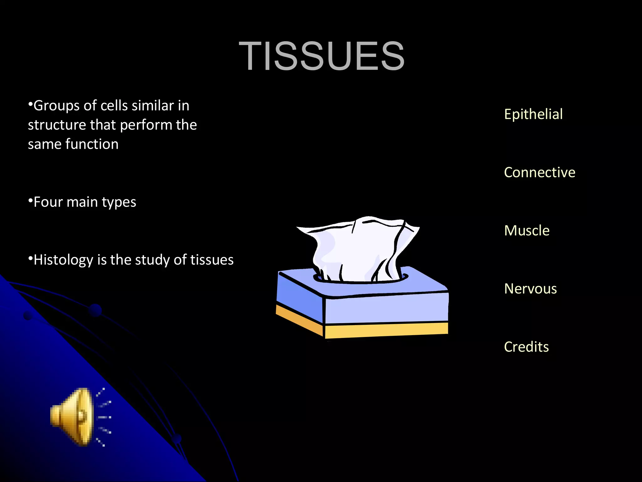
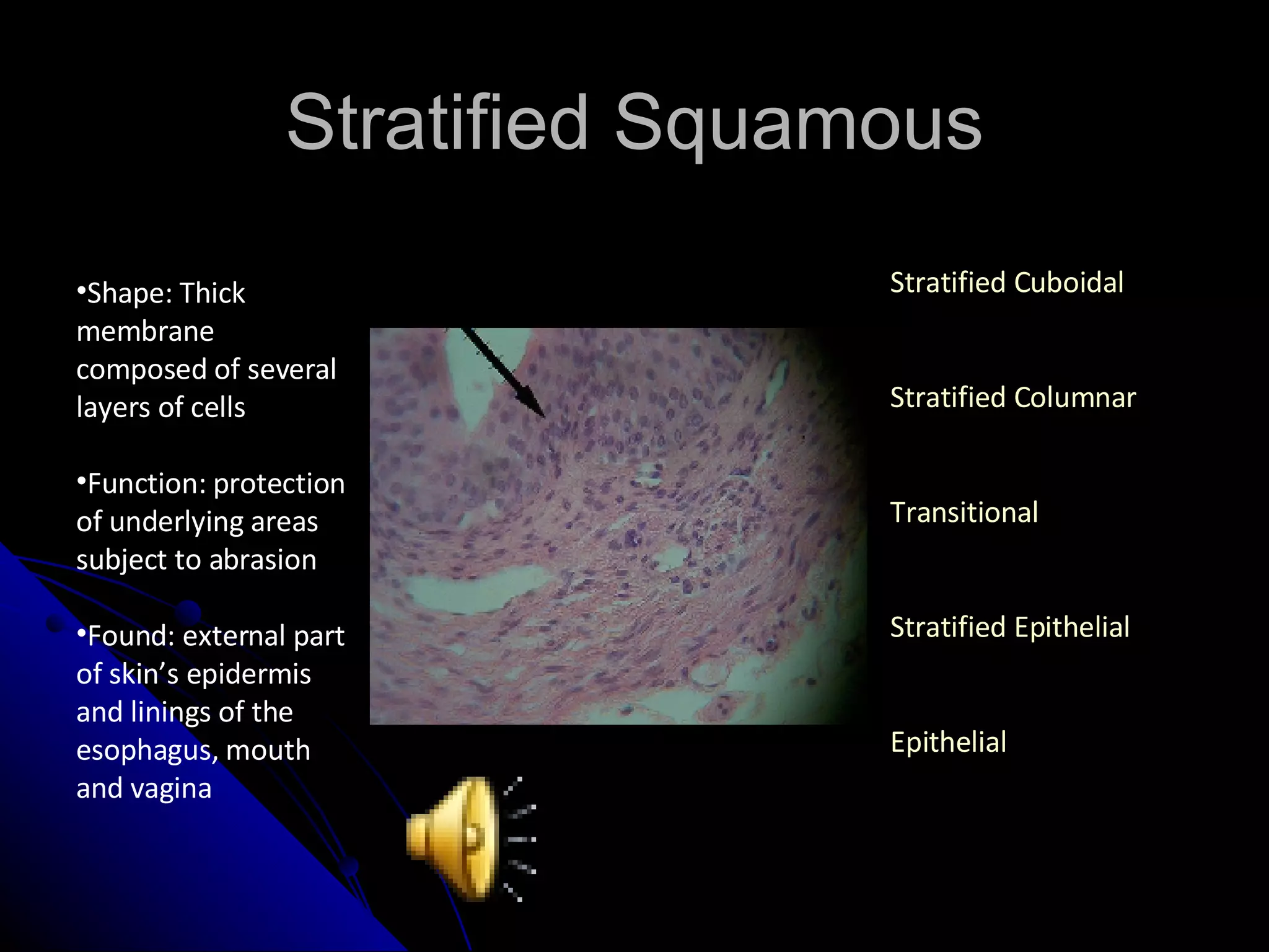
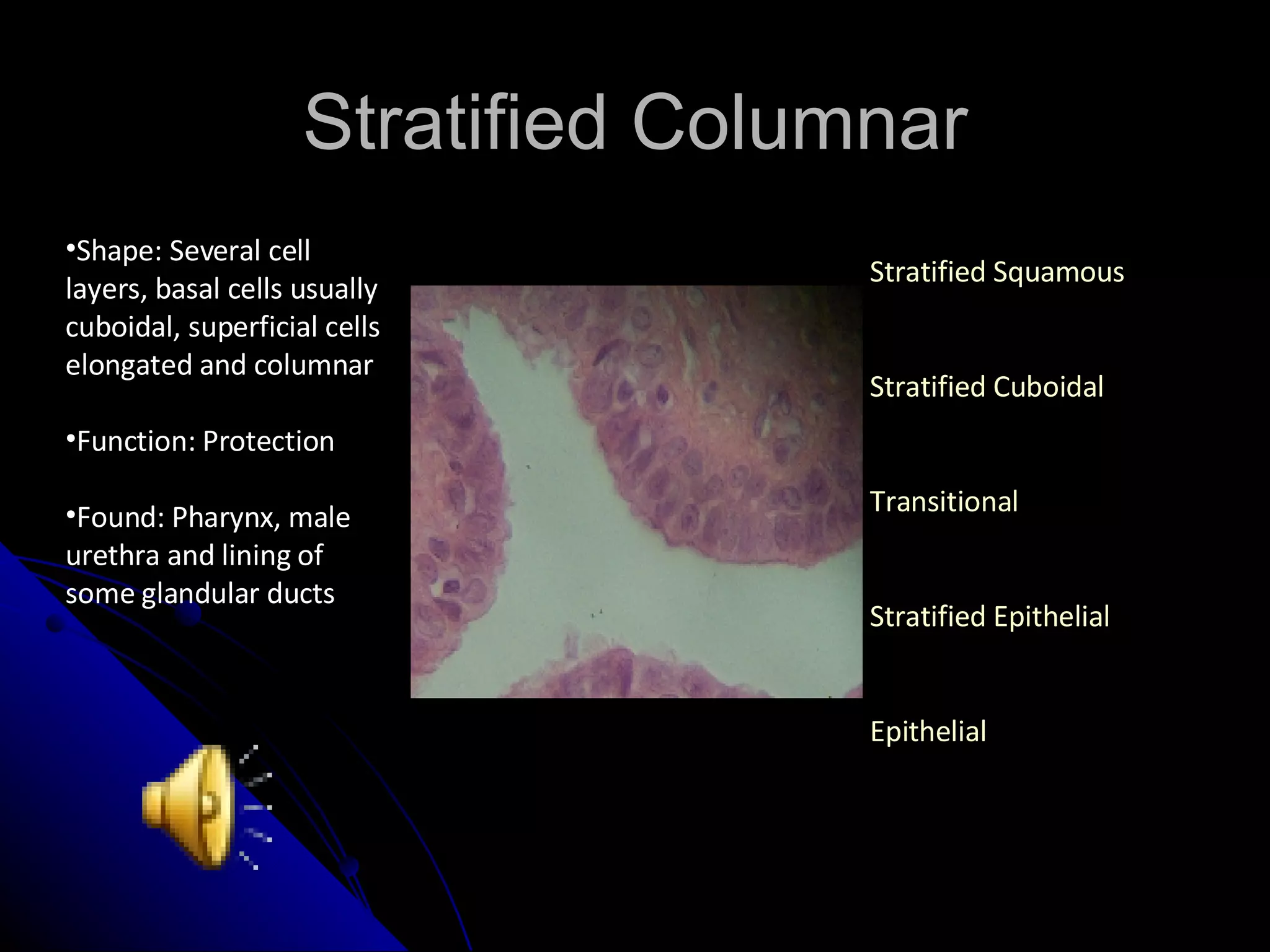
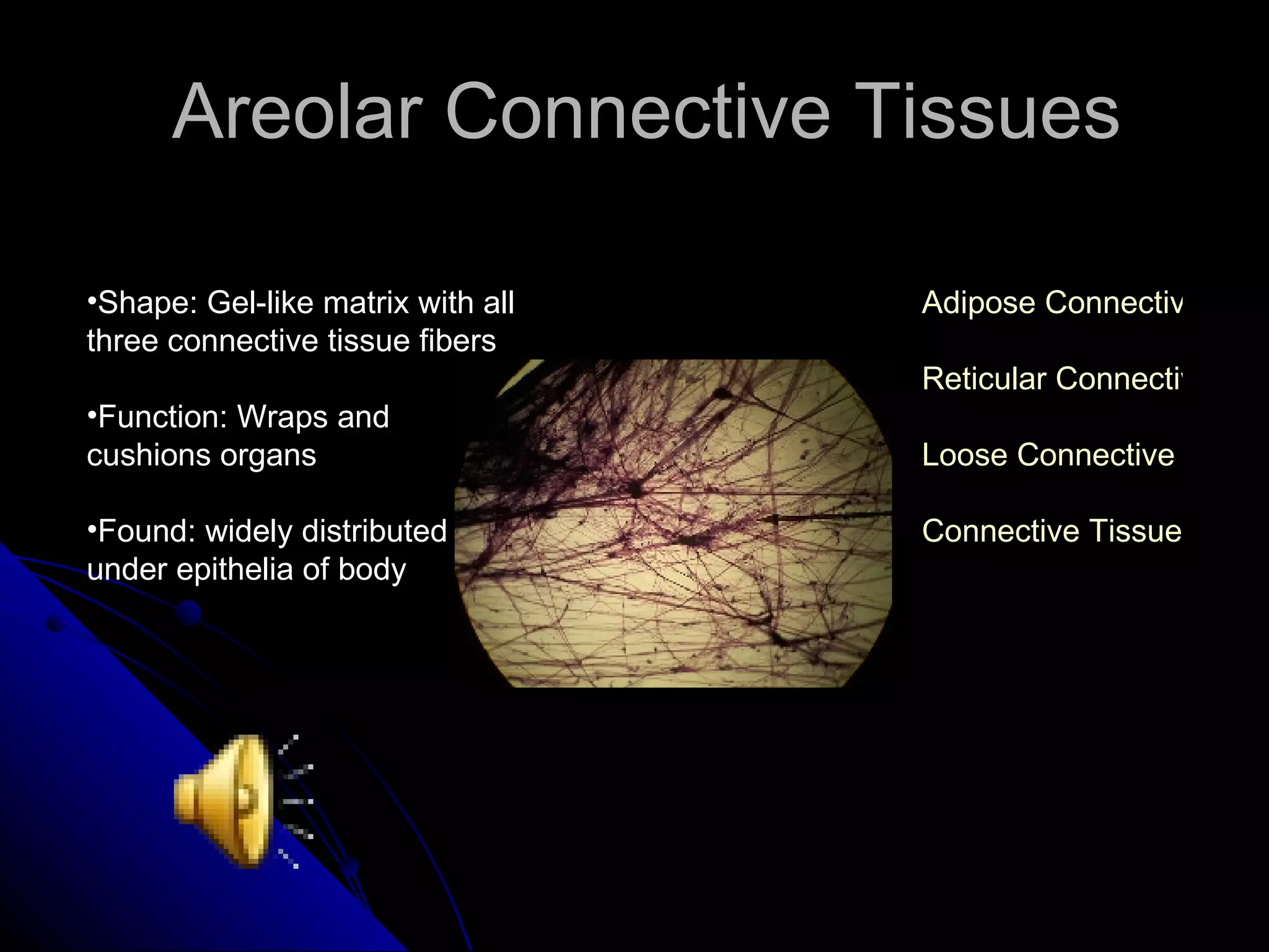
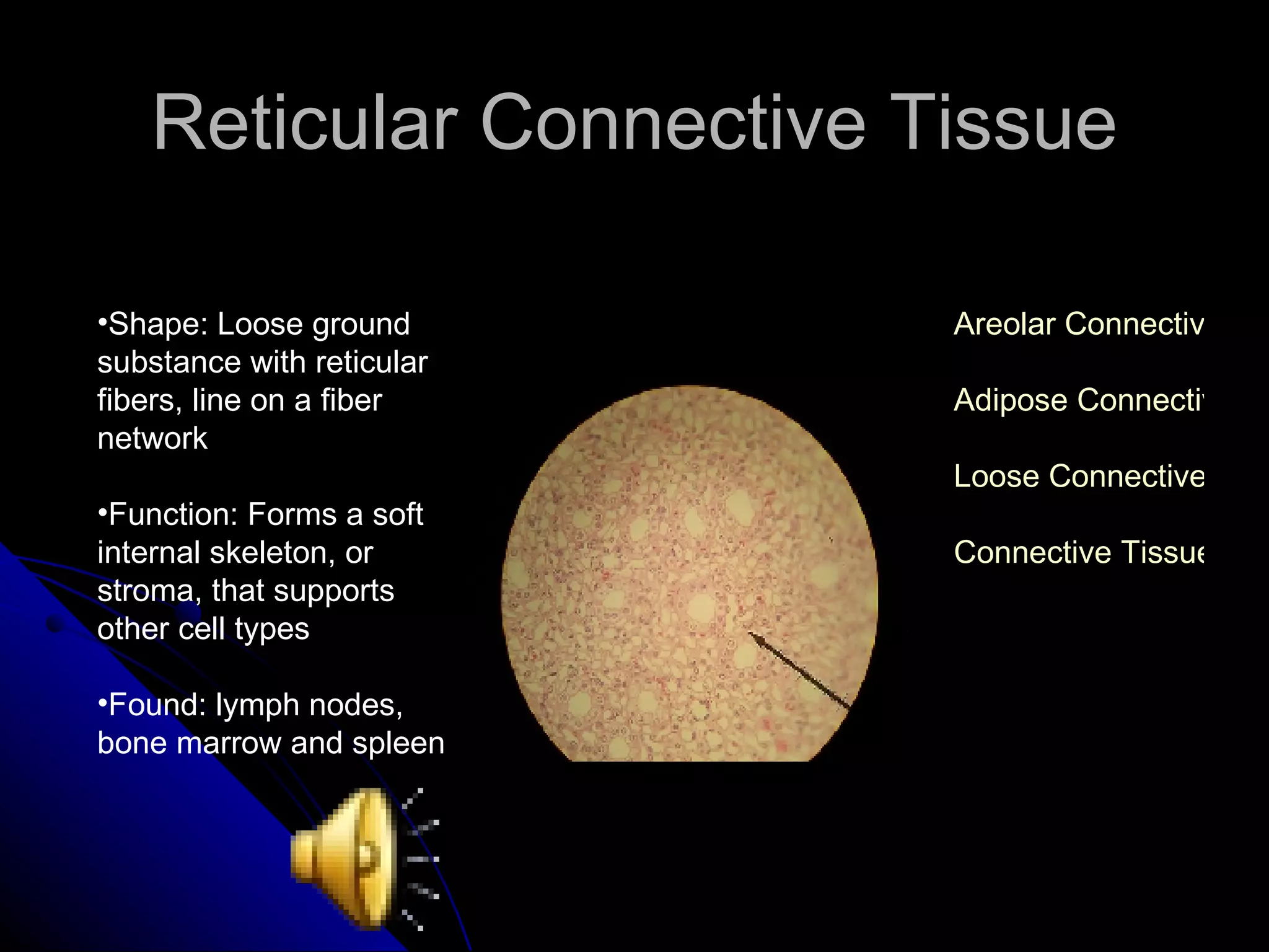
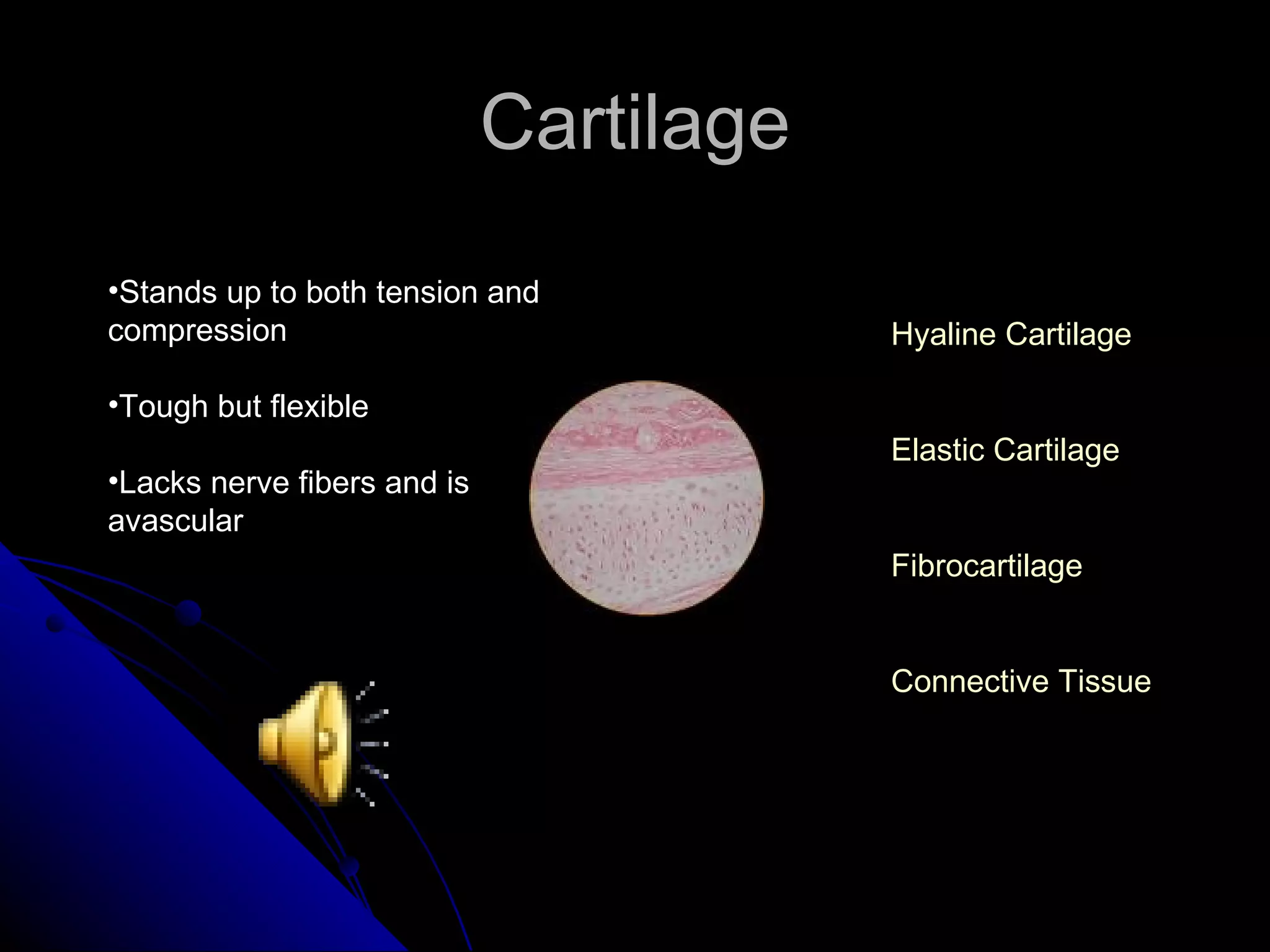
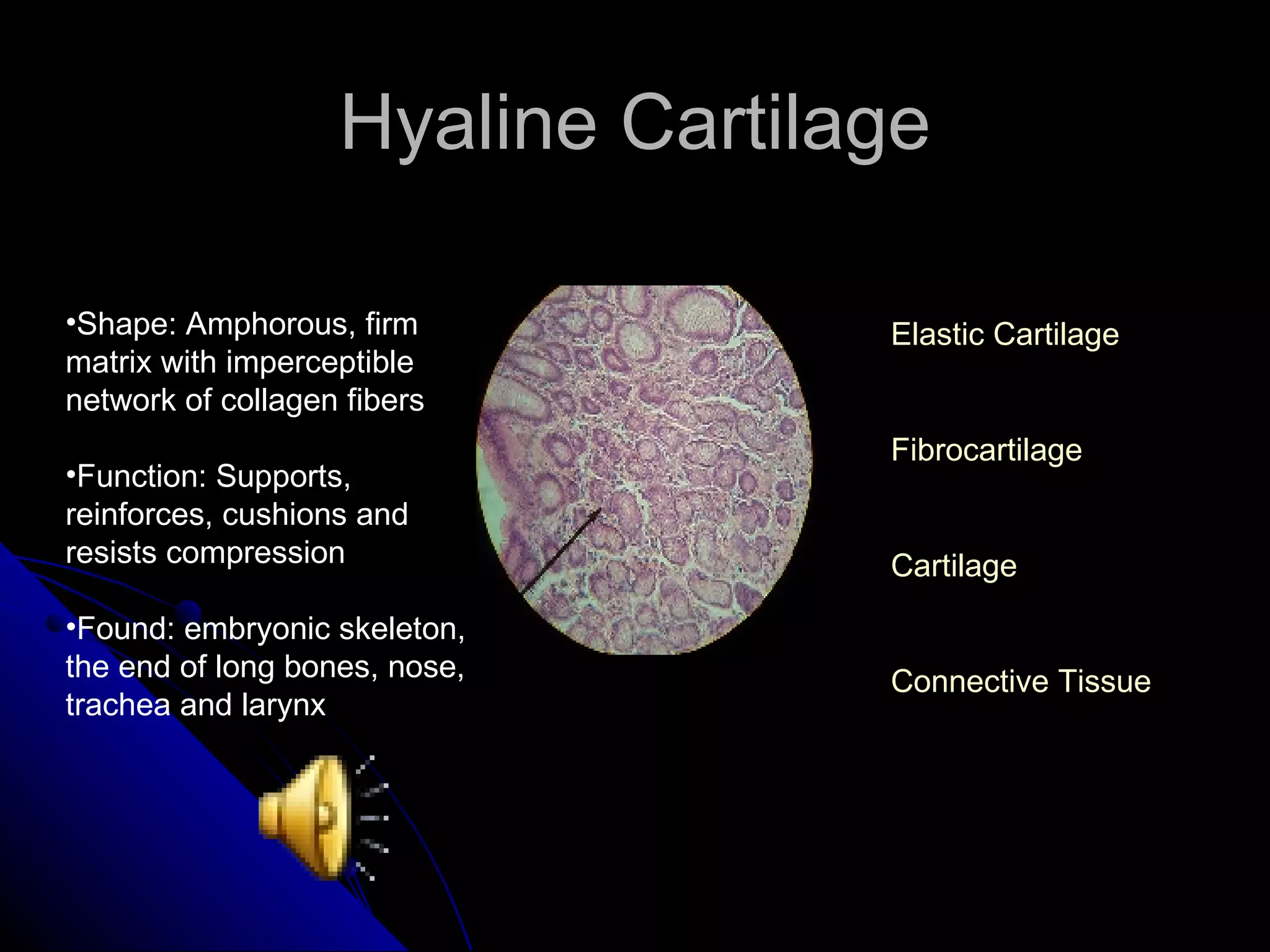
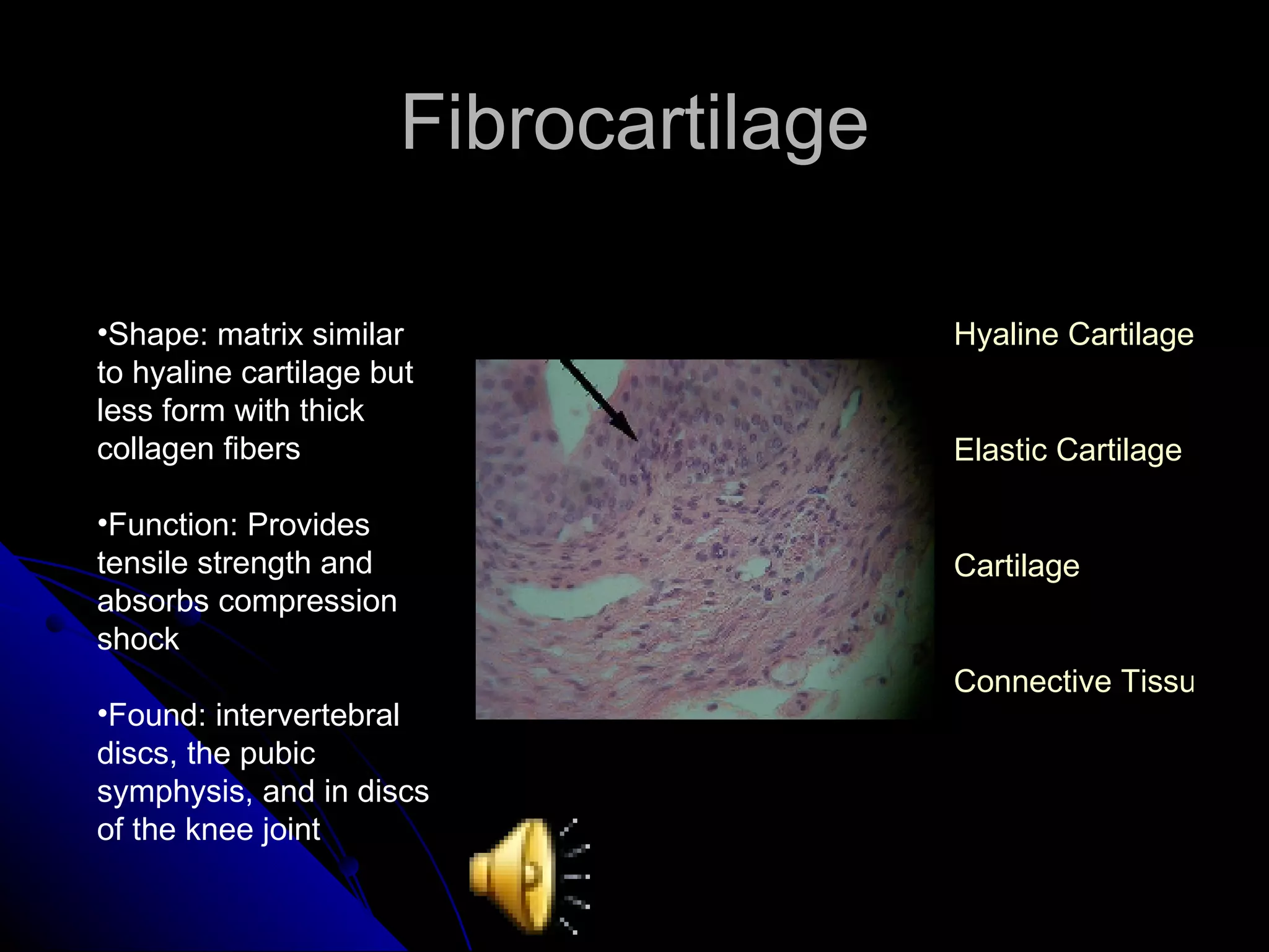
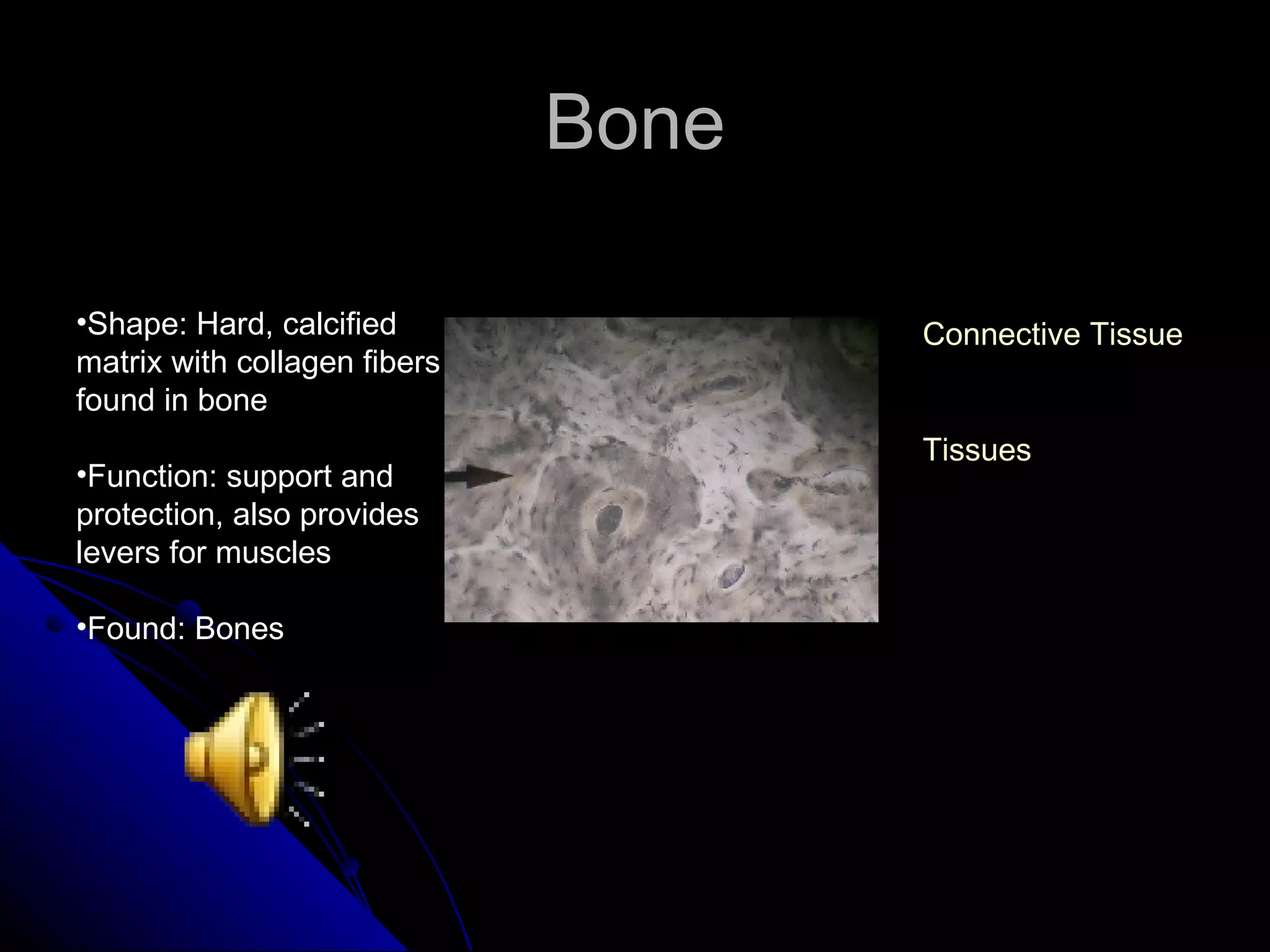
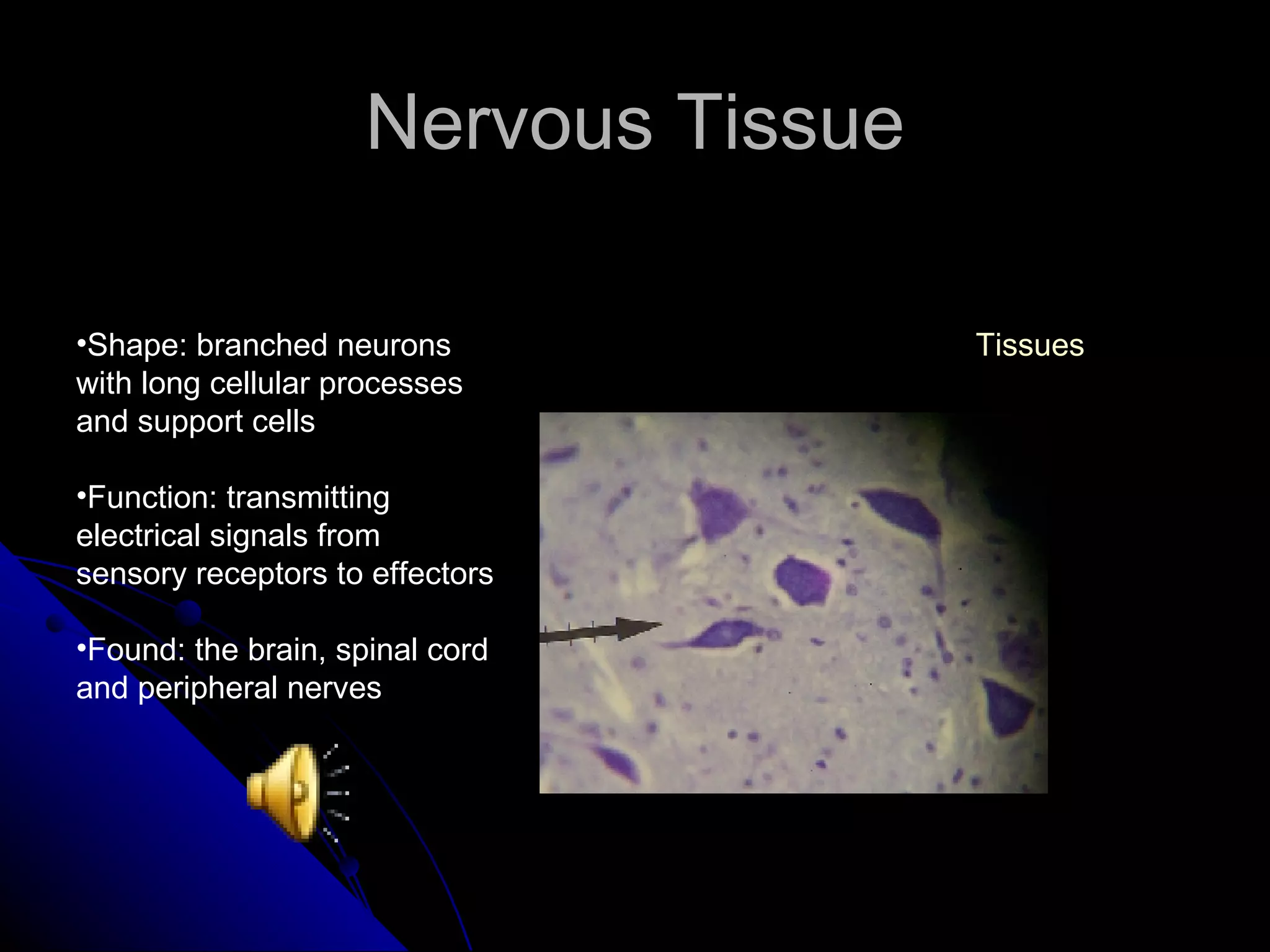
TISSUES Groups of cells similar in  structure that perform the  same function Four main types Histology is the study of tissues Epithelial Connective Muscle Nervous Credits
Epithelial Plays a role in protection, filtration, absorption, excretion, secretion and sensory reception Simple Stratified Tissues
Simple Epithelial Made up of single layer cells Used for absorption and filtration Six irregular sides Simple Squamous Simple Cuboidal Simple Columnar Pseudostratified Columnar Epithelial
Stratified Epithelial Having two or more cell layers Stack on top of one another Used for protection Stratified Squamous Stratified Cuboidal Stratified Columnar Transitional Epithelial
Simple Squamous Shape: Flattened cells with disc-shaped nuclei and sparse cytoplasm Function: Diffusion and Filtration Found: Kidney glomeruli, air sacs of lungs, lining of heart, blood vessels Simple Cuboidal Simple Columnar Pseudostratified Columnar Simple Epithelial Epithelial
Simple Cuboidal Shape: Single layer of cube-like cells with large, spherical central nuclei Function: Secretion and Absorption Found: Kidney tubules, ovary surface, ducts and secretory portions of small glands Simple Squamous Simple Columnar Pseudostratified Columnar Simple Epithelial Epithelial
Simple Columnar Shape: Single layer of tall cells with oval nuclei, may contain cilia Function: Absorption, Secretion Found: Nonciliated type lines most of the digestive tract, gallbladder, excretory ducts of some glands, and some regions of uterus Simple Squamous Simple Cuboidal Pseudostratified Columnar Simple Epithelial Epithelial
Pseudostratified Columnar Shape: Single layer of cells with different heights, some do not reach free surface Function: Secretion and Propulsion of mucus Found: Male Sperm and Trachea Simple Squamous Simple Cuboidal Simple Columnar Simple Epithelial Epithelial
Stratified Squamous Shape: Thick membrane composed of several layers of cells Function: protection of underlying areas subject to abrasion Found: external part of skin’s epidermis and linings of the esophagus, mouth and vagina Stratified Cuboidal Stratified Columnar Transitional Stratified Epithelial Epithelial
Stratified Cuboidal Shape: Generally to layers of cube-like cells Function: Protection Found: Largest ducts of sweat glands and salivary glands Stratified Squamous Stratified Columnar Transitional Stratified Epithelial Epithelial
Stratified Columnar Shape: Several cell layers, basal cells usually cuboidal, superficial cells elongated and columnar Function: Protection Found: Pharynx, male urethra and lining of some glandular ducts Stratified Squamous Stratified Cuboidal Transitional Stratified Epithelial Epithelial
Transitional Shape: Several cell layers. Basal cells are cuboidal, surface cells are dome shaped Function: Stretches to permit the distension of the urinary bladder Found: Lining of urinary bladder, ureters and part of urethra Stratified Squamous Stratified Cuboidal Stratified Columnar Stratified Epithelial Epithelial
Connective Tissues Found everywhere in body Four main classes Functions include binding and support, protection, and insulation Connective Tissue Proper Cartilage Bone Blood Tissues
Connective Tissue Proper Contain cells, fibers and a ground Loose and dense tissues proper Loose Connective Tissues Dense Connective Tissues Tissues
Loose Connective Tissues Often used to link epithelia Found in epidermis Distinguished by vascularity and cellularity Areolar Connective Tissue Adipose Connective Tissue Reticular Connective Tissue Connective Tissue Tissue
Dense Connective Tissues Has collagen fibers as main matrix element Contains Fibroblasts Forms tendons and ligaments Dense Regular Dense Irregular Connective Tissue Proper Connective Tissues
Areolar Connective Tissues Shape: Gel-like matrix with all three connective tissue fibers Function: Wraps and cushions organs Found: widely distributed under epithelia of body Adipose Connective Tissue Reticular Connective Tissue Loose Connective Tissue Connective Tissue
Adipose Connective Tissue Shape: Matrix similar to aerolar with closely packed adipocytes Function:Reserves food stores, insulates heat loss, supports and protects Found: under skin, around kidneys, within abdomen and in breast Areolar Connective Tissue Reticular Connective Tissue Loose Connective Tissue Connective Tissue
Reticular Connective Tissue Shape: Loose ground substance with reticular fibers, line on a fiber network Function: Forms a soft internal skeleton, or stroma, that supports other cell types Found: lymph nodes, bone marrow and spleen Areolar Connective Tissue Adipose Connective Tissue Loose Connective Tissue Connective Tissue
Dense Regular Connective Tissue Shape: Parallel collagen fibers with a few elastic fibers Function: Attaches muscles to bone or to other muscles and bone to bone Found: tendons, ligaments, aponeuroses Dense Irregular Connective Tissue Dense Connective Tissue Connective Tissue
Dense Irregular Connective Tissue Shape: Irregularly arranged collagen fibers with some elastic fibers Function: Withstand tension in many directions providing structural strength Found: dermis, submucosa of the digestive tract, and fibrous organ capsules Dense Regular Connective Tissue Dense Connective Tissue Connective Tissue
Cartilage Stands up to both tension and compression Tough but flexible Lacks nerve fibers and is avascular Hyaline Cartilage Elastic Cartilage Fibrocartilage Connective Tissue
Hyaline Cartilage Shape: Amphorous, firm matrix with imperceptible network of collagen fibers Function: Supports, reinforces, cushions and resists compression Found: embryonic skeleton, the end of long bones, nose, trachea and larynx Elastic Cartilage Fibrocartilage Cartilage Connective Tissue
Elastic Cartilage Shape: Similar to hyaline cartilage but with more elastic fibers Function: Maintains shape structure while allowing flexibility Found: support of external ear and the epiglottis Hyaline Cartilage Fibrocartilage Cartilage Connective Tissue
Fibrocartilage Shape: matrix similar to hyaline cartilage but less form with thick collagen fibers Function: Provides tensile strength and absorbs compression shock Found: intervertebral discs, the pubic symphysis, and in discs of the knee joint Hyaline Cartilage Elastic Cartilage Cartilage Connective Tissue
Bone Shape: Hard, calcified matrix with collagen fibers found in bone Function: support and protection, also provides levers for muscles  Found: Bones Connective Tissues Tissues
Blood Shape: Red and white blood cells in a fluid matrix called plasma Function: transport of respiratory gases, nutrients and wastes Found: blood vessels Connective Tissues Tissues
Nervous Tissue Shape: branched neurons with long cellular processes and support cells Function: transmitting electrical signals from sensory receptors to effectors Found: the brain, spinal cord and peripheral nerves Tissues
Muscle Tissues Highly textured Well vascularized Responsible for most types of body movement Skeletal Muscle Cardiac Muscle Smooth Muscle Tissues
Skeletal Muscle Tissue Shape: long, cylindrical, multinucleated cells with obvious striation Function: Initiates and controls voluntary movement Found: skeletal muscles that attach to bones or skin Cardiac Muscle Smooth Muscle Muscle Tissues
Cardiac Muscle Tissue Shape: Branching, striated, uninucleated cells interlocking at intercalated  Function: Propels blood into the circulation Found: walls of the heart Skeletal Muscle Smooth Muscle Muscle Tissues
Smooth Muscle Tissue Shape: sheet of spindle-shaped cells with central nuclei that have no striation Function: propels substances along internal passageways called peristalsis. Found: walls of hollow organs Skeletal Muscle Cardiac Muscle Muscle Tissues
Credits http://www.innvista.com/health/anatomy/connect.htm http://www.mc.vanderbilt.edu/histology/labmanual2002/labsection1/Connective Tissue03.htm http://biology.about.come/library/weekly/aa011801a.htm http://cellbio.utmb.edu/microanatomy/connective_tissue/loose_connective_tissue.htm   Human Anatomy and Physiology Google Images Flickr images Heather and Olivia pictures Inc.

Makinson Tissues

  • 1.
    TISSUES Groups ofcells similar in structure that perform the same function Four main types Histology is the study of tissues Epithelial Connective Muscle Nervous Credits
  • 2.
    Epithelial Plays arole in protection, filtration, absorption, excretion, secretion and sensory reception Simple Stratified Tissues
  • 3.
    Simple Epithelial Madeup of single layer cells Used for absorption and filtration Six irregular sides Simple Squamous Simple Cuboidal Simple Columnar Pseudostratified Columnar Epithelial
  • 4.
    Stratified Epithelial Havingtwo or more cell layers Stack on top of one another Used for protection Stratified Squamous Stratified Cuboidal Stratified Columnar Transitional Epithelial
  • 5.
    Simple Squamous Shape:Flattened cells with disc-shaped nuclei and sparse cytoplasm Function: Diffusion and Filtration Found: Kidney glomeruli, air sacs of lungs, lining of heart, blood vessels Simple Cuboidal Simple Columnar Pseudostratified Columnar Simple Epithelial Epithelial
  • 6.
    Simple Cuboidal Shape:Single layer of cube-like cells with large, spherical central nuclei Function: Secretion and Absorption Found: Kidney tubules, ovary surface, ducts and secretory portions of small glands Simple Squamous Simple Columnar Pseudostratified Columnar Simple Epithelial Epithelial
  • 7.
    Simple Columnar Shape:Single layer of tall cells with oval nuclei, may contain cilia Function: Absorption, Secretion Found: Nonciliated type lines most of the digestive tract, gallbladder, excretory ducts of some glands, and some regions of uterus Simple Squamous Simple Cuboidal Pseudostratified Columnar Simple Epithelial Epithelial
  • 8.
    Pseudostratified Columnar Shape:Single layer of cells with different heights, some do not reach free surface Function: Secretion and Propulsion of mucus Found: Male Sperm and Trachea Simple Squamous Simple Cuboidal Simple Columnar Simple Epithelial Epithelial
  • 9.
    Stratified Squamous Shape:Thick membrane composed of several layers of cells Function: protection of underlying areas subject to abrasion Found: external part of skin’s epidermis and linings of the esophagus, mouth and vagina Stratified Cuboidal Stratified Columnar Transitional Stratified Epithelial Epithelial
  • 10.
    Stratified Cuboidal Shape:Generally to layers of cube-like cells Function: Protection Found: Largest ducts of sweat glands and salivary glands Stratified Squamous Stratified Columnar Transitional Stratified Epithelial Epithelial
  • 11.
    Stratified Columnar Shape:Several cell layers, basal cells usually cuboidal, superficial cells elongated and columnar Function: Protection Found: Pharynx, male urethra and lining of some glandular ducts Stratified Squamous Stratified Cuboidal Transitional Stratified Epithelial Epithelial
  • 12.
    Transitional Shape: Severalcell layers. Basal cells are cuboidal, surface cells are dome shaped Function: Stretches to permit the distension of the urinary bladder Found: Lining of urinary bladder, ureters and part of urethra Stratified Squamous Stratified Cuboidal Stratified Columnar Stratified Epithelial Epithelial
  • 13.
    Connective Tissues Foundeverywhere in body Four main classes Functions include binding and support, protection, and insulation Connective Tissue Proper Cartilage Bone Blood Tissues
  • 14.
    Connective Tissue ProperContain cells, fibers and a ground Loose and dense tissues proper Loose Connective Tissues Dense Connective Tissues Tissues
  • 15.
    Loose Connective TissuesOften used to link epithelia Found in epidermis Distinguished by vascularity and cellularity Areolar Connective Tissue Adipose Connective Tissue Reticular Connective Tissue Connective Tissue Tissue
  • 16.
    Dense Connective TissuesHas collagen fibers as main matrix element Contains Fibroblasts Forms tendons and ligaments Dense Regular Dense Irregular Connective Tissue Proper Connective Tissues
  • 17.
    Areolar Connective TissuesShape: Gel-like matrix with all three connective tissue fibers Function: Wraps and cushions organs Found: widely distributed under epithelia of body Adipose Connective Tissue Reticular Connective Tissue Loose Connective Tissue Connective Tissue
  • 18.
    Adipose Connective TissueShape: Matrix similar to aerolar with closely packed adipocytes Function:Reserves food stores, insulates heat loss, supports and protects Found: under skin, around kidneys, within abdomen and in breast Areolar Connective Tissue Reticular Connective Tissue Loose Connective Tissue Connective Tissue
  • 19.
    Reticular Connective TissueShape: Loose ground substance with reticular fibers, line on a fiber network Function: Forms a soft internal skeleton, or stroma, that supports other cell types Found: lymph nodes, bone marrow and spleen Areolar Connective Tissue Adipose Connective Tissue Loose Connective Tissue Connective Tissue
  • 20.
    Dense Regular ConnectiveTissue Shape: Parallel collagen fibers with a few elastic fibers Function: Attaches muscles to bone or to other muscles and bone to bone Found: tendons, ligaments, aponeuroses Dense Irregular Connective Tissue Dense Connective Tissue Connective Tissue
  • 21.
    Dense Irregular ConnectiveTissue Shape: Irregularly arranged collagen fibers with some elastic fibers Function: Withstand tension in many directions providing structural strength Found: dermis, submucosa of the digestive tract, and fibrous organ capsules Dense Regular Connective Tissue Dense Connective Tissue Connective Tissue
  • 22.
    Cartilage Stands upto both tension and compression Tough but flexible Lacks nerve fibers and is avascular Hyaline Cartilage Elastic Cartilage Fibrocartilage Connective Tissue
  • 23.
    Hyaline Cartilage Shape:Amphorous, firm matrix with imperceptible network of collagen fibers Function: Supports, reinforces, cushions and resists compression Found: embryonic skeleton, the end of long bones, nose, trachea and larynx Elastic Cartilage Fibrocartilage Cartilage Connective Tissue
  • 24.
    Elastic Cartilage Shape:Similar to hyaline cartilage but with more elastic fibers Function: Maintains shape structure while allowing flexibility Found: support of external ear and the epiglottis Hyaline Cartilage Fibrocartilage Cartilage Connective Tissue
  • 25.
    Fibrocartilage Shape: matrixsimilar to hyaline cartilage but less form with thick collagen fibers Function: Provides tensile strength and absorbs compression shock Found: intervertebral discs, the pubic symphysis, and in discs of the knee joint Hyaline Cartilage Elastic Cartilage Cartilage Connective Tissue
  • 26.
    Bone Shape: Hard,calcified matrix with collagen fibers found in bone Function: support and protection, also provides levers for muscles Found: Bones Connective Tissues Tissues
  • 27.
    Blood Shape: Redand white blood cells in a fluid matrix called plasma Function: transport of respiratory gases, nutrients and wastes Found: blood vessels Connective Tissues Tissues
  • 28.
    Nervous Tissue Shape:branched neurons with long cellular processes and support cells Function: transmitting electrical signals from sensory receptors to effectors Found: the brain, spinal cord and peripheral nerves Tissues
  • 29.
    Muscle Tissues Highlytextured Well vascularized Responsible for most types of body movement Skeletal Muscle Cardiac Muscle Smooth Muscle Tissues
  • 30.
    Skeletal Muscle TissueShape: long, cylindrical, multinucleated cells with obvious striation Function: Initiates and controls voluntary movement Found: skeletal muscles that attach to bones or skin Cardiac Muscle Smooth Muscle Muscle Tissues
  • 31.
    Cardiac Muscle TissueShape: Branching, striated, uninucleated cells interlocking at intercalated Function: Propels blood into the circulation Found: walls of the heart Skeletal Muscle Smooth Muscle Muscle Tissues
  • 32.
    Smooth Muscle TissueShape: sheet of spindle-shaped cells with central nuclei that have no striation Function: propels substances along internal passageways called peristalsis. Found: walls of hollow organs Skeletal Muscle Cardiac Muscle Muscle Tissues
  • 33.
    Credits http://www.innvista.com/health/anatomy/connect.htm http://www.mc.vanderbilt.edu/histology/labmanual2002/labsection1/ConnectiveTissue03.htm http://biology.about.come/library/weekly/aa011801a.htm http://cellbio.utmb.edu/microanatomy/connective_tissue/loose_connective_tissue.htm Human Anatomy and Physiology Google Images Flickr images Heather and Olivia pictures Inc.