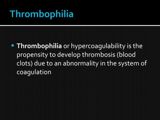
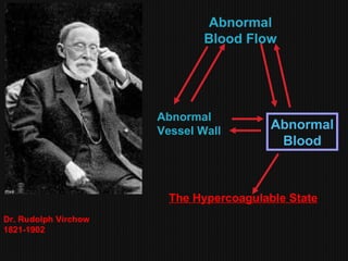
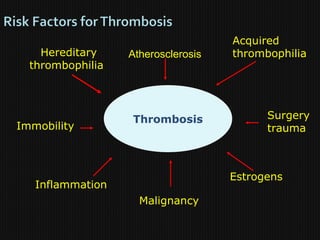
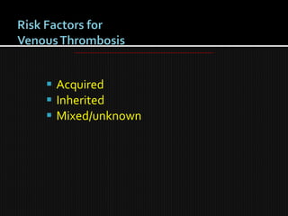
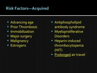
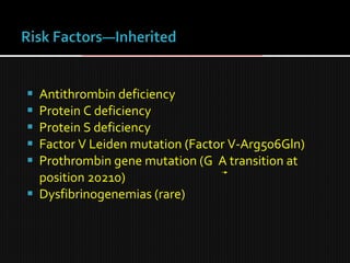
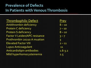
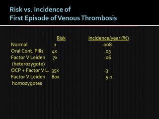
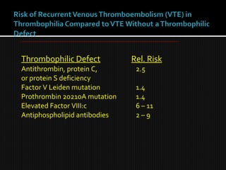
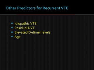
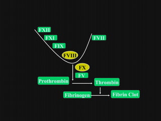
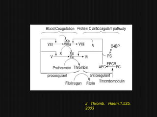
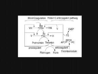
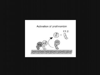
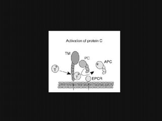
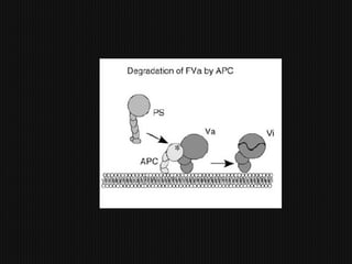
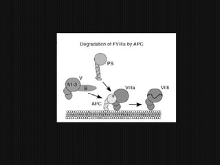
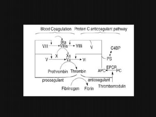
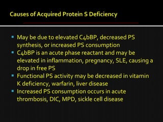
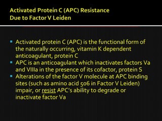
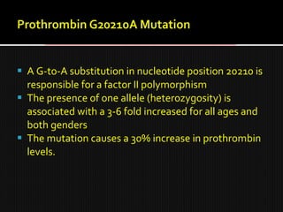
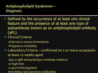
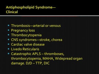
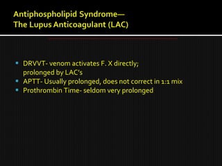
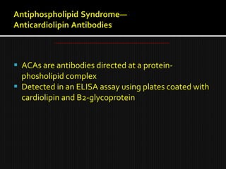
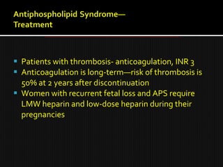
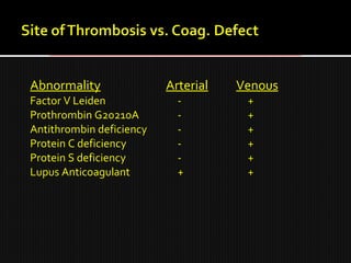
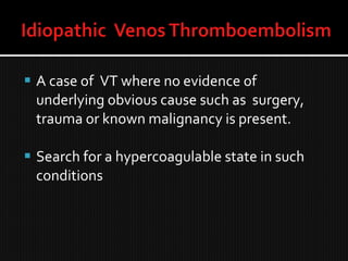
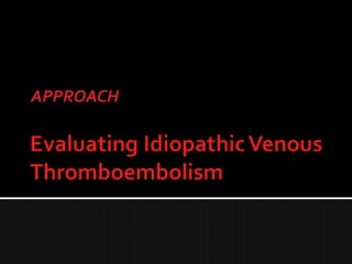
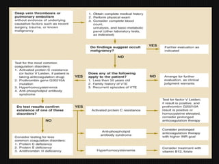
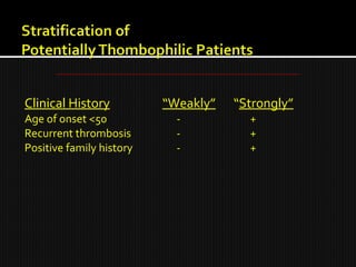
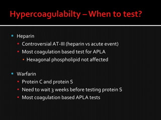
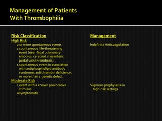
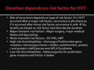
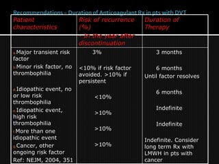
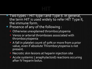
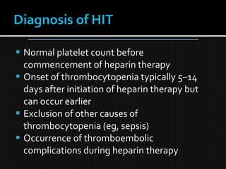
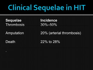
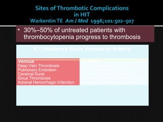
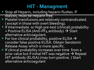
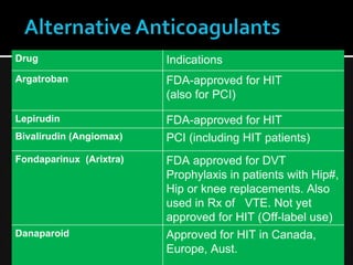
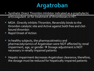
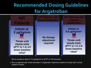
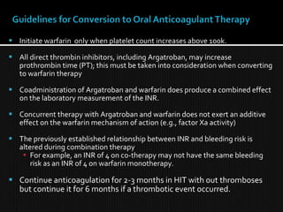
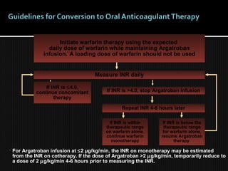
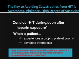
6. Advancing age
Prior Thrombosis
Immobilization
Major surgery
Malignancy
Estrogens
Antiphospholipid
antibody syndrome
Myeloproliferative
Disorders
Heparin-induced
thrombocytopenia
(HIT)
Prolonged air travel
7. Antithrombin deficiency
Protein C deficiency
Protein S deficiency
Factor V Leiden mutation (Factor V-Arg506Gln)
Prothrombin gene mutation (G A transition at
position 20210)
Dysfibrinogenemias (rare)
8. High levels of factor VIII
Acquired Protein C resistance in the absence of
Factor V Leiden
High levels of Factor IX, XI
9.
10. Thrombophilic Defect Prev
Antithrombin deficiency 8 – 10
Protein C deficiency 7 – 10
Protein S deficiency 8 – 10
Factor V Leiden/APC resisance 3 – 7
Prothrombin 20210 A muation 3
Elevated Factor VIII 2 – 11
Lupus Anticoagulant 11
Anticardiolipin antibodies 1.6-3.2
Mild hyperhomocysteinemia 2.5
11. Risk Incidence/year (%)
Normal 1 .008
Oral Cont. Pills 4x .03
Factor V Leiden 7x .06
(heterozygote)
OCP + Factor V L. 35x .3
Factor V Leiden 80x .5-1
homozygotes
12. Thrombophilic Defect Rel. Risk
Antithrombin, protein C, 2.5
or protein S deficiency
Factor V Leiden mutation 1.4
Prothrombin 20210A mutation 1.4
Elevated Factor VIII:c 6 – 11
Antiphospholipid antibodies 2 – 9
16. Also known as Antithrombin III
Inhibits coagulation by irreversibly binding the
thrombogenic proteins thrombin (IIa), IXa, Xa, XIa
and XIIa
Antithrombin’s binding reaction is amplified 1000-
fold by heparin, which binds to antithrombin to
cause a conformational change which more avidly
binds thrombin and the other serine proteases
17. Protein C is a vitamin K dependent glycoprotein produced in the liver
In the activation of protein C, thrombin binds to thrombomodulin, a
structural protein on the endothelial cell surface
This complex then converts protein C to activated protein C (APC), which
degrades factors Va and VIIIa, limiting thrombin production
For protein C to bind, cleave and degrade factors Va and VIIIa, protein S
must be available
Protein C deficiency, whether inherited or acquired, may cause
thrombosis when levels drop to 50% or below
Protein C deficiency also occurs with surgery, trauma, pregnancy, OCP,
liver or renal failure, DIC,or warfarin
18.
19.
20.
21.
22.
23.
24. Protein S is an essential cofactor in the protein C
pathway
Protein S exists in a free and bound state
60-70% of protein S circulates bound to C4b binding
proten
The remaining protein S, called free PS, is the
functionally active form of protein S
Inherited PS deficiency is an autosomal dominant
disorder, causing thrombosis when levels drop to
50% or lower
25. May be due to elevated C4bBP, decreased PS
synthesis, or increased PS consumption
C4bBP is an acute phase reactant and may be
elevated in inflammation, pregnancy, SLE, causing a
drop in free PS
Functional PS activity may be decreased in vitamin
K deficiency, warfarin, liver disease
Increased PS consumption occurs in acute
thrombosis, DIC, MPD, sickle cell disease
26. Activated protein C (APC) is the functional form of
the naturally occurring, vitamin K dependent
anticoagulant, protein C
APC is an anticoagulant which inactivates factors Va
and VIIIa in the presence of its cofactor, protein S
Alterations of the factor V molecule at APC binding
sites (such as amino acid 506 in Factor V Leiden)
impair, or resist APC’s ability to degrade or
inactivate factor Va
27. A G-to-A substitution in nucleotide position 20210 is
responsible for a factor II polymorphism
The presence of one allele (heterozygosity) is
associated with a 3-6 fold increased for all ages and
both genders
The mutation causes a 30% increase in prothrombin
levels.
28.
29. Defined by the occurrence of at least one clinical
feature and the presence of at least one type of
autoantibody known as an antiphospholipid antibody
(aPL).
Clinical Criteria
-Arterial or venous thrombosis
-Pregnancy morbidity
Laboratory Criteria – confirmed on 2 or more occassions
at least 12 weeks apart
-IgG or IgM anticardiolipin antibody-medium
or high titer
-Lupus Anticoagulant
- Anti-Beta2 Glycoprotein antibodies
30. Thrombosis—arterial or venous
Pregnancy loss
Thrombocytopenia
CNS syndromes—stroke, chorea
Cardiac valve disease
Livedo Reticularis
Catastrophic APLS – thromboses,
thrombocytopenia, MAHA, Widepread organ
damage. D/D – TTP, DIC
31. DRVVT- venom activates F. X directly;
prolonged by LAC’s
APTT- Usually prolonged, does not correct in 1:1 mix
Prothrombin Time- seldom very prolonged
32. ACAs are antibodies directed at a protein-
phosholipid complex
Detected in an ELISA assay using plates coated with
cardiolipin and B2-glycoprotein
33. Patients with thrombosis- anticoagulation, INR 3
Anticoagulation is long-term—risk of thrombosis is
50% at 2 years after discontinuation
Women with recurrent fetal loss and APS require
LMW heparin and low-dose heparin during their
pregnancies
34.
35. Abnormality Arterial Venous
Factor V Leiden - +
Prothrombin G20210A - +
Antithrombin deficiency - +
Protein C deficiency - +
Protein S deficiency - +
Lupus Anticoagulant + +
36. A case of VT where no evidence of
underlying obvious cause such as surgery,
trauma or known malignancy is present.
Search for a hypercoagulable state in such
conditions
39. Clinical History “Weakly” “Strongly”
Age of onset <50 - +
Recurrent thrombosis - +
Positive family history - +
40. Pro
Improve understanding of pathogenesis of thrombosis
Identify and counsel affected family members
Obviate expensive diagnostic testing (e.g. CT scans) looking for
a malignancy
Con
Infrequent identification of patients with defects whose
management would change
Potential for overaggressive management
Insurance implications
Cost of testing
41. Routine screening of patients with VTE for an underlying
thrombophilic defect “is not justified”
However, the risk of subsequent thrombosis over 5 years in
men with idiopathic VTE is 30%
Any additional defect adds to risk and to possible need for
prolongation of anticoagulation
Furthermore, women with a history of VTE who wish to
become pregnant will be treated differently if a defect were
found
42. Test for Factor V Leiden
Genetic test for prothrombin gene mutation 20210A
Functional assay of antithrombin III
Functional assay of protein C
Functional assay of protein S
Testing for Anti-phospholipid antibody syndrome :
Clotting test for lupus anticoagulant ( mixing studies,
DRVVT, STACLOT-LA) /ELISA for cardiolipin antibodies.
A complete hypercoagulability work – up as above is recommended in
strongly thrombophilic patients.
43. Test for Factor V Leiden
Genetic test for prothrombin gene mutation G20210A
Clotting assay for lupus anticoagulant/ELISA for
cardiolipin antibodies
LIMITED THROMBOPHILIA W/U AS ABOVE IN “Weakly”
thrombophilic patients.
Eg. for a “weakly” thrombophilic case : one time
spontaneous thrombosis in a patient age > 50 years with
no family history of thrombophilia.
44. Hyperhomocystinemia in Thrombophilia
In the past, homocysteine levels were recommended in
thrombophilic patients.
Measurement of fasting total plasma homocysteine is no longer
recommended
There are no data supporting a change in the duration or type of
therapy for a patient with hyperhomocysteinemia and a past history
of VTE, since hyperhomocysteinemia may be a marker of thrombotic
disease rather than a cause.
Results from the Leiden MEGA study indicate that the presence of
methylenetetrahydrofolate reductase (MTHFR) mutation which
mildly increases homocysteine levels, is not associated with an
increased risk for venous thrombosis.
Hence, there is no clinical rationale for measurement of plasma
homocysteine levels or for assaying for presence of the MTHFR
mutation when screening for the risk of VTE.
45. Clues –
Bilateral DVTs, Arterial and venous thrombosis
and Warfarin refractory thrombosis.
Follow age – specific screening
recommendations and clinical history to select
appropriate investigations when cancer
associated thromboses are suscpected.
46. Heparin
Controversial AT-III (heparin vs acute event)
Most coagulation based test for APLA
▪ Hexagonal phospholipid not affected
Warfarin
Protein C and protein S
Need to wait 3 weeks before testing protein S
Most coagulation based APLA tests
48. Risk Classification Management
High Risk
2 or more spontaneous events Indefinite Anticoagulation
1 spontaneous life-threatening
event (near-fatal pulmonary
embolus, cerebral, mesenteric,
portal vein thrombosis)
1 spontaneous event in association
with antiphospholipid antibody
syndrome, antithrombin deficiency,
or more than 1 genetic defect
Moderate Risk
1 event with a known provocative Vigorous prophylaxis in
stimulus high-risk settings
Asymptomatic
49. Risk of recurrence depends on type of risk factor. If 1st
DVT
occurred after a major risk factor, recurrence is 3% where as
if it occurred after minor risk factor recurrence is 10% So,
stratify pts based on risk factor and then decide duration
Major transient risk factors : Major surgery, major medical
illness and leg casting.
Minor transient risk factors : OC Pills, HRT
High risk thrombophilias : Homzygos Prothrombin gene
mutation, Homozygos Factor v leiden, antithrombin, protein
c and protein s deficiencies and APLA Syndrome
Low risk thrombophilias : Heterozygosity for prothrobin
gene mutation and Factor V leiden
50. Patient
Patient
characteristics
characteristics
Risk of recurrence
Risk of recurrence
(%)
(%)
- In the year after
- In the year after
discontinuation
discontinuation
Duration of
Duration of
Therapy
Therapy
a.
a.Major transient risk
Major transient risk
factor
factor
b.
b.Minor risk factor, no
Minor risk factor, no
thrombophilia
thrombophilia
d.
d.Idiopathic event, no
Idiopathic event, no
or low risk
or low risk
thrombophilia
thrombophilia
e.
e.Idiopathic event,
Idiopathic event,
high risk
high risk
thrombophilia
thrombophilia
f.
f.More than one
More than one
idiopathic event
idiopathic event
g.
g.Cancer, other
Cancer, other
ongoing risk factor
ongoing risk factor
Ref: NEJM, 2004, 351
3%
3%
<10% if risk factor
<10% if risk factor
avoided. >10% if
avoided. >10% if
persistent
persistent
<10%
<10%
>10%
>10%
>10%
>10%
>10%
>10%
3 months
3 months
6 months
6 months
Until factor resolves
6 months
6 months
Indefinite
Indefinite
Indefinite
Indefinite
Indefinite. Consider
long term Rx with
long term Rx with
LMWH in pts with
LMWH in pts with
cancer
cancer
52. More than 1 trillion units heparin used yearly in
US; 1/3 of hospitalized exposed (12 million).
Unfractionated heparin – 3 - 5% incidence;
Heart surgery 2.5% incidence
LMWHeparin, Catheter-flushes -- ~0.5%
Frequency of thromboemboli : 30%–50% of patients
with untreated HIT will have a thrombotic
complication within 30 days ( Warkentin TE Am J Med.
1996;101:502–507) Based on increased morbidity and
mortality, heparin cessation alone is inadequate in HIT
management
54. HIT
Two types – HIT type I and Type II. In general,
the term HIT is used widely to refer HIT Type II,
the immune form.
Presence of any of the following :
Otherwise unexplained thrombocytopenia
Venos or arterial thromboses associated with
thrombocytopenia
A fall in platelet count of 50% or more from a prior
value, even if absolute Thrombocytopenia is not
present.
Necrotic skin lesions at heparin injection site
Acute systemic ( anaphylactoid) reactions occuring
after IV heparin bolus.
55. Normal platelet count before
commencement of heparin therapy
Onset of thrombocytopenia typically 5–14
days after initiation of heparin therapy but
can occur earlier
Exclusion of other causes of
thrombocytopenia (eg, sepsis)
Occurrence of thromboembolic
complications during heparin therapy
57. • 30%–50% of untreated patients with
thrombocytopenia progress to thrombosis
4:1 Incidence Ratio Venous to Arterial
Arterial
Aortic/Ileofemoral Thrombosis
Acute Thrombotic Stroke
Myocardial Infarction, Mural
thrombosis, Thrombi in upper limb,
mesenteric, renal and spinal arts.
Venous
Deep Vein Thrombosis
Pulmonary Embolism
Cerebral Dural
Sinus Thrombosis
Adrenal Hemorrhagic Infarction
58. HIT Temporal Variants
Day 1 Day 4 Day 14
Day 30
Delayed-onset
HIT
(9–40 days)
Rapid-onset
HIT
(hours–days)
Typical HIT
Mean Day 9
(5–14 days)
Heparin (re) Exposure
THROMBOCYTOPENIA (± THROMBOSIS)
59. Thrombocytopenia
Platelet count fall > 50% and nadir greater than 20k : 2 points
Platelet count fall 30 to 50% or nadir 10 to 19k : 1 point
Platelet count fall < 30% or nadir < 10k : 0 points
Timing of platelet count fall
Clear onset b/w days 5 to 10 or platelet count fall at ≤1 day if prior
heparin exposure within the last 30 days: 2 points
Consistent with fall at 5 to 10 days but not clear (eg, missing platelet counts) or
onset after day 10 or fall ≤1 day with prior heparin exposure within the last 30 to
100 days: 1 point
Platelet count fall at <4 days without recent exposure: 0 points
Thrombosis or other sequelae
Confirmed new thrombosis, skin necrosis, or acute systemic reaction after
intravenous unfractionated heparin bolus: 2 points
Progressive or recurrent thrombosis, non-necrotizing (erythematous) skin
lesions, or suspected thrombosis which has not been proven: 1 point
None: zero points
Other causes for thrombocytopenia present —
None apparent: 2 points
Possible: 1 point , Definite: zero points The 5th
T : The TEST
60. A score is determined for each of the four
above categories, resulting in a total score
from zero to 8.
Pretest probabilities for HIT are, as follows:
▪ zero to 3: Low probability
▪ 4 to 5: Intermediate probability
▪ 6 to 8: High probability
Laboratory tests are ordered to confirm HIT.
61. Test Advantages Disadvantages
SRA Sensitivity >95%, Technically demanding
Specific > 95% Low predictive value
HIPA Rapid, available Variable sensitivity (30% – 80%);
Technique-dependent
ELISA High sensitivity High cost, less specificity,
> 95% 10% false-negative tests
There is no Gold Standard in diagnostic testing; HIT
requires a clinical diagnosis .
62. Stop all Heparin, including heparin flushes. If
dialysis, must be Heparin free.
Platelet transfusions are relatively contraindicated.
( except in those with overt bleeding).
If Intermediate or High pre-test (clinical) probability
+ Positive ELISA (Anti-PF4 antibody) Start
alternative anticoagulant.
For low clinical probability, positive ELISA
consider false positive ELISA. Obtain Serotonin
Release Assay which is more specific.
If clinical probability increases over time from a
prior value but if initial HIT was negative Repeat
HIT antibody (ELISA) (may turn positive. ) Start
alternative anticoagulant
63. Drug Indications
Argatroban FDA-approved for HIT
(also for PCI)
Lepirudin FDA-approved for HIT
Bivalirudin (Angiomax) PCI (including HIT patients)
Fondaparinux (Arixtra) FDA approved for DVT
Prophylaxis in patients with Hip#,
Hip or knee replacements. Also
used in Rx of VTE. Not yet
approved for HIT (Off-label use)
Danaparoid Approved for HIT in Canada,
Europe, Aust.
64. Synthetic Direct Thrombin Inhibitor indicated as a prophylactic
anticoagulant or for treatment of thromboses in HIT.
MOA : Directly inhibits Thrombin, Reversibly binds to the
thrombin catalytic site and Active against both free and clot-
bound thrombin
Rapid Onset of Action
In healthy subjects, the pharmacokinetics and
pharmacodynamics of Argatroban were NOT affected by renal
impairment, age, or gender Dosage adjustment is NOT
necessary in renally impaired patients
Hepatic impairment decreases Argatroban clearance; therefore,
the dosage must be reduced for hepatically impaired patients
65. HIT
Patients
HIT Patients with
Renal Impairment
HIT Patients with
Hepatic
Impairment
*
Not to exceed a dose of 10 µg/kg/min or aPTT of 100 seconds
† Due to approximate 4-fold decrease in Argatroban clearance relative to those with normal
hepatic function
Initiate at
2 µg/kg/min
Titrate until
steady-state
aPTT is 1.5–3.0
times baseline
value*
No dosage
adjustment
required
Initiate at
0.5 µg/kg/min†
Titrate until
steady-state
aPTT is 1.5–3.0
times baseline
value*
66. Initiate warfarin only when platelet count increases above 100k.
All direct thrombin inhibitors, including Argatroban, may increase
prothrombin time (PT); this must be taken into consideration when converting
to warfarin therapy
Coadministration of Argatroban and warfarin does produce a combined effect
on the laboratory measurement of the INR.
Concurrent therapy with Argatroban and warfarin does not exert an additive
effect on the warfarin mechanism of action (e.g., factor Xa activity)
The previously established relationship between INR and bleeding risk is
altered during combination therapy
For example, an INR of 4 on co-therapy may not have the same bleeding
risk as an INR of 4 on warfarin monotherapy.
Continue anticoagulation for 2-3 months in HIT with out thromboses
but continue it for 6 months if a thrombotic event occurred.
67. If INR is below the
therapeutic range
for warfarin alone,
resume Argatroban
therapy
If INR is >4.0, stop Argatroban infusion
Initiate warfarin therapy using the expected
daily dose of warfarin while maintaining Argatroban
infusion.*
A loading dose of warfarin should not be used
If INR is within
therapeutic range
on warfarin alone,
continue warfarin
monotherapy
If INR is ≤4.0,
continue concomitant
therapy
Repeat INR 4-6 hours later
Measure INR daily
*
For Argatroban infusion at ≤2 µg/kg/min, the INR on monotherapy may be estimated
from the INR on cotherapy. If the dose of Argatroban >2 µg/kg/min, temporarily reduce to
a dose of 2 µg/kg/min 4-6 hours prior to measuring the INR.
68. When a patient...
☞ experiences a drop in platelet counts
☞ develops thrombosis
Consider HIT during/soon after
heparin exposure*
* Heparin exposure may be through virtually any preparation
(including LMWH), any dose, or any route of heparin
(including flushes and coated lines)