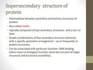
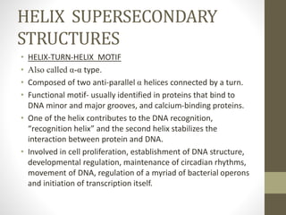
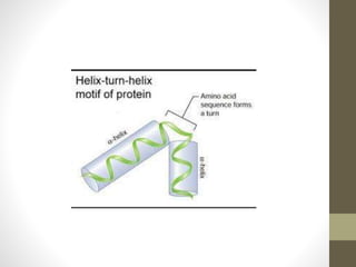
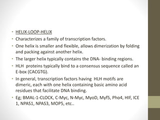
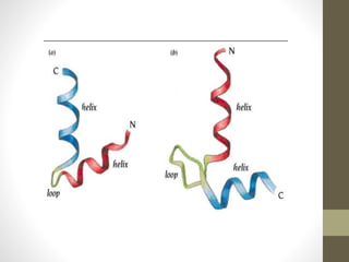
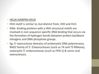
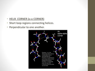
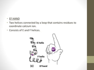
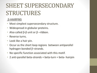
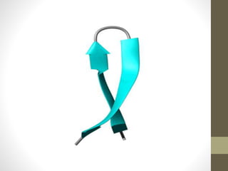
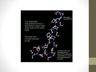
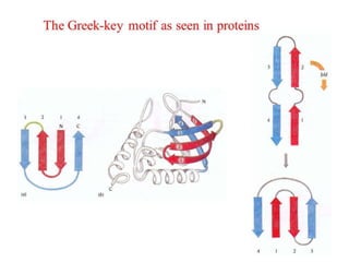
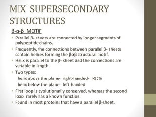
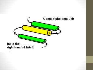
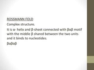
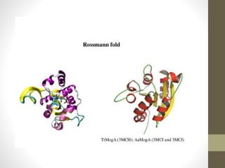
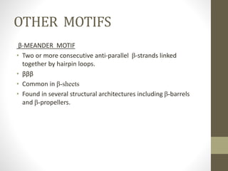
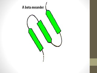
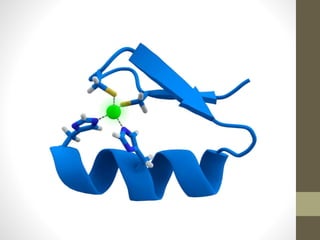
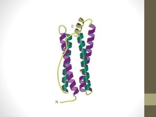
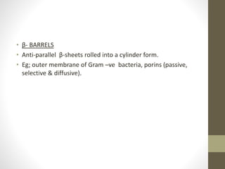
SUPERSECONDARY
STRUCTURE OF
PROTEIN
Mary Theresa
S1; MSc. Microbiology
Supersecondary structure of
protein
• Intermediate between secondary and tertiary structures of
protein.
• Also called motifs.
• Typically composed of two secondary structures and a turn or
loop.
• Simple combinations of few secondary structure elements
with a specific geometric arrangement – occur frequently in
protein structures.
• Can be associated with particular function- DNA binding;
others have no biological function alone but are part of larger
structural and functional assemblies.
SUPERSECONDARY STURUCTURES
Helix Sheet Mix
helix-turn-helix β-hairpins β-α-β
helix-loop-helix β-β corner Rossmann fold
helix-hairpin-helix Greek key motif
α-α corner
HELIX SUPERSECONDARY
STRUCTURES
• HELIX-TURN-HELIX MOTIF
• Also called α-α type.
• Composed of two anti-parallel α helices connected by a turn.
• Functional motif- usually identified in proteins that bind to
DNA minor and major grooves, and calcium-binding proteins.
• One of the helix contributes to the DNA recognition,
“recognition helix” and the second helix stabilizes the
interaction between protein and DNA.
• Involved in cell proliferation, establishment of DNA structure,
developmental regulation, maintenance of circadian rhythms,
movement of DNA, regulation of a myriad of bacterial operons
and initiation of transcription itself.
• HELIX-LOOP-HELIX
• Characterizes a family of transcription factors.
• One helix is smaller and flexible, allows dimerization by folding
and packing against another helix.
• The larger helix typically contains the DNA- binding regions.
• HLH proteins typically bind to a consensus sequence called an
E-box (CACGTG).
• In general, transcription factors having HLH motifs are
dimeric, each with one helix containing basic amino acid
residues that facilitate DNA binding.
• Eg; BMAL-1-CLOCK, C-Myc, N-Myc, MyoD, Myf5, Pho4, HIF, ICE
1, NPAS1, NPAS3, MOP5, etc..
• HELIX-HAIRPIN-HELIX
• HhH motif is similar to, but distinct from, HtH and HLH.
• DNA- binding proteins with a HhH structural motifs are
involved in non-sequence specific DNA binding that occurs via
the formation of hydrogen bonds between protein backbone
nitrogens and DNA phosphate groups.
• Eg; 5’-exonuclease domains of prokaryotic DNA polymerases,
RAD2 family of 5’-3’exonucleases (such as T4 and T5 RNAase),
eukaryotic 5’ endonucleases (such as FEN-1) & some viral
exonucleases.
• HELIX CORNER (α-α CORNER)
• Short loop regions connecting helices.
• Perpendicular to one another.
• EF HAND
• Two helices connected by a loop that contains residues to
coordinate calcium ion.
• Consists of E and F helices.
SHEET SUPERSECONDARY
STRUCTURES
β-HAIRPINS
• Most simplest supersecondary structure.
• Widespread in globular proteins.
• Also called β-β unit or β –ribbon.
• Reverse turns.
• Look like a hair pin.
• Occur as the short loop regions between antiparallel
hydrogen bonded β–strands.
• No specific function associated with this motif.
• 2 anti-parallel beta-strands + beta-turn = beta- hairpin
• β-β CORNER ( β CORNER)
• Consists of two anti-parallel beta strands.
• Can change the direction abruptly.
• The angle of change of direction is about 90°.
• The abrupt angle change is achieved by one strand having a
glycine residue and the other hand having a beta bulge.
• No known function.
• GREEK KEY MOTIF
• formed by four sequentially connected β-strands adjacent to
each other (geometrically aligned to each other)- ββββ
• Alternate strands runs in the opposite direction.
• The first strand (N-terminal strand) and the last strand (C-
terminal strand) are adjacent to each other and hydrogen
bonds exist between them.
• Connecting loops can be long and include other secondary
structures.
• Eg; prealbumin, PapD (a chaperon), nitrite reductase, bacterial
cellulase, spherical virus capsid proteins.
•
MIX SUPERSECONDARY
STRUCTURES
β-α-β MOTIF
• Parallel β- sheets are connected by longer segments of
polypeptide chains.
• Frequently, the connections between parallel β- sheets
contain helices forming the βαβ structural motif.
• Helix is parallel to the β- sheet and the connections are
variable in length.
• Two types:
helix above the plane- right-handed- >95%
helix below the plane- left-handed
• First loop is evolutionarily conserved, whereas the second
loop rarely has a known function.
• Found in most proteins that have a parallel β-sheet.
ROSSMANN FOLD
Complex structure.
It is α- helix and β-sheet connected with βαβ motif
with the middle β shared between the two units
and it binds to nucleotides.
βαβαβ
OTHER MOTIFS
β-MEANDER MOTIF
• Two or more consecutive anti-parallel β-strands linked
together by hairpin loops.
• βββ
• Common in β-sheets
• Found in several structural architectures including β-barrels
and β-propellers.
ψ-LOOP MOTIF
• Consists of two anti-parallel strands with one strand in
between that is connected to both by hydrogen bonds.
• Four possible strand topologies for single ψ–loops.
• Rare one – formation seems unlikely to occur during protein
folding.
• First identified in the aspartic protease family.
ZINC FINGER MOTIF
• Consists of a segment of α-helix bound to a loop by a zinc ion.
• The zinc ion is held in place by two cysteine and two histidine
R groups.
• Motifs are often repeated in clusters.
TRANS MEMBRANE MOTIFS
• HELIX BUNDLES
• It is long streches of a polar aminoacids, fold into trans
membrane α-helices.
• Eg; cell surface receptors, ion channels, active and passive
transporters.
• β- BARRELS
• Anti-parallel β-sheets rolled into a cylinder form.
• Eg; outer membrane of Gram –ve bacteria, porins (passive,
selective & diffusive).
REFERENCES
• Blanco F. J., G. Rivas, L. Serrano. A short linear polypeptide
that folds into a native β- hairpin in aqueous solution. Natural
Structural Biology, 1994; 1(9): 584-590.
• WWW.andrew.cmuedu/course/03-
231/LecF05/Lec09/Lec09.pdf
• Swift.cmbi.ru.nl/gv/students/mtom/sup_2.html
• https://WWW.acsu.buffalo.edu/~sjpark6/pednotes/Motifs.pdf
• https://WWW.uibk.ac.at/organic/de/teaching/gruber_karp_2.
pdf
• Faculty.ksu.edu.sa/77379/Documents/(14)_tertiary_prot.pdf
• https://WWW.youtube.com/watch?v=9ypbWROaaLU
• Rao S. T, M. G. Rassmann. Comparison of supersecondary
structure in protein. Journal of Molecular Biology, 1973; 76:
241-256.
• Janin J., C. Chothia. Packing of α-helices onto β-pleated sheets
and the anatomy of α/β proteins,1980; 143: 95-128.
• Creighton E. Proteins- Structures and Molecular Proteins. 2
edition. W.H. Freeman and Company, New York,1993: 227-
228.
Supersecondary structure ppt

Supersecondary structure ppt

  • 1.
  • 2.
    Supersecondary structure of protein •Intermediate between secondary and tertiary structures of protein. • Also called motifs. • Typically composed of two secondary structures and a turn or loop. • Simple combinations of few secondary structure elements with a specific geometric arrangement – occur frequently in protein structures. • Can be associated with particular function- DNA binding; others have no biological function alone but are part of larger structural and functional assemblies.
  • 3.
    SUPERSECONDARY STURUCTURES Helix SheetMix helix-turn-helix β-hairpins β-α-β helix-loop-helix β-β corner Rossmann fold helix-hairpin-helix Greek key motif α-α corner
  • 4.
    HELIX SUPERSECONDARY STRUCTURES • HELIX-TURN-HELIXMOTIF • Also called α-α type. • Composed of two anti-parallel α helices connected by a turn. • Functional motif- usually identified in proteins that bind to DNA minor and major grooves, and calcium-binding proteins. • One of the helix contributes to the DNA recognition, “recognition helix” and the second helix stabilizes the interaction between protein and DNA. • Involved in cell proliferation, establishment of DNA structure, developmental regulation, maintenance of circadian rhythms, movement of DNA, regulation of a myriad of bacterial operons and initiation of transcription itself.
  • 6.
    • HELIX-LOOP-HELIX • Characterizesa family of transcription factors. • One helix is smaller and flexible, allows dimerization by folding and packing against another helix. • The larger helix typically contains the DNA- binding regions. • HLH proteins typically bind to a consensus sequence called an E-box (CACGTG). • In general, transcription factors having HLH motifs are dimeric, each with one helix containing basic amino acid residues that facilitate DNA binding. • Eg; BMAL-1-CLOCK, C-Myc, N-Myc, MyoD, Myf5, Pho4, HIF, ICE 1, NPAS1, NPAS3, MOP5, etc..
  • 8.
    • HELIX-HAIRPIN-HELIX • HhHmotif is similar to, but distinct from, HtH and HLH. • DNA- binding proteins with a HhH structural motifs are involved in non-sequence specific DNA binding that occurs via the formation of hydrogen bonds between protein backbone nitrogens and DNA phosphate groups. • Eg; 5’-exonuclease domains of prokaryotic DNA polymerases, RAD2 family of 5’-3’exonucleases (such as T4 and T5 RNAase), eukaryotic 5’ endonucleases (such as FEN-1) & some viral exonucleases.
  • 10.
    • HELIX CORNER(α-α CORNER) • Short loop regions connecting helices. • Perpendicular to one another.
  • 11.
    • EF HAND •Two helices connected by a loop that contains residues to coordinate calcium ion. • Consists of E and F helices.
  • 12.
    SHEET SUPERSECONDARY STRUCTURES β-HAIRPINS • Mostsimplest supersecondary structure. • Widespread in globular proteins. • Also called β-β unit or β –ribbon. • Reverse turns. • Look like a hair pin. • Occur as the short loop regions between antiparallel hydrogen bonded β–strands. • No specific function associated with this motif. • 2 anti-parallel beta-strands + beta-turn = beta- hairpin
  • 14.
    • β-β CORNER( β CORNER) • Consists of two anti-parallel beta strands. • Can change the direction abruptly. • The angle of change of direction is about 90°. • The abrupt angle change is achieved by one strand having a glycine residue and the other hand having a beta bulge. • No known function.
  • 16.
    • GREEK KEYMOTIF • formed by four sequentially connected β-strands adjacent to each other (geometrically aligned to each other)- ββββ • Alternate strands runs in the opposite direction. • The first strand (N-terminal strand) and the last strand (C- terminal strand) are adjacent to each other and hydrogen bonds exist between them. • Connecting loops can be long and include other secondary structures. • Eg; prealbumin, PapD (a chaperon), nitrite reductase, bacterial cellulase, spherical virus capsid proteins. •
  • 18.
    MIX SUPERSECONDARY STRUCTURES β-α-β MOTIF •Parallel β- sheets are connected by longer segments of polypeptide chains. • Frequently, the connections between parallel β- sheets contain helices forming the βαβ structural motif. • Helix is parallel to the β- sheet and the connections are variable in length. • Two types: helix above the plane- right-handed- >95% helix below the plane- left-handed • First loop is evolutionarily conserved, whereas the second loop rarely has a known function. • Found in most proteins that have a parallel β-sheet.
  • 20.
    ROSSMANN FOLD Complex structure. Itis α- helix and β-sheet connected with βαβ motif with the middle β shared between the two units and it binds to nucleotides. βαβαβ
  • 22.
    OTHER MOTIFS β-MEANDER MOTIF •Two or more consecutive anti-parallel β-strands linked together by hairpin loops. • βββ • Common in β-sheets • Found in several structural architectures including β-barrels and β-propellers.
  • 24.
    ψ-LOOP MOTIF • Consistsof two anti-parallel strands with one strand in between that is connected to both by hydrogen bonds. • Four possible strand topologies for single ψ–loops. • Rare one – formation seems unlikely to occur during protein folding. • First identified in the aspartic protease family.
  • 25.
    ZINC FINGER MOTIF •Consists of a segment of α-helix bound to a loop by a zinc ion. • The zinc ion is held in place by two cysteine and two histidine R groups. • Motifs are often repeated in clusters.
  • 27.
    TRANS MEMBRANE MOTIFS •HELIX BUNDLES • It is long streches of a polar aminoacids, fold into trans membrane α-helices. • Eg; cell surface receptors, ion channels, active and passive transporters.
  • 29.
    • β- BARRELS •Anti-parallel β-sheets rolled into a cylinder form. • Eg; outer membrane of Gram –ve bacteria, porins (passive, selective & diffusive).
  • 31.
    REFERENCES • Blanco F.J., G. Rivas, L. Serrano. A short linear polypeptide that folds into a native β- hairpin in aqueous solution. Natural Structural Biology, 1994; 1(9): 584-590. • WWW.andrew.cmuedu/course/03- 231/LecF05/Lec09/Lec09.pdf • Swift.cmbi.ru.nl/gv/students/mtom/sup_2.html • https://WWW.acsu.buffalo.edu/~sjpark6/pednotes/Motifs.pdf • https://WWW.uibk.ac.at/organic/de/teaching/gruber_karp_2. pdf • Faculty.ksu.edu.sa/77379/Documents/(14)_tertiary_prot.pdf
  • 32.
    • https://WWW.youtube.com/watch?v=9ypbWROaaLU • RaoS. T, M. G. Rassmann. Comparison of supersecondary structure in protein. Journal of Molecular Biology, 1973; 76: 241-256. • Janin J., C. Chothia. Packing of α-helices onto β-pleated sheets and the anatomy of α/β proteins,1980; 143: 95-128. • Creighton E. Proteins- Structures and Molecular Proteins. 2 edition. W.H. Freeman and Company, New York,1993: 227- 228.