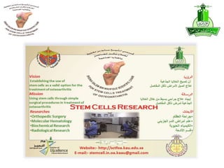
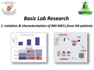
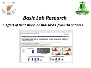
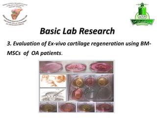
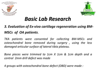
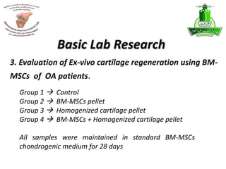
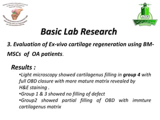
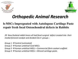
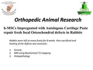
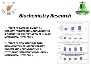
Stem Cell Research
DR.MOHAMMAD ABBAS
Assistant Professor
& Orthopedic Consultant
Faculty of Medicine
King Abdulaziz University
 Basic Lab Research
 Orthopedic Surgery
 Molecular Hematology
 Biochemistry
 Radiology
Research Groups
1. Isolation & characterization of BM-MSCs from OA patients
Basic Lab Research
Basic Lab Research
• Primary cultures of BM aspirate from OA patients
showed characteristic spindle shaped cells which
expressed MSCs related CD surface markers
• BM- MSCs demonstrated good viability , increased
proliferation rate and differentiation into :
ADIPOCYTES , CHONDROCYTE , OSTEOCYTE
1. Isolation & characterization of BM-MSCs from OA patients
Basic Lab Research
Collagen secretion and Alkaline phosphates
activity where increased with chondrogenic and
osteogenic differentiation
Isolation & characterization were successful in
both the OR stem cell Lab. & stem cells unit in
KFMRC
1. Isolation & characterization of BM-MSCs from OA patients
2. Effect of Heat shock on BM- MSCs from OA patients
Basic Lab Research
After MSCs characterization and differentiation BM-
MSCs were exposed to illuminated Arthroscope either
as cell suspension or cell pellet to 37,45,55 degrees
for 10,20 & 30 minutes followed by cell proliferation
assay for 72 hrs which showed :
Basic Lab Research
2. Effect of Heat shock on BM- MSCs from OA patients
The study concluded that BM-MSCs cell pellet appears better
protected from temperature alterations compared to cell
suspension
1- 63% cell prolifration in the cell suspension group
2- 62 – 68 % in cell prolifration in the cell pellet group
Basic Lab Research
2. Effect of Heat shock on BM- MSCs from OA patients
Results :
Transplantation of BM-MSCs as pellet rather than as
a single cells suspension to the site of cartilage
defect would therefore support their viability and
aid cartilage prolifration
Basic Lab Research
2. Effect of Heat shock on BM- MSCs from OA patients
Conclusion :
3. Evaluation of Ex-vivo cartilage regeneration using BM-
MSCs of OA patients.
Basic Lab Research
Basic Lab Research
TKA patients were consented for collecting BM-MSCs and
osteochondral bone removed during surgery , using the less
damaged articular surface of lateral tibia plateau.
Bone pieces were trimmed to 1cm X 1cm & 1cm depth and a
central 2mm drill defect was made
4 groups with osteochondral bone defect (OBD) were made :
3. Evaluation of Ex-vivo cartilage regeneration using BM-
MSCs of OA patients.
Basic Lab Research
Group 1  Control
Group 2  BM-MSCs pellet
Group 3  Homogenized cartilage pellet
Group 4  BM-MSCs + Homogenized cartilage pellet
All samples were maintained in standard BM-MSCs
chondrogenic medium for 28 days
3. Evaluation of Ex-vivo cartilage regeneration using BM-
MSCs of OA patients.
Basic Lab Research
Results :
•Light microscopy showed cartilagenus filling in group 4 with
full OBD closure with more mature matrix revealed by
H&E staining .
•Group 1 & 3 showed no filling of defect
•Group2 showed partial filling of OBD with immture
cartilagenus matrix
3. Evaluation of Ex-vivo cartilage regeneration using BM-
MSCs of OA patients.
Basic Lab Research
Conclusion :
The study concludes that adding cartilage
fragments to MSCs provide better formation
in Ex – vivo models
3. Evaluation of Ex-vivo cartilage regeneration using BM-
MSCs of OA patients.
1- Impact f cartilage paste
impregnated with MSCs on
regeneration of focal articular
cartilage defects in rabbits
Orthopedic Animal Research
20 New Zealand rabbit knees all had focal surgical defect created into their
medial femoral condyle and divided into 4 groups :
Group 1  Control (untreated)
Group 2  Human umbilical Cord MSCs
Group 3  Human umbilical MSCs + Commercial fibrin sealant scaffold .
Group 4  Human umbilical MSCs + Minced cartilage paste.
h-MSCs Impregnated with Autologous Cartilage Paste
repair fresh focal Osteochondral defects in Rabbits
Orthopedic Animal Research
Rabbits were left to move freely for 8 weeks then sacrificed and
healing of the defects was assessed :
1. Grossly
2. MRI using Biochemical T2 mapping
3. Histopathology
h-MSCs Impregnated with Autologous Cartilage Paste
repair fresh focal Osteochondral defects in Rabbits
Orthopedic Animal Research
Group 1  showed no cartilage filling of defect.
Group 2
Group 3
Showed partial filling of the defect but was a bit better
in group 3 ( MOCART score 5 points )
Group 4  - Complete filling of defect
-Intact cartilage surface
-Complete integration with adjacent cartilage as seen
in Histopathology & T2 mapping MRI ( MOCART score 80 points )
h-MSCs Impregnated with Autologous Cartilage Paste
repair fresh focal Osteochondral defects in Rabbits
Results :
Orthopedic Animal Research
Conclusions:
Repair of focal osteochondral defects in rabbit knees using
human umbilical cord MSCs impregnated with autologous
cartilage paste appears to be successful as proven
clinically, radiological, as well Pathologically
h-MSCs Impregnated with Autologous Cartilage Paste
repair fresh focal Osteochondral defects in Rabbits
‫صور‬ ‫من‬ ‫يستعمل‬ ‫انسجه‬ ‫و‬‫د‬‫ولرنين‬ ‫عادية‬ ‫للركبة‬ ‫صور‬ ‫حازم‬
2- Impact of hylofast scaffold
impregnated with human MSCs &
cartilage paste on surgically
induced total arthritis in rabbits
knees
Orthopedic Animal Research
‫صورة‬‫هيلو‬‫فاست‬
2- Impact of hylofast scaffold impregnated with human MSCs & cartilage
paste on surgically induced total arthritis in rabbits knees
Orthopedic Animal Research
16 newzland rabbit knees were used having surgically induced
total arthritis and divided into 4 groups
Group 1  control (untreated)
Group 2  MSCs + cartilage paste
Group 3 MSCs + hylofast scaffold
Group 4  MSCs + hylofast scaffold +cartilage paste
Study still in progress
- Darweish
- Chaudary
- Hamdi
Orthopedic Colleagues
1. Genetic mapping of osteoarthritis
2. Epigenetic analysis of osteoarthritis patient
3. Exome sequence analysis for osteoarthritis
patients
4. Molecular regulation of chondrogenic human
induced pluripotent stem cells
Molecular Hematology Research
Genetic mapping of osteoarthritis
Molecular Hematology Research
Epigenetic analysis of osteoarthritis patient
Molecular Hematology Research
Exome sequence analysis for osteoarthritis
patients
Molecular Hematology Research
Molecular regulation of chondrogenic human
induced pluripotent stem cells
Molecular Hematology Research
Molecular regulation of chondrogenic human
induced pluripotent stem cells
Molecular Hematology Research
Purpose
Human induced pleuripotent stem cells (hiPSC) are a promising source for
chondrogenic stem cells. Sequential
differentiation of hiPSC provides a platform for dissecting the molecular
pathways associated with
chondrogenesis in vivo and could reveal targets for better control of
chondrocyte fate for cartilage repair
applications. The aim of this study was to use nextgeneration
sequencing (NGS) to investigate the transcriptome
of chondrogenic hiPSCs.
Molecular regulation of chondrogenic human
induced pluripotent stem cells
Molecular Hematology Research
Methods and Materials
The hiPSC line (C19) was derived through reprogramming of human dermal
fibroblasts using viral vectors
expressing Oct4, Sox2 and Klf4. A protocol of sequential growth factors including
Activin A, FGF2
and BMP4
was used to drive the formation of chondroprogenitors directly from hiPSC colonies.
The chondrogenic hiPSCs
were characterised exhaustively by tissue engineering, histochemical analysis and
biochemical analysis. The
transcriptome of undifferentiated hiPSCs, hiPSCderived
Molecular regulation of chondrogenic human
induced pluripotent stem cells
Molecular Hematology Research
Results
Differential gene expression revealed the induction of several collagen genes including
type 1 to type 12, type 14
and type 18 during transition from the pluripotent state to the chondrogenic state (Figure
1). Collagenregulatory
genes such as PCOLCE which drives the endopeptidase cleavage of procollagen as well as
regulators of
collagen glycosylation were upregulated. The expression of various fibroblasts growth
factors (FGFs) including
FGF11,
FGFR2 and insulin growth factor2
Molecular regulation of chondrogenic human
induced pluripotent stem cells
Molecular Hematology Research
Conclusion
NGS analysis demonstrated the recapitulation of early events in cartilage
development during hiPSC
chondrogenesis. The upregulation of many members of the collagen family
indicate the intricate nature of
collagen expression during chondrogenesis. Further analysis of
nonchondrogenic
targets may reveal novel
pathways for controlling the fate of chondrogenic hiPSCs.
1- EFFECT OF CATECHOLAMINES ON
VIABILITY, PROLIFERATION,CHONDROGENIC
& OSTEOGENIC DIFFERETIATION OF HUMAN
MESENCHMAL STEM CELLS.
2- EFFECT OF NON STEROIDAL ANTI-
INFLAMMATORY DRUGS ON VIABILITY,
PROLIFERATION,CHONDROGENIC &
OSTEOGENIC DIFFERETIATION OF HUMAN
MESENCHMAL STEM CELLS.
Biochemistry Research
1. From Dr. Qutb)
Radiological Research
 One oral presentation
 One poster
ICRS 2015 Chicago
 One poster
EULAR Conference
- Not yet
- Planning on high tibial ostetomy for
unicompartmental osteoarthritis
Human Research

Stem cell research

  • 1.
    Stem Cell Research DR.MOHAMMADABBAS Assistant Professor & Orthopedic Consultant Faculty of Medicine King Abdulaziz University
  • 4.
     Basic LabResearch  Orthopedic Surgery  Molecular Hematology  Biochemistry  Radiology Research Groups
  • 5.
    1. Isolation &characterization of BM-MSCs from OA patients Basic Lab Research
  • 6.
    Basic Lab Research •Primary cultures of BM aspirate from OA patients showed characteristic spindle shaped cells which expressed MSCs related CD surface markers • BM- MSCs demonstrated good viability , increased proliferation rate and differentiation into : ADIPOCYTES , CHONDROCYTE , OSTEOCYTE 1. Isolation & characterization of BM-MSCs from OA patients
  • 7.
    Basic Lab Research Collagensecretion and Alkaline phosphates activity where increased with chondrogenic and osteogenic differentiation Isolation & characterization were successful in both the OR stem cell Lab. & stem cells unit in KFMRC 1. Isolation & characterization of BM-MSCs from OA patients
  • 8.
    2. Effect ofHeat shock on BM- MSCs from OA patients Basic Lab Research
  • 9.
    After MSCs characterizationand differentiation BM- MSCs were exposed to illuminated Arthroscope either as cell suspension or cell pellet to 37,45,55 degrees for 10,20 & 30 minutes followed by cell proliferation assay for 72 hrs which showed : Basic Lab Research 2. Effect of Heat shock on BM- MSCs from OA patients
  • 10.
    The study concludedthat BM-MSCs cell pellet appears better protected from temperature alterations compared to cell suspension 1- 63% cell prolifration in the cell suspension group 2- 62 – 68 % in cell prolifration in the cell pellet group Basic Lab Research 2. Effect of Heat shock on BM- MSCs from OA patients Results :
  • 11.
    Transplantation of BM-MSCsas pellet rather than as a single cells suspension to the site of cartilage defect would therefore support their viability and aid cartilage prolifration Basic Lab Research 2. Effect of Heat shock on BM- MSCs from OA patients Conclusion :
  • 12.
    3. Evaluation ofEx-vivo cartilage regeneration using BM- MSCs of OA patients. Basic Lab Research
  • 13.
    Basic Lab Research TKApatients were consented for collecting BM-MSCs and osteochondral bone removed during surgery , using the less damaged articular surface of lateral tibia plateau. Bone pieces were trimmed to 1cm X 1cm & 1cm depth and a central 2mm drill defect was made 4 groups with osteochondral bone defect (OBD) were made : 3. Evaluation of Ex-vivo cartilage regeneration using BM- MSCs of OA patients.
  • 14.
    Basic Lab Research Group1  Control Group 2  BM-MSCs pellet Group 3  Homogenized cartilage pellet Group 4  BM-MSCs + Homogenized cartilage pellet All samples were maintained in standard BM-MSCs chondrogenic medium for 28 days 3. Evaluation of Ex-vivo cartilage regeneration using BM- MSCs of OA patients.
  • 15.
    Basic Lab Research Results: •Light microscopy showed cartilagenus filling in group 4 with full OBD closure with more mature matrix revealed by H&E staining . •Group 1 & 3 showed no filling of defect •Group2 showed partial filling of OBD with immture cartilagenus matrix 3. Evaluation of Ex-vivo cartilage regeneration using BM- MSCs of OA patients.
  • 16.
    Basic Lab Research Conclusion: The study concludes that adding cartilage fragments to MSCs provide better formation in Ex – vivo models 3. Evaluation of Ex-vivo cartilage regeneration using BM- MSCs of OA patients.
  • 17.
    1- Impact fcartilage paste impregnated with MSCs on regeneration of focal articular cartilage defects in rabbits Orthopedic Animal Research
  • 18.
    20 New Zealandrabbit knees all had focal surgical defect created into their medial femoral condyle and divided into 4 groups : Group 1  Control (untreated) Group 2  Human umbilical Cord MSCs Group 3  Human umbilical MSCs + Commercial fibrin sealant scaffold . Group 4  Human umbilical MSCs + Minced cartilage paste. h-MSCs Impregnated with Autologous Cartilage Paste repair fresh focal Osteochondral defects in Rabbits Orthopedic Animal Research
  • 19.
    Orthopedic Animal Research Rabbitswere left to move freely for 8 weeks then sacrificed and healing of the defects was assessed : 1. Grossly 2. MRI using Biochemical T2 mapping 3. Histopathology h-MSCs Impregnated with Autologous Cartilage Paste repair fresh focal Osteochondral defects in Rabbits
  • 20.
    Orthopedic Animal Research Group1  showed no cartilage filling of defect. Group 2 Group 3 Showed partial filling of the defect but was a bit better in group 3 ( MOCART score 5 points ) Group 4  - Complete filling of defect -Intact cartilage surface -Complete integration with adjacent cartilage as seen in Histopathology & T2 mapping MRI ( MOCART score 80 points ) h-MSCs Impregnated with Autologous Cartilage Paste repair fresh focal Osteochondral defects in Rabbits Results :
  • 21.
    Orthopedic Animal Research Conclusions: Repairof focal osteochondral defects in rabbit knees using human umbilical cord MSCs impregnated with autologous cartilage paste appears to be successful as proven clinically, radiological, as well Pathologically h-MSCs Impregnated with Autologous Cartilage Paste repair fresh focal Osteochondral defects in Rabbits ‫صور‬ ‫من‬ ‫يستعمل‬ ‫انسجه‬ ‫و‬‫د‬‫ولرنين‬ ‫عادية‬ ‫للركبة‬ ‫صور‬ ‫حازم‬
  • 22.
    2- Impact ofhylofast scaffold impregnated with human MSCs & cartilage paste on surgically induced total arthritis in rabbits knees Orthopedic Animal Research ‫صورة‬‫هيلو‬‫فاست‬
  • 23.
    2- Impact ofhylofast scaffold impregnated with human MSCs & cartilage paste on surgically induced total arthritis in rabbits knees Orthopedic Animal Research 16 newzland rabbit knees were used having surgically induced total arthritis and divided into 4 groups Group 1  control (untreated) Group 2  MSCs + cartilage paste Group 3 MSCs + hylofast scaffold Group 4  MSCs + hylofast scaffold +cartilage paste Study still in progress
  • 24.
    - Darweish - Chaudary -Hamdi Orthopedic Colleagues
  • 25.
    1. Genetic mappingof osteoarthritis 2. Epigenetic analysis of osteoarthritis patient 3. Exome sequence analysis for osteoarthritis patients 4. Molecular regulation of chondrogenic human induced pluripotent stem cells Molecular Hematology Research
  • 26.
    Genetic mapping ofosteoarthritis Molecular Hematology Research
  • 27.
    Epigenetic analysis ofosteoarthritis patient Molecular Hematology Research
  • 28.
    Exome sequence analysisfor osteoarthritis patients Molecular Hematology Research
  • 29.
    Molecular regulation ofchondrogenic human induced pluripotent stem cells Molecular Hematology Research
  • 30.
    Molecular regulation ofchondrogenic human induced pluripotent stem cells Molecular Hematology Research Purpose Human induced pleuripotent stem cells (hiPSC) are a promising source for chondrogenic stem cells. Sequential differentiation of hiPSC provides a platform for dissecting the molecular pathways associated with chondrogenesis in vivo and could reveal targets for better control of chondrocyte fate for cartilage repair applications. The aim of this study was to use nextgeneration sequencing (NGS) to investigate the transcriptome of chondrogenic hiPSCs.
  • 31.
    Molecular regulation ofchondrogenic human induced pluripotent stem cells Molecular Hematology Research Methods and Materials The hiPSC line (C19) was derived through reprogramming of human dermal fibroblasts using viral vectors expressing Oct4, Sox2 and Klf4. A protocol of sequential growth factors including Activin A, FGF2 and BMP4 was used to drive the formation of chondroprogenitors directly from hiPSC colonies. The chondrogenic hiPSCs were characterised exhaustively by tissue engineering, histochemical analysis and biochemical analysis. The transcriptome of undifferentiated hiPSCs, hiPSCderived
  • 32.
    Molecular regulation ofchondrogenic human induced pluripotent stem cells Molecular Hematology Research Results Differential gene expression revealed the induction of several collagen genes including type 1 to type 12, type 14 and type 18 during transition from the pluripotent state to the chondrogenic state (Figure 1). Collagenregulatory genes such as PCOLCE which drives the endopeptidase cleavage of procollagen as well as regulators of collagen glycosylation were upregulated. The expression of various fibroblasts growth factors (FGFs) including FGF11, FGFR2 and insulin growth factor2
  • 33.
    Molecular regulation ofchondrogenic human induced pluripotent stem cells Molecular Hematology Research Conclusion NGS analysis demonstrated the recapitulation of early events in cartilage development during hiPSC chondrogenesis. The upregulation of many members of the collagen family indicate the intricate nature of collagen expression during chondrogenesis. Further analysis of nonchondrogenic targets may reveal novel pathways for controlling the fate of chondrogenic hiPSCs.
  • 34.
    1- EFFECT OFCATECHOLAMINES ON VIABILITY, PROLIFERATION,CHONDROGENIC & OSTEOGENIC DIFFERETIATION OF HUMAN MESENCHMAL STEM CELLS. 2- EFFECT OF NON STEROIDAL ANTI- INFLAMMATORY DRUGS ON VIABILITY, PROLIFERATION,CHONDROGENIC & OSTEOGENIC DIFFERETIATION OF HUMAN MESENCHMAL STEM CELLS. Biochemistry Research
  • 35.
    1. From Dr.Qutb) Radiological Research
  • 36.
     One oralpresentation  One poster ICRS 2015 Chicago
  • 37.
  • 38.
    - Not yet -Planning on high tibial ostetomy for unicompartmental osteoarthritis Human Research

Editor's Notes

  • #23 صورة هيلو فاست