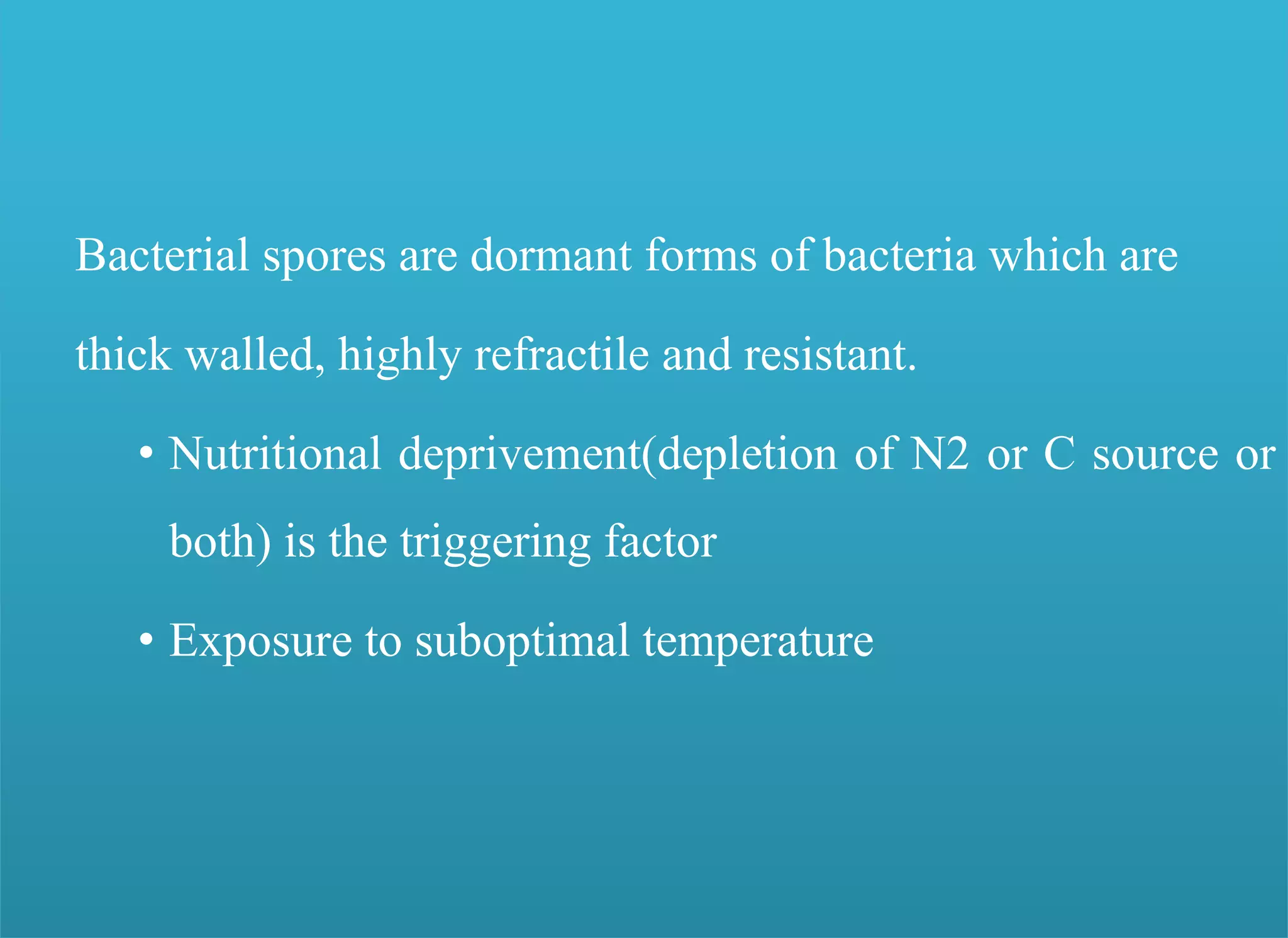
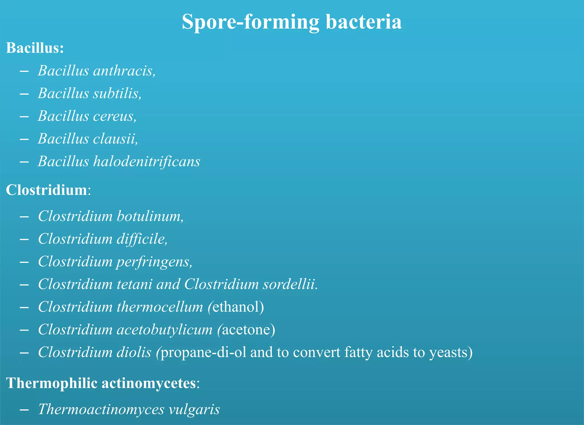
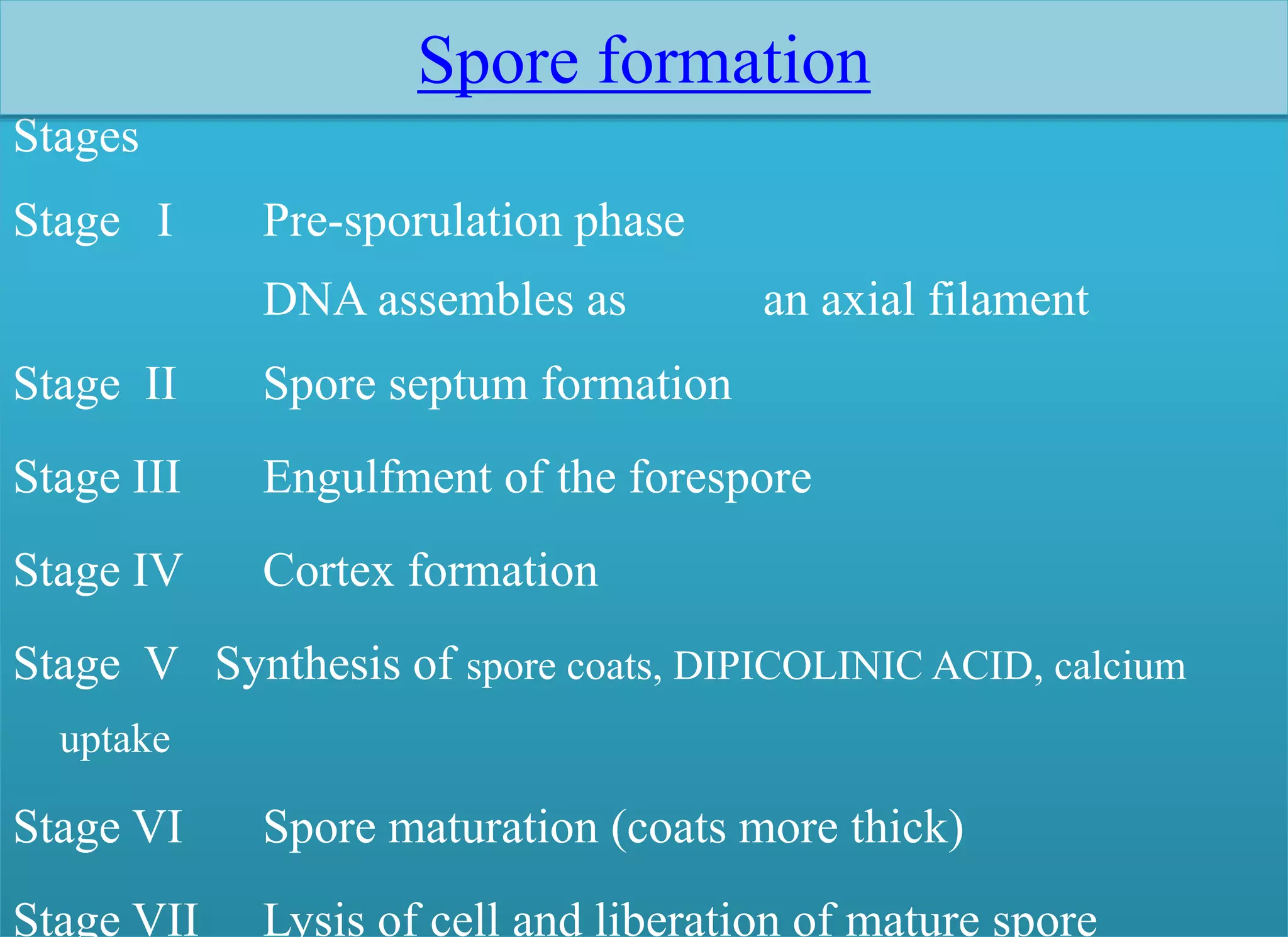
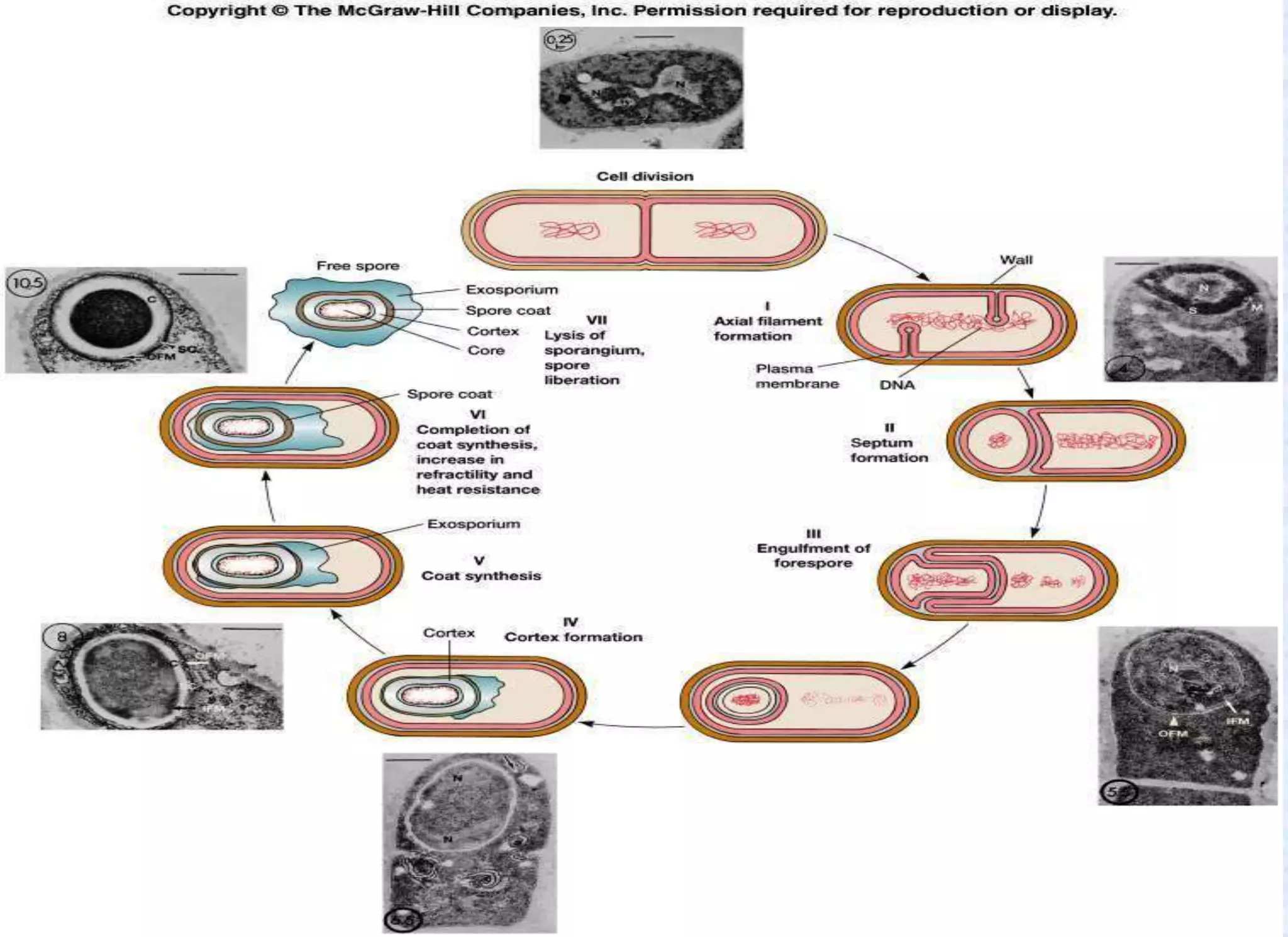
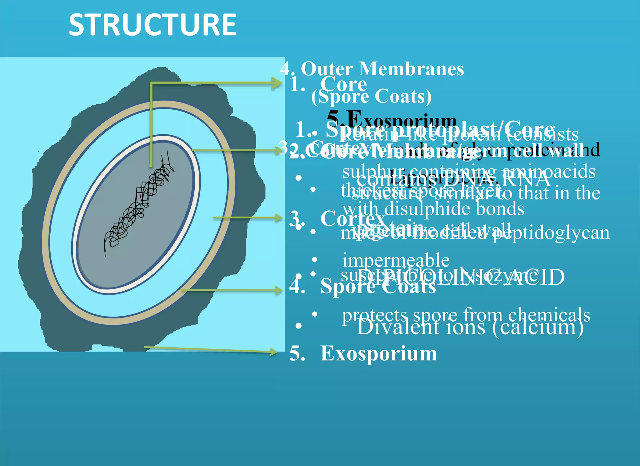
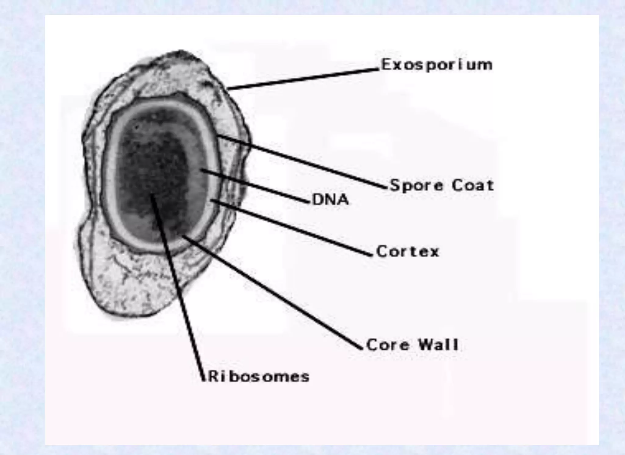
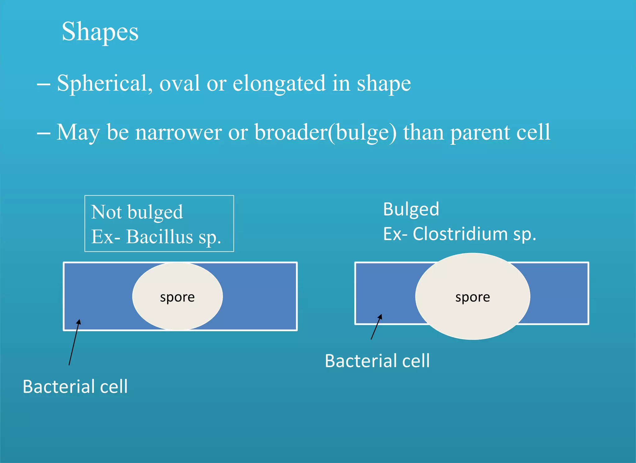
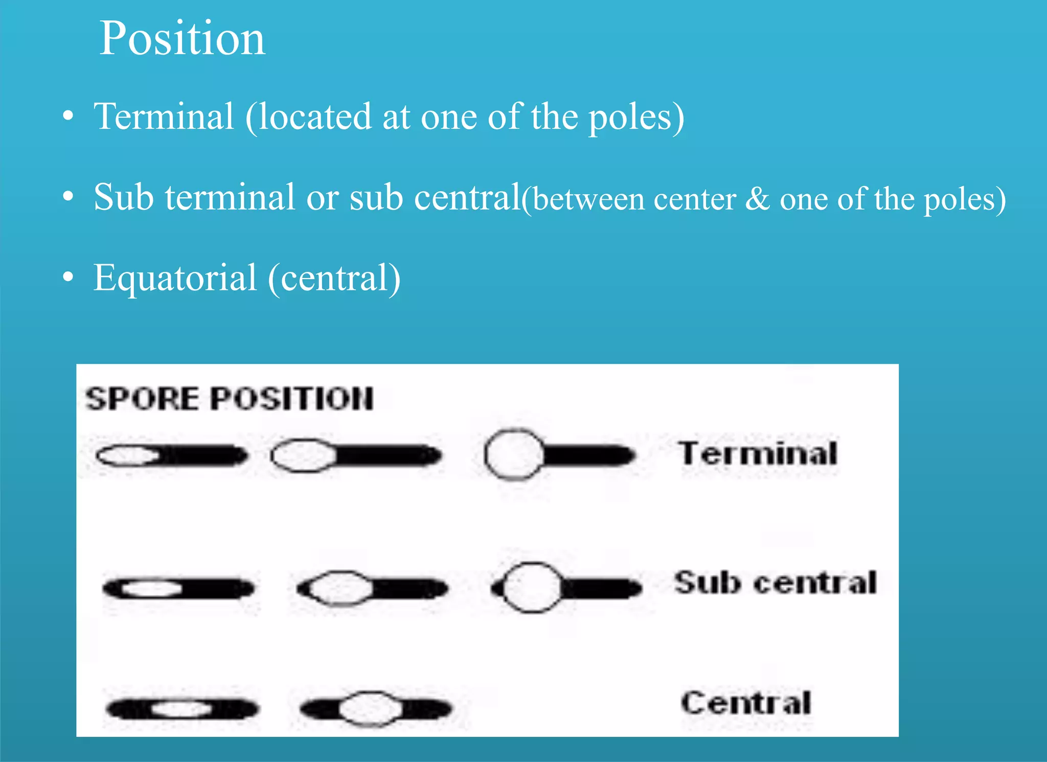
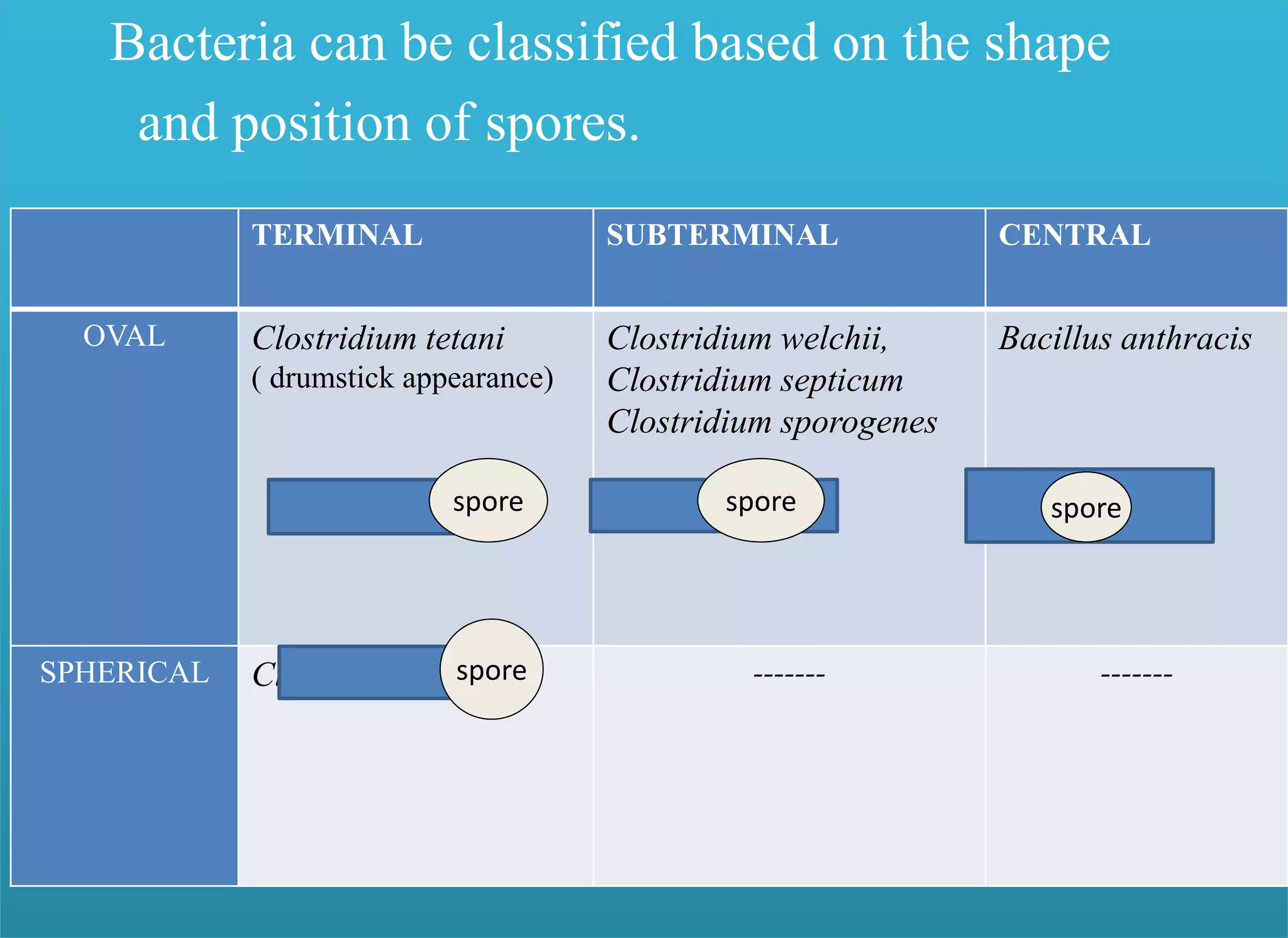
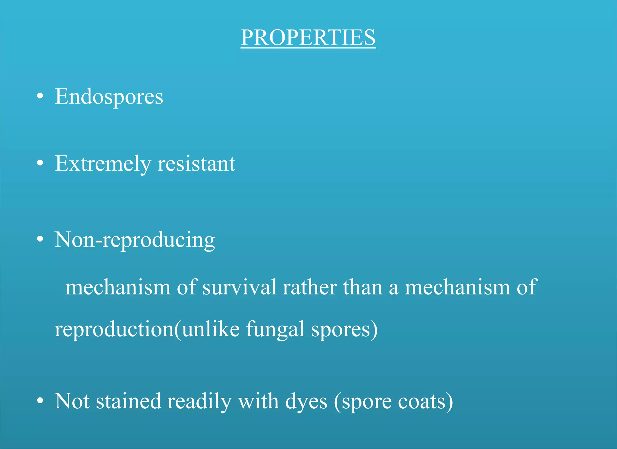
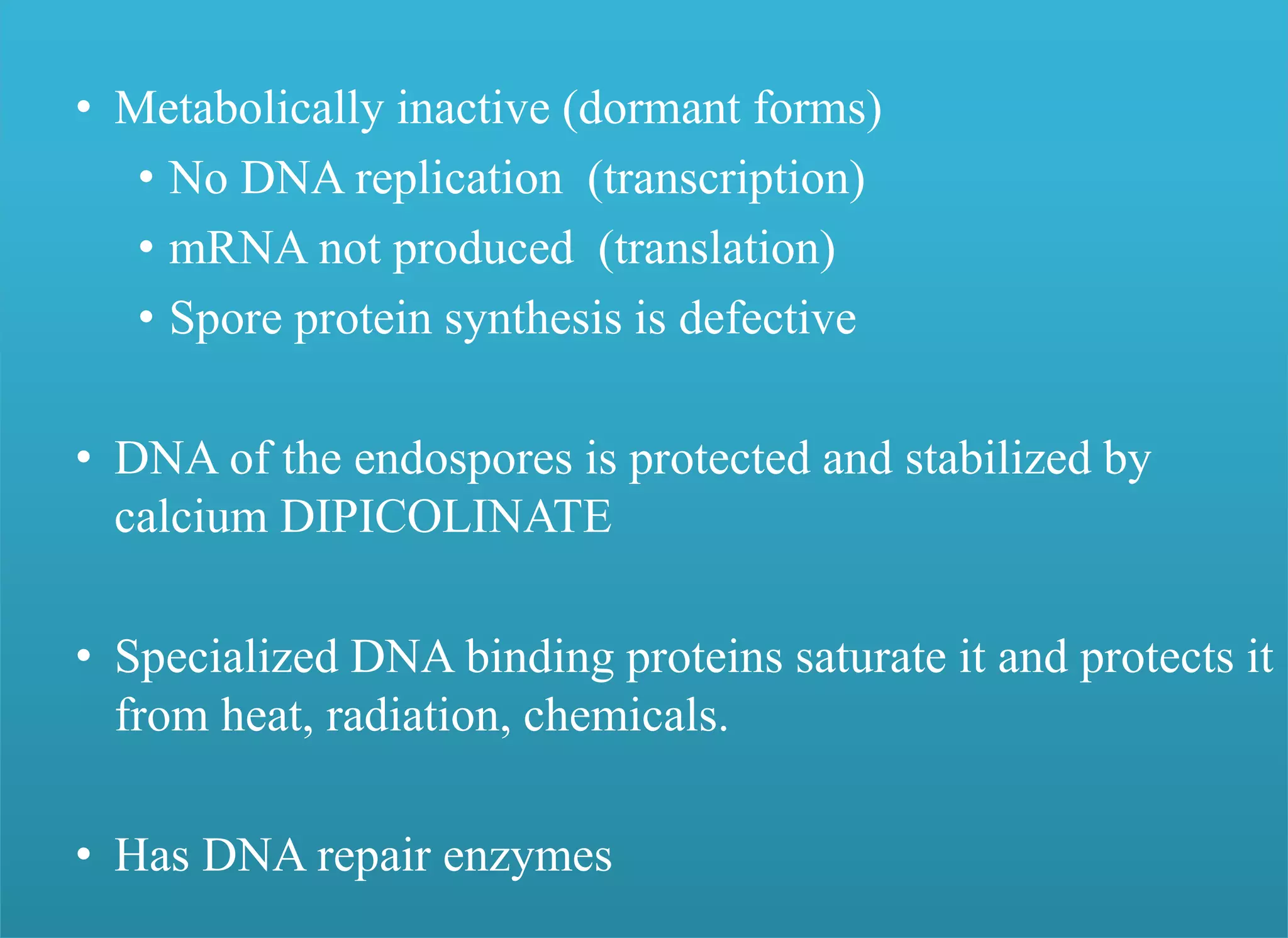
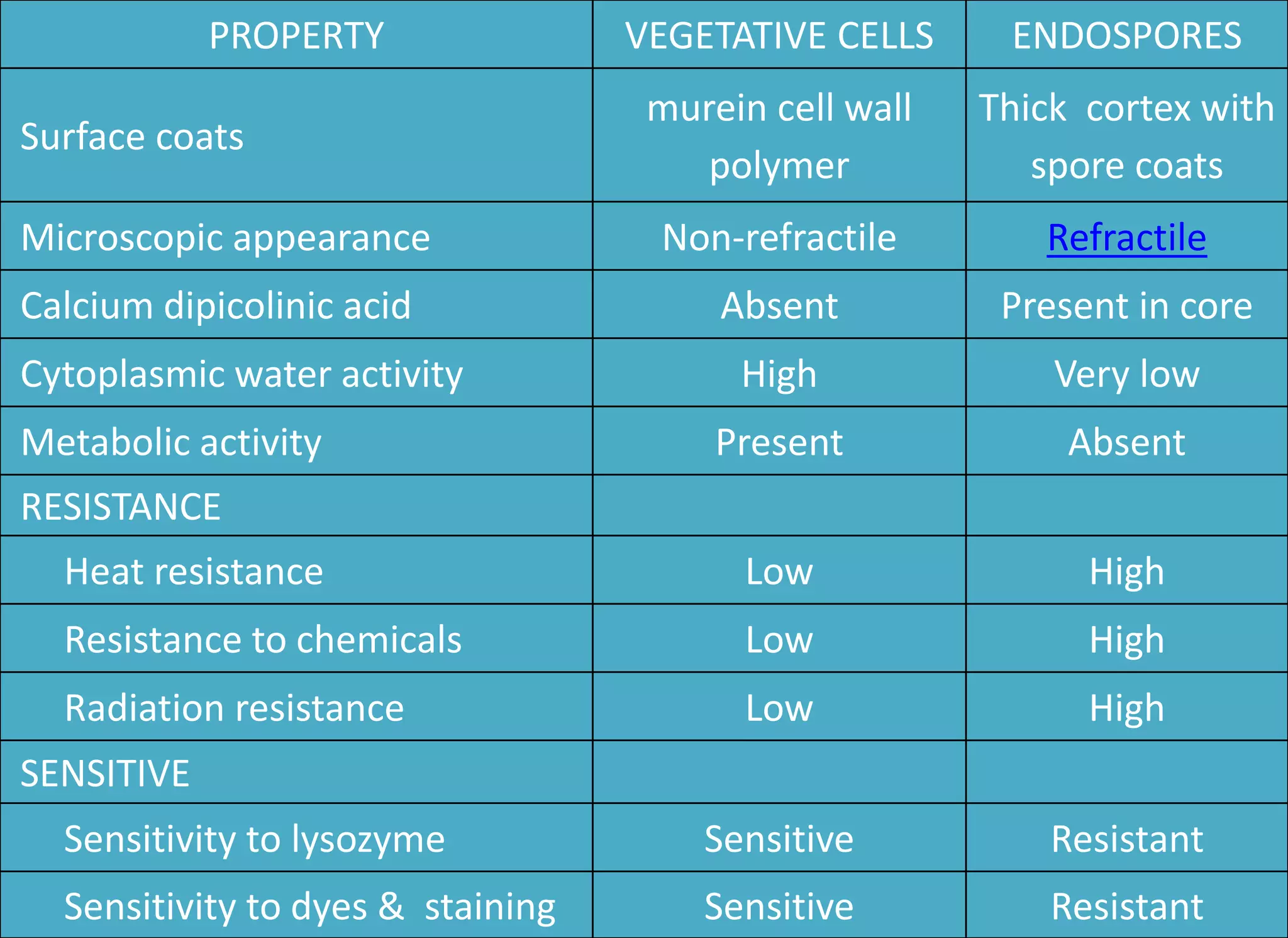
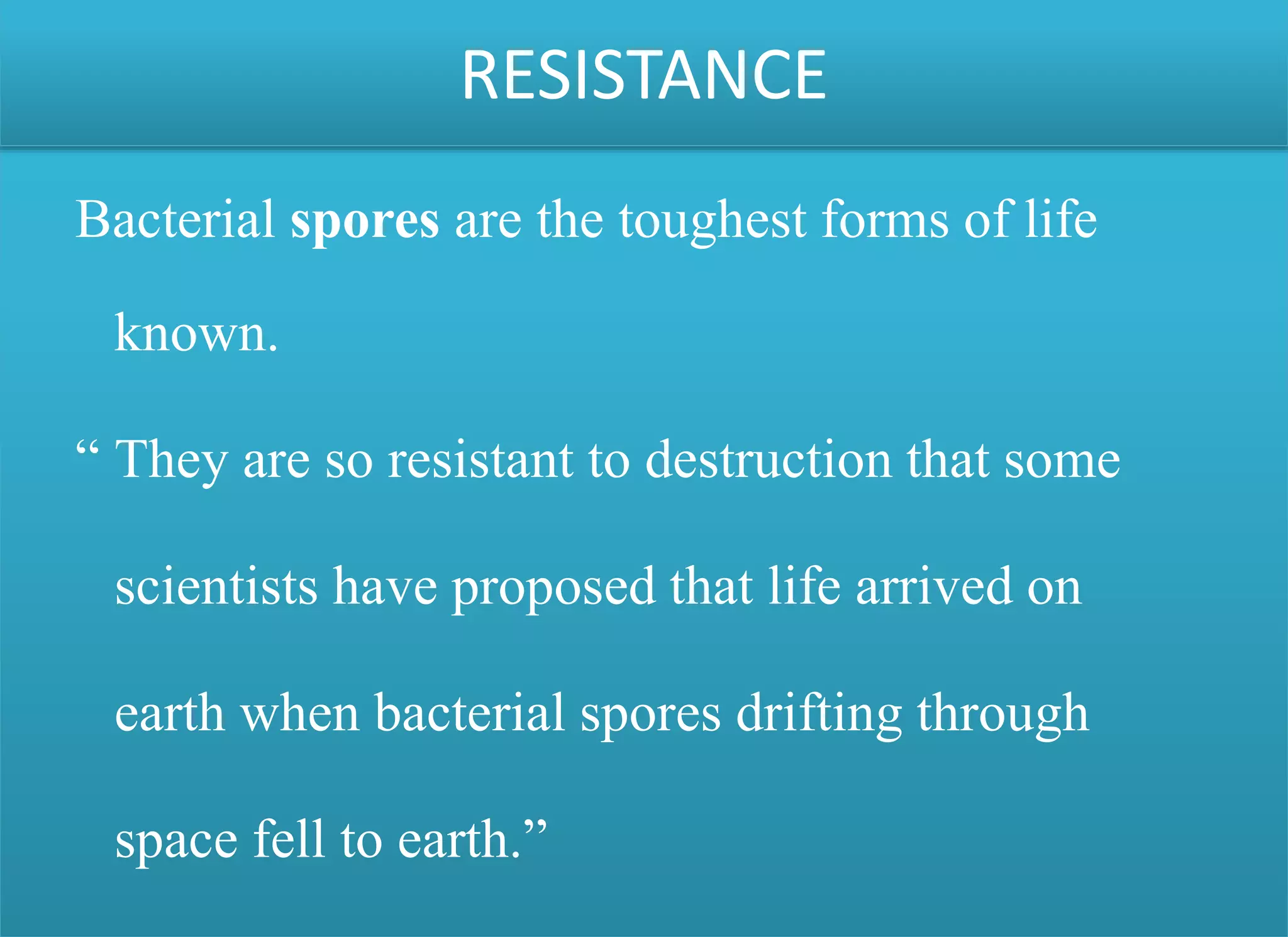
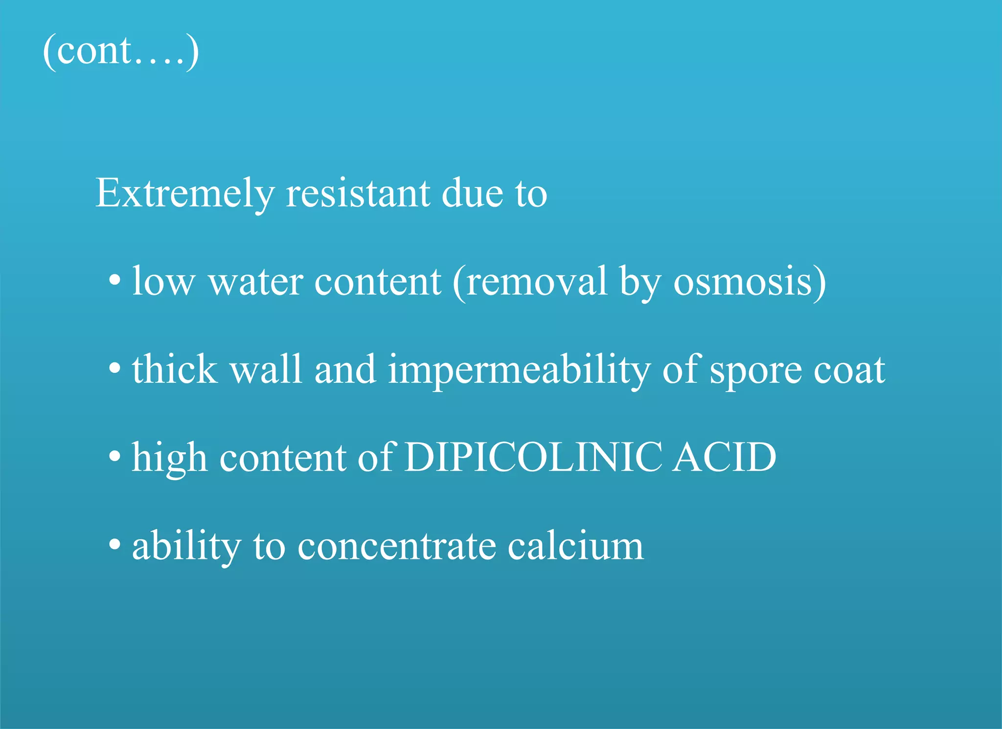
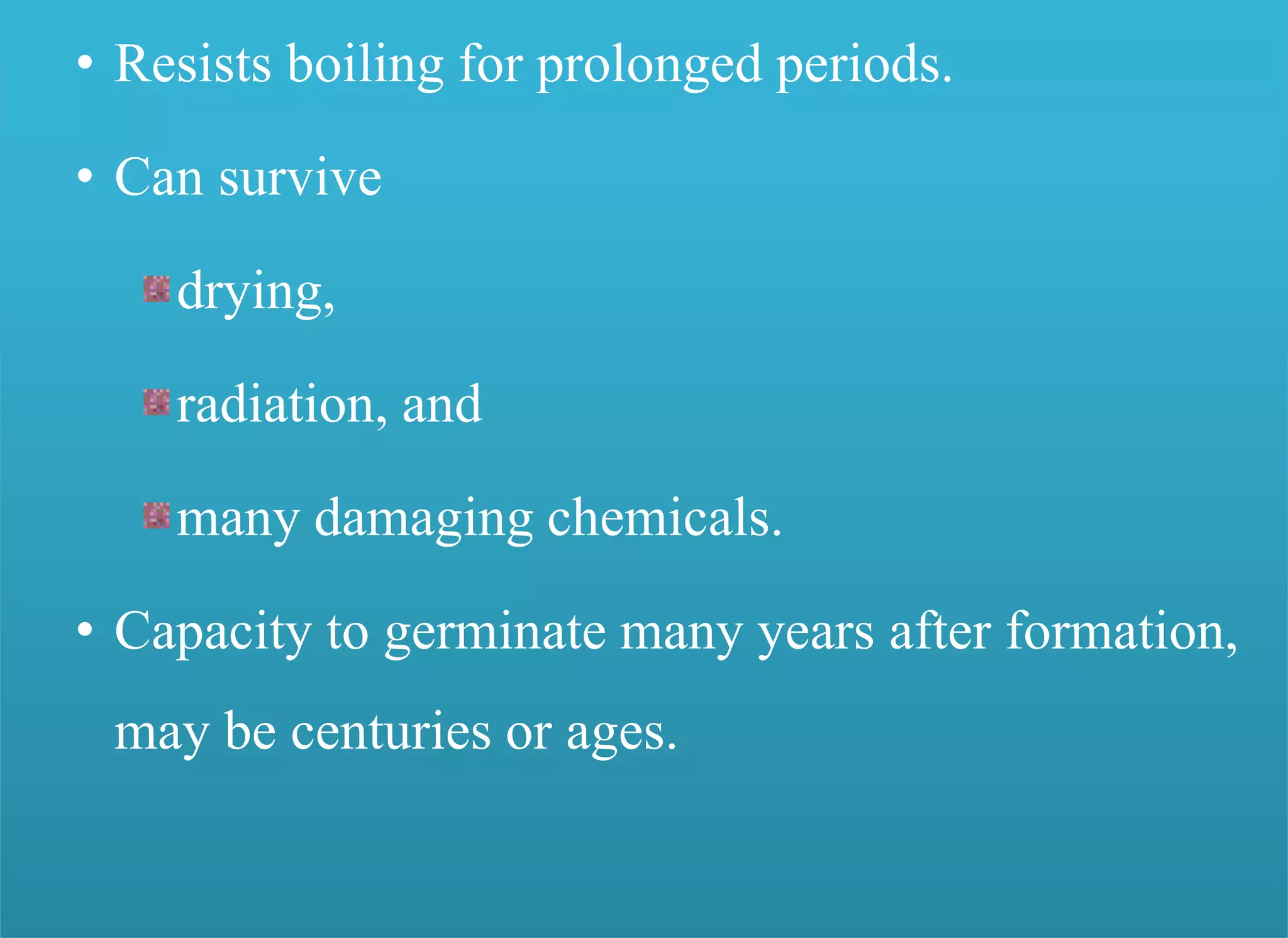
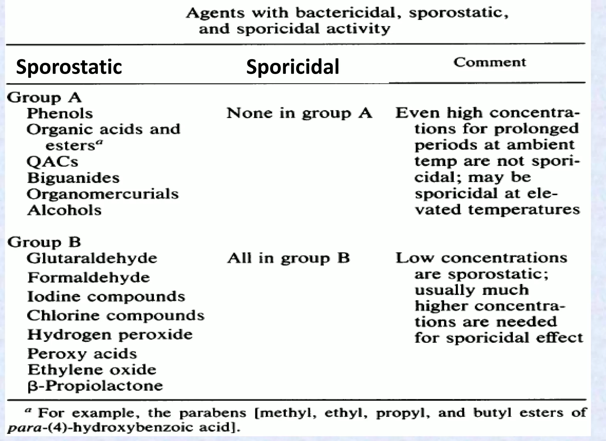
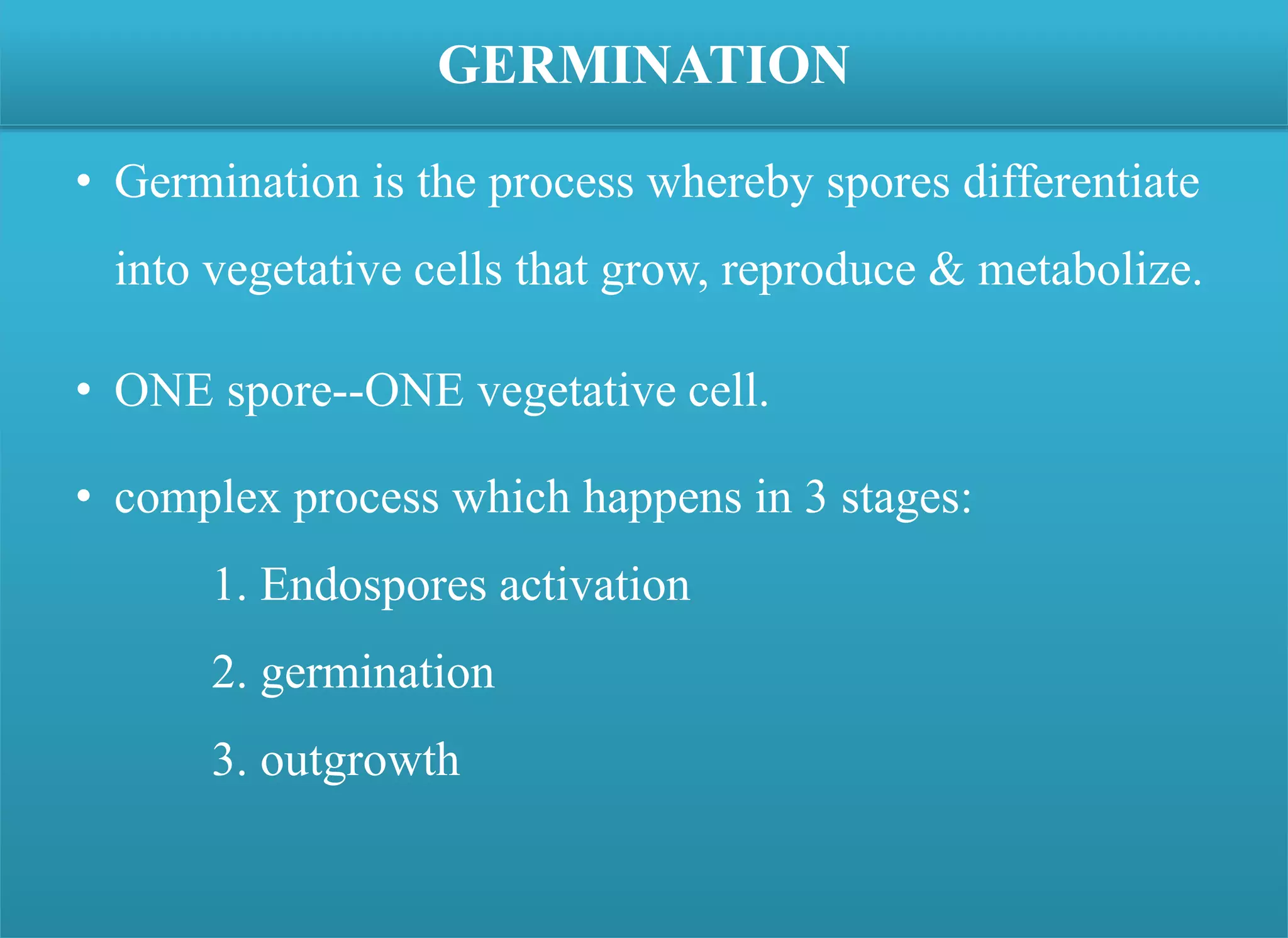
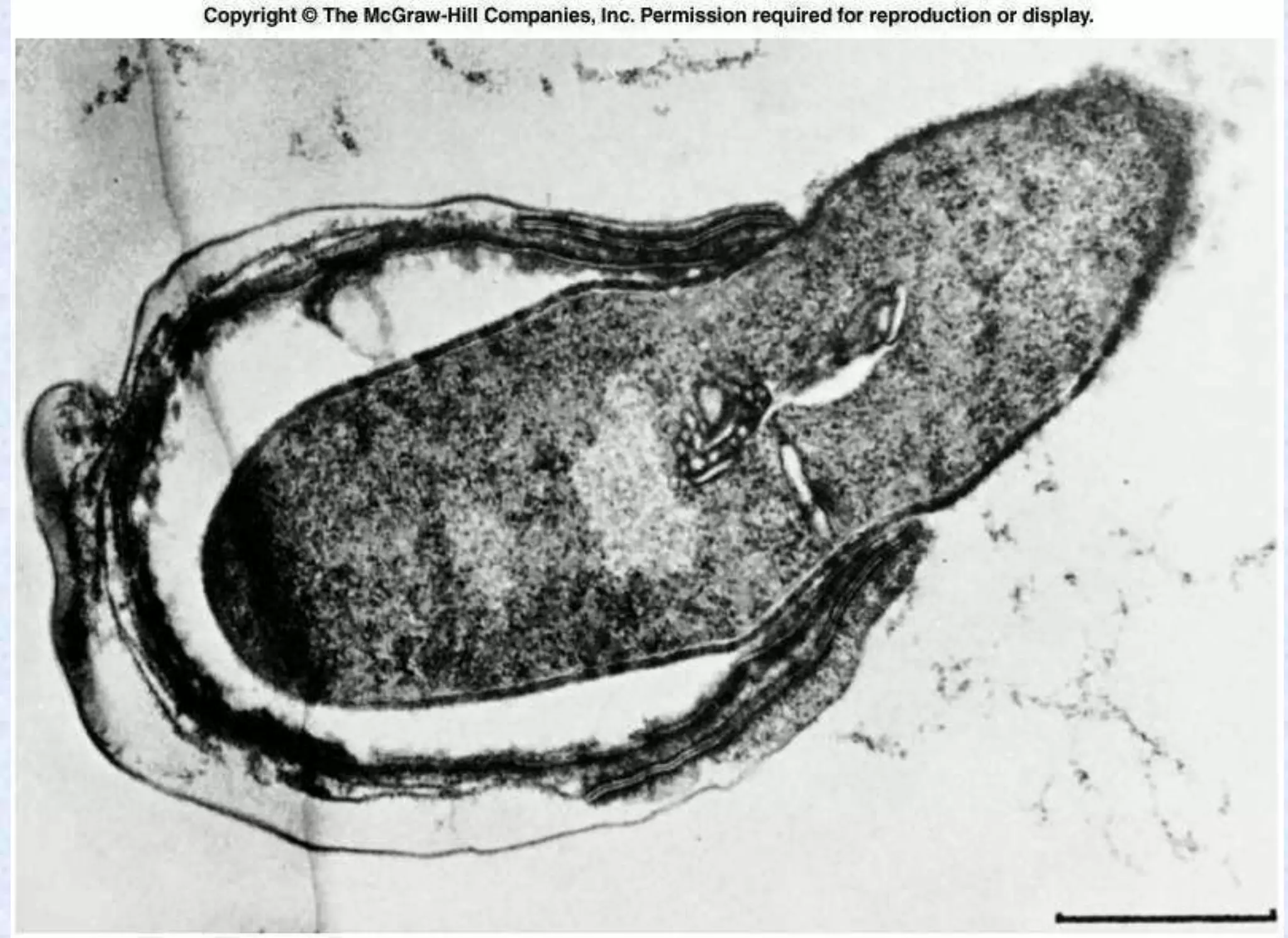
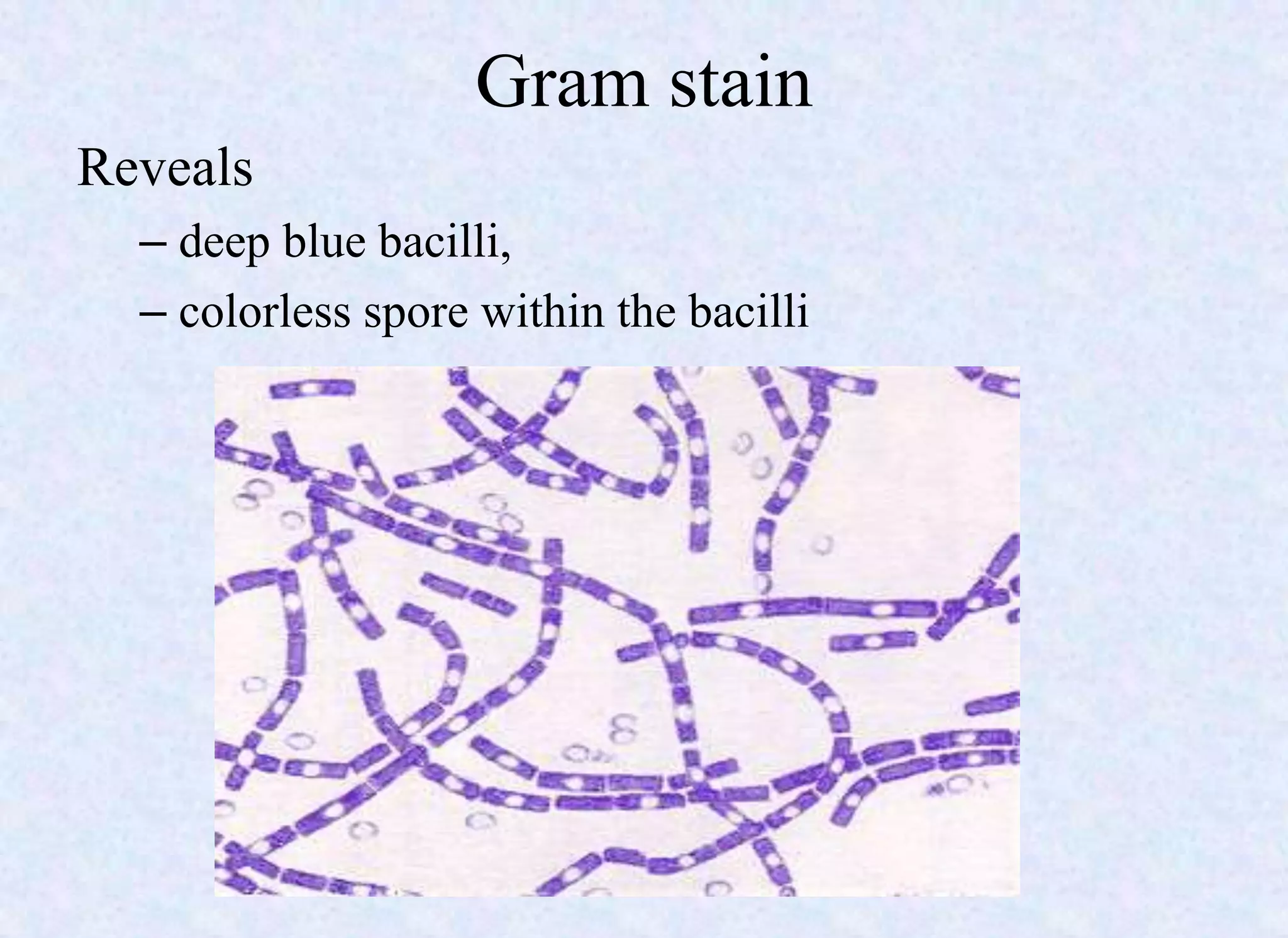
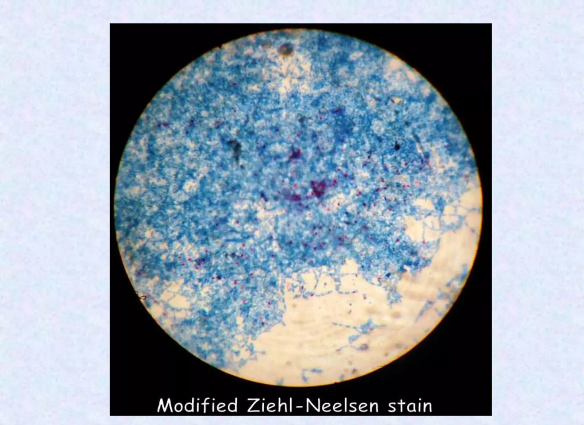
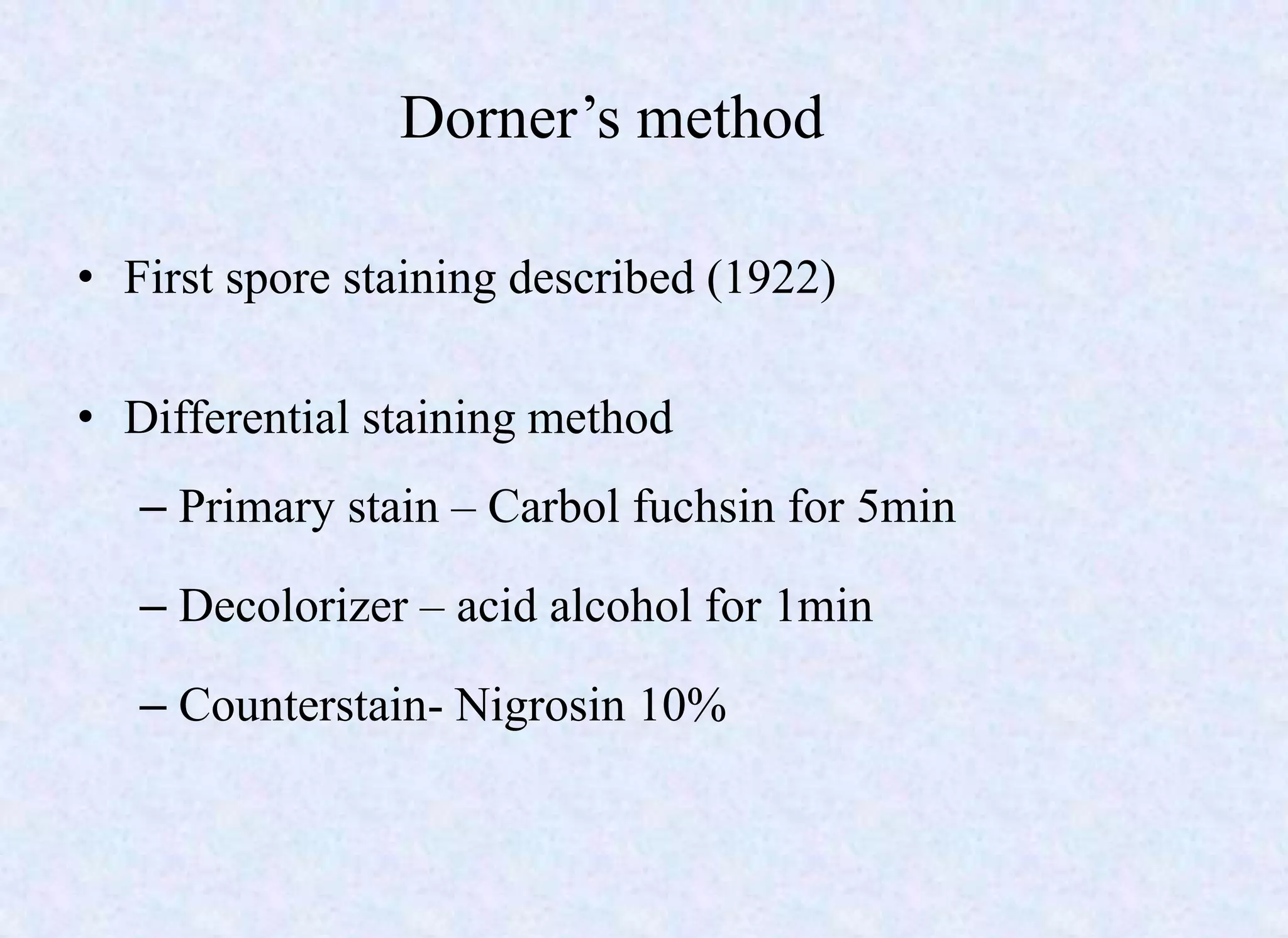
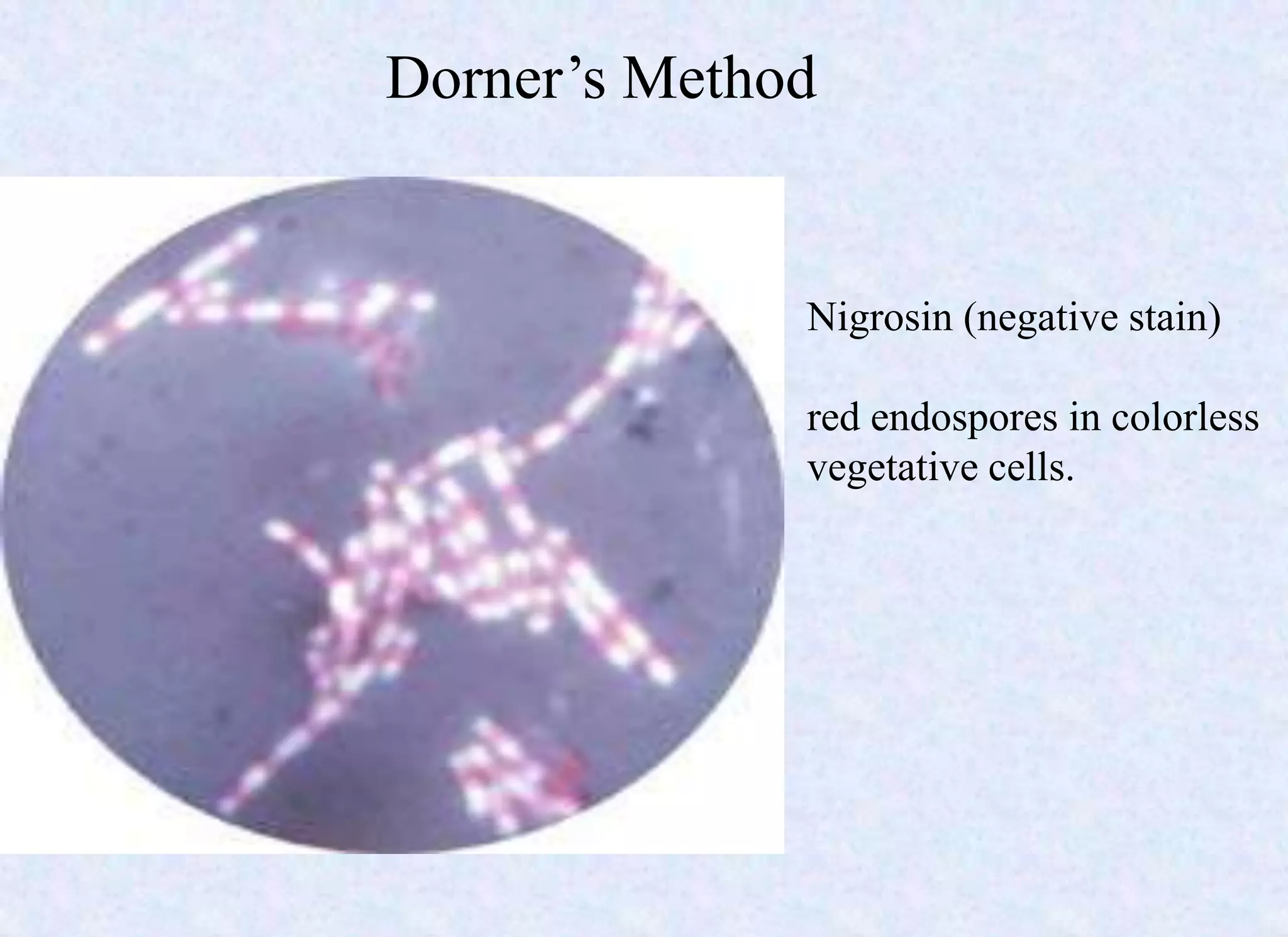
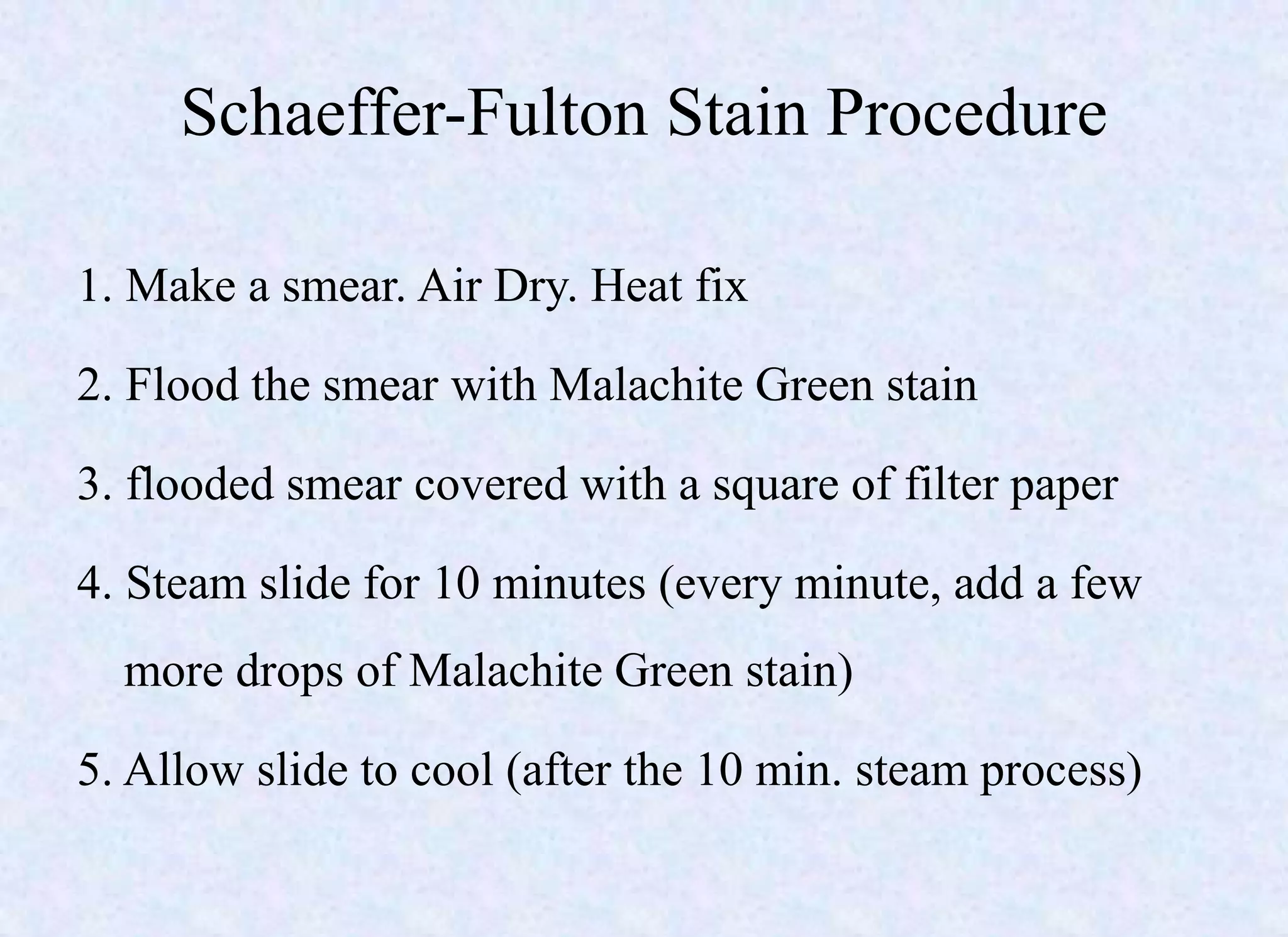
Download link:
http://www.authorstream.com/Presentation/ashrafnamaji-1900223-bacterial-spores-
ashraf/
Definition, history
List of medically important spore forming bacteria
Spore formation/ Sporulation
Structure
Classification based on shape and position
Properties
Resistance and destruction
Germination
Demonstration of spores
Uses of spores
Endospores were reported by numerous scientists
Perty 1852;
Pasteur 1869;
Koch, 1876 and
Cohn, 1872.
Bacterial spores are dormant forms of bacteria which are
thick walled, highly refractile and resistant.
• Nutritional deprivement(depletion of N2 or C source or
both) is the triggering factor
• Exposure to suboptimal temperature
Spore-forming bacteria
Bacillus:
– Bacillus anthracis,
– Bacillus subtilis,
– Bacillus cereus,
– Bacillus clausii,
– Bacillus halodenitrificans
Clostridium:
– Clostridium botulinum,
– Clostridium difficile,
– Clostridium perfringens,
– Clostridium tetani and Clostridium sordellii.
– Clostridium thermocellum (ethanol)
– Clostridium acetobutylicum (acetone)
– Clostridium diolis (propane-di-ol and to convert fatty acids to yeasts)–
Thermophilic actinomycetes:
– Thermoactinomyces vulgaris
Stages
Stage I Pre-sporulation phase
DNA assembles as an axial filament
Stage II Spore septum formation
Stage III Engulfment of the forespore
Stage IV Cortex formation
Stage V Synthesis of spore coats, DIPICOLINIC ACID, calcium
uptake
Stage VI Spore maturation (coats more thick)
Stage VII Lysis of cell and liberation of mature spore
Spore formation
STRUCTURE
5.Exosporium
made of glycoprotein and
Lipoproteins.
4. Outer Membranes
(Spore Coats)
• keratin-like protein (consists
sulphur containing aminoacids
with disulphide bonds
• impermeable
• protects spore from chemicals
3. Cortex
• thickest spore layer,
• made of modified peptidoglycan
• susceptible to lysozyme
2.Core Membrane/germ cell wall
structure similar to that in the
vegetative cell wall
1. Spore protoplast/Core
• contains DNA,RNA
• protein
• DIPICOLINIC ACID
• Divalent ions (calcium)
1. Core
2. Core Membrane
3. Cortex
4. Spore Coats
5. Exosporium
SHAPES
AND
POSITION
Shapes
– Spherical, oval or elongated in shape
– May be narrower or broader(bulge) than parent cell
spore
Bacterial cell
spore
Bacterial cell
Bulged
Ex- Clostridium sp.
Not bulged
Ex- Bacillus sp.
Position
• Terminal (located at one of the poles)
• Sub terminal or sub central(between center & one of the poles)
• Equatorial (central)
Bacteria can be classified based on the shape
and position of spores.
TERMINAL SUBTERMINAL CENTRAL
OVAL Clostridium tetani
( drumstick appearance)
Clostridium welchii,
Clostridium septicum
Clostridium sporogenes
Bacillus anthracis
SPHERICAL Clostridium tertium ------- -------
spore spore spore
spore
PROPERTIES
• Endospores
• Extremely resistant
• Non-reproducing
mechanism of survival rather than a mechanism of
reproduction(unlike fungal spores)
• Not stained readily with dyes (spore coats)
• Metabolically inactive (dormant forms)
• No DNA replication (transcription)
• mRNA not produced (translation)
• Spore protein synthesis is defective
• DNA of the endospores is protected and stabilized by
calcium DIPICOLINATE
• Specialized DNA binding proteins saturate it and protects it
from heat, radiation, chemicals.
• Has DNA repair enzymes
PROPERTY VEGETATIVE CELLS ENDOSPORES
Surface coats
murein cell wall
polymer
Thick cortex with
spore coats
Microscopic appearance Non-refractile Refractile
Calcium dipicolinic acid Absent Present in core
Cytoplasmic water activity High Very low
Metabolic activity Present Absent
RESISTANCE
Heat resistance Low High
Resistance to chemicals Low High
Radiation resistance Low High
SENSITIVE
Sensitivity to lysozyme Sensitive Resistant
Sensitivity to dyes & staining Sensitive Resistant
Bacterial spores are the toughest forms of life
known.
“ They are so resistant to destruction that some
scientists have proposed that life arrived on
earth when bacterial spores drifting through
space fell to earth.”
RESISTANCE
(cont….)
Extremely resistant due to
• low water content (removal by osmosis)
• thick wall and impermeability of spore coat
• high content of DIPICOLINIC ACID
• ability to concentrate calcium
• Resists boiling for prolonged periods.
• Can survive
drying,
radiation, and
many damaging chemicals.
• Capacity to germinate many years after formation,
may be centuries or ages.
Sporostatic Sporicidal
• Germination is the process whereby spores differentiate
into vegetative cells that grow, reproduce & metabolize.
• ONE spore--ONE vegetative cell.
• complex process which happens in 3 stages:
1. Endospores activation
2. germination
3. outgrowth
GERMINATION
• Activation- breaks the dormancy in spores,
• most spore properties retained
• Reversible
• Activators(triggering agents)
Heating at 60°C for more than 1 hour
Decreasing the pH
Treatment with mercaptoethanol
• Conformational change in spore macromolecules
• No morphological changes
2. Germination:
• Germination of activated spores occurs when exposed
to certain substances(nutrients)
– aminoacids
– sugars
– nucleosides
• Irreversible
• Removal of cortex (enzyme Ƀ-N acetyl glucosaminidase)- renders it
susceptible to chemical agents
• Loss of refractility
• In vitro germination
incubated at 37°C, 5% CO2, FBS (fetal bovine serum)
– Dulbecco’s modified Eagle’s medium(DMEM)+10% FBS
– Roswell Park Memorial Institute(RPMI)1640 medium +10%FBS,
(b) development of
phase dark forms
c) Germination completes
with full conversion to phase
dark cells
(a) mature, phase-bright
(REFRACTILE) spores
3. Outgrowth
• The core of the endospores leave the old spore coat to
develop into a fully functional vegetative cell.
• First visible change is swelling of cell--water uptake
• Molecular biosynthetic process starts
RNA synthesis– first
Protein
DNA synthesis
• Cell wall synthesis coincides with swelling
DEMONSTRATION OF SPORES
Stained preparation
•Gram stain
•ZN method (modified)
•Dorner’s method
•Malachite green stain
1. Schaeffer and fulton method
2. Ashby modification
Microscopy:
Unstained preparation
– Using Phase contrast microscope
– Morphology best observed
– large, refractile, oval or spherical bodies within mother
cells
Stained preparation
Gram stain
Reveals
– deep blue bacilli,
– colorless spore within the bacilli
Modified Ziehl-Neelsen technique
• 0.25% sulphuric acid used as decolorizer
• Spores- pink
Bacteria - blue
Dorner’s method
• First spore staining described (1922)
• Differential staining method
– Primary stain – Carbol fuchsin for 5min
– Decolorizer – acid alcohol for 1min
– Counterstain- Nigrosin 10%
Dorner’s Method
Nigrosin (negative stain)
red endospores in colorless
vegetative cells.
Malachite green staining
Schaeffer-Fulton Stain Procedure
1. Make a smear. Air Dry. Heat fix
2. Flood the smear with Malachite Green stain
3. flooded smear covered with a square of filter paper
4. Steam slide for 10 minutes (every minute, add a few
more drops of Malachite Green stain)
5. Allow slide to cool (after the 10 min. steam process)
Schaeffer-Fulton Stain Procedure (continued)
6. Drain slide and rinse for 30 seconds with DI water
(discard filter paper)
7. Put slide on steam rack
8. Flood smear with Safranin (counter stain). This stains
the vegetative cell. (Leave for 1 minute)
9. Drain the slide and rinse with DI water
10. Blot Dry
11. Use oil immersion objective to view
Schaeffer-fulton Method
Spore: green Vegetative cell: red
http://www.arches.uga.edu/~howie/MVC-052endoS.JPG
http://homepages.wmich.edu/~rossbach/bios312/LabProcedures/endospore.jpg
Uses of spores
• Sterilization control
Bacillus stearothermophilus
• Nanobiotechnology
– substrate for delivery of biomolecules
– vaccine vehicles spores
– source for understanding unknown self-assembling
molecules.
Anthrax bioterrorism
• Concentrated anthrax spores were used for bioterrorism
in the 2001, in U.S.
• Spores mailed in the form of powder
• led to 11 cases of cutaneous anthrax and 11 cases of
inhalational anthrax, with five deaths in the latter.
• Used as agent for biological warfare by germans.
References
• Topley & Wilson’s principles of bacteriology, virology and immunity, 8th
edition. Vol-1
• Mackie & McCartney Practical Medical Microbiology, 14th edition
• A. D. RUSSELL “Bacterial Spores and Chemical Sporicidal Agents”
Clinical microbiology reviews, ASM, Apr. 1990, p. 99-119.
• en.wikipedia.org/wiki/2001_anthrax_attacks
• Emerging Applications of Bacterial Spores in Nanobiotechnology, Journal of
Nanobiotechnology http://www.jnanobiotechnology.com/content/1/1/6
SPORE ashraf

SPORE ashraf

  • 1.
  • 3.
    Definition, history List ofmedically important spore forming bacteria Spore formation/ Sporulation Structure Classification based on shape and position Properties Resistance and destruction Germination Demonstration of spores Uses of spores
  • 4.
    Endospores were reportedby numerous scientists Perty 1852; Pasteur 1869; Koch, 1876 and Cohn, 1872. Bacterial spores are dormant forms of bacteria which are thick walled, highly refractile and resistant. • Nutritional deprivement(depletion of N2 or C source or both) is the triggering factor • Exposure to suboptimal temperature
  • 5.
    Spore-forming bacteria Bacillus: – Bacillusanthracis, – Bacillus subtilis, – Bacillus cereus, – Bacillus clausii, – Bacillus halodenitrificans Clostridium: – Clostridium botulinum, – Clostridium difficile, – Clostridium perfringens, – Clostridium tetani and Clostridium sordellii. – Clostridium thermocellum (ethanol) – Clostridium acetobutylicum (acetone) – Clostridium diolis (propane-di-ol and to convert fatty acids to yeasts)– Thermophilic actinomycetes: – Thermoactinomyces vulgaris
  • 6.
    Stages Stage I Pre-sporulationphase DNA assembles as an axial filament Stage II Spore septum formation Stage III Engulfment of the forespore Stage IV Cortex formation Stage V Synthesis of spore coats, DIPICOLINIC ACID, calcium uptake Stage VI Spore maturation (coats more thick) Stage VII Lysis of cell and liberation of mature spore Spore formation
  • 8.
    STRUCTURE 5.Exosporium made of glycoproteinand Lipoproteins. 4. Outer Membranes (Spore Coats) • keratin-like protein (consists sulphur containing aminoacids with disulphide bonds • impermeable • protects spore from chemicals 3. Cortex • thickest spore layer, • made of modified peptidoglycan • susceptible to lysozyme 2.Core Membrane/germ cell wall structure similar to that in the vegetative cell wall 1. Spore protoplast/Core • contains DNA,RNA • protein • DIPICOLINIC ACID • Divalent ions (calcium) 1. Core 2. Core Membrane 3. Cortex 4. Spore Coats 5. Exosporium
  • 10.
  • 11.
    Shapes – Spherical, ovalor elongated in shape – May be narrower or broader(bulge) than parent cell spore Bacterial cell spore Bacterial cell Bulged Ex- Clostridium sp. Not bulged Ex- Bacillus sp.
  • 12.
    Position • Terminal (locatedat one of the poles) • Sub terminal or sub central(between center & one of the poles) • Equatorial (central)
  • 13.
    Bacteria can beclassified based on the shape and position of spores. TERMINAL SUBTERMINAL CENTRAL OVAL Clostridium tetani ( drumstick appearance) Clostridium welchii, Clostridium septicum Clostridium sporogenes Bacillus anthracis SPHERICAL Clostridium tertium ------- ------- spore spore spore spore
  • 14.
    PROPERTIES • Endospores • Extremelyresistant • Non-reproducing mechanism of survival rather than a mechanism of reproduction(unlike fungal spores) • Not stained readily with dyes (spore coats)
  • 15.
    • Metabolically inactive(dormant forms) • No DNA replication (transcription) • mRNA not produced (translation) • Spore protein synthesis is defective • DNA of the endospores is protected and stabilized by calcium DIPICOLINATE • Specialized DNA binding proteins saturate it and protects it from heat, radiation, chemicals. • Has DNA repair enzymes
  • 16.
    PROPERTY VEGETATIVE CELLSENDOSPORES Surface coats murein cell wall polymer Thick cortex with spore coats Microscopic appearance Non-refractile Refractile Calcium dipicolinic acid Absent Present in core Cytoplasmic water activity High Very low Metabolic activity Present Absent RESISTANCE Heat resistance Low High Resistance to chemicals Low High Radiation resistance Low High SENSITIVE Sensitivity to lysozyme Sensitive Resistant Sensitivity to dyes & staining Sensitive Resistant
  • 17.
    Bacterial spores arethe toughest forms of life known. “ They are so resistant to destruction that some scientists have proposed that life arrived on earth when bacterial spores drifting through space fell to earth.” RESISTANCE
  • 18.
    (cont….) Extremely resistant dueto • low water content (removal by osmosis) • thick wall and impermeability of spore coat • high content of DIPICOLINIC ACID • ability to concentrate calcium
  • 19.
    • Resists boilingfor prolonged periods. • Can survive drying, radiation, and many damaging chemicals. • Capacity to germinate many years after formation, may be centuries or ages.
  • 20.
  • 21.
    • Germination isthe process whereby spores differentiate into vegetative cells that grow, reproduce & metabolize. • ONE spore--ONE vegetative cell. • complex process which happens in 3 stages: 1. Endospores activation 2. germination 3. outgrowth GERMINATION
  • 22.
    • Activation- breaksthe dormancy in spores, • most spore properties retained • Reversible • Activators(triggering agents) Heating at 60°C for more than 1 hour Decreasing the pH Treatment with mercaptoethanol • Conformational change in spore macromolecules • No morphological changes
  • 23.
    2. Germination: • Germinationof activated spores occurs when exposed to certain substances(nutrients) – aminoacids – sugars – nucleosides • Irreversible • Removal of cortex (enzyme Ƀ-N acetyl glucosaminidase)- renders it susceptible to chemical agents • Loss of refractility • In vitro germination incubated at 37°C, 5% CO2, FBS (fetal bovine serum) – Dulbecco’s modified Eagle’s medium(DMEM)+10% FBS – Roswell Park Memorial Institute(RPMI)1640 medium +10%FBS,
  • 24.
    (b) development of phasedark forms c) Germination completes with full conversion to phase dark cells (a) mature, phase-bright (REFRACTILE) spores
  • 25.
    3. Outgrowth • Thecore of the endospores leave the old spore coat to develop into a fully functional vegetative cell. • First visible change is swelling of cell--water uptake • Molecular biosynthetic process starts RNA synthesis– first Protein DNA synthesis • Cell wall synthesis coincides with swelling
  • 27.
  • 28.
    Stained preparation •Gram stain •ZNmethod (modified) •Dorner’s method •Malachite green stain 1. Schaeffer and fulton method 2. Ashby modification Microscopy: Unstained preparation – Using Phase contrast microscope – Morphology best observed – large, refractile, oval or spherical bodies within mother cells Stained preparation
  • 29.
    Gram stain Reveals – deepblue bacilli, – colorless spore within the bacilli
  • 30.
    Modified Ziehl-Neelsen technique •0.25% sulphuric acid used as decolorizer • Spores- pink Bacteria - blue
  • 32.
    Dorner’s method • Firstspore staining described (1922) • Differential staining method – Primary stain – Carbol fuchsin for 5min – Decolorizer – acid alcohol for 1min – Counterstain- Nigrosin 10%
  • 33.
    Dorner’s Method Nigrosin (negativestain) red endospores in colorless vegetative cells.
  • 34.
  • 35.
    Schaeffer-Fulton Stain Procedure 1.Make a smear. Air Dry. Heat fix 2. Flood the smear with Malachite Green stain 3. flooded smear covered with a square of filter paper 4. Steam slide for 10 minutes (every minute, add a few more drops of Malachite Green stain) 5. Allow slide to cool (after the 10 min. steam process)
  • 36.
    Schaeffer-Fulton Stain Procedure(continued) 6. Drain slide and rinse for 30 seconds with DI water (discard filter paper) 7. Put slide on steam rack 8. Flood smear with Safranin (counter stain). This stains the vegetative cell. (Leave for 1 minute) 9. Drain the slide and rinse with DI water 10. Blot Dry 11. Use oil immersion objective to view
  • 37.
    Schaeffer-fulton Method Spore: greenVegetative cell: red http://www.arches.uga.edu/~howie/MVC-052endoS.JPG http://homepages.wmich.edu/~rossbach/bios312/LabProcedures/endospore.jpg
  • 38.
    Uses of spores •Sterilization control Bacillus stearothermophilus • Nanobiotechnology – substrate for delivery of biomolecules – vaccine vehicles spores – source for understanding unknown self-assembling molecules.
  • 39.
    Anthrax bioterrorism • Concentratedanthrax spores were used for bioterrorism in the 2001, in U.S. • Spores mailed in the form of powder • led to 11 cases of cutaneous anthrax and 11 cases of inhalational anthrax, with five deaths in the latter. • Used as agent for biological warfare by germans.
  • 41.
    References • Topley &Wilson’s principles of bacteriology, virology and immunity, 8th edition. Vol-1 • Mackie & McCartney Practical Medical Microbiology, 14th edition • A. D. RUSSELL “Bacterial Spores and Chemical Sporicidal Agents” Clinical microbiology reviews, ASM, Apr. 1990, p. 99-119. • en.wikipedia.org/wiki/2001_anthrax_attacks • Emerging Applications of Bacterial Spores in Nanobiotechnology, Journal of Nanobiotechnology http://www.jnanobiotechnology.com/content/1/1/6

Editor's Notes

  • #5 primarily described them as refractile bodies
  • #6 - Found in soils. Bacillus: - aerobic, Gram +ve bacilli - express extra-cellular degradative enzymes - used as a means of biological control of insects. - anaerobic, Gram +ve bacilli - grow in canned foods producing toxin Sporolactobacillus Sporolactobacillus is a group of anaerobic, rod-shaped, spore forming bacteria that include Sporolactobacillus dextrus, Sporolactobacillus inulinus, Sporolactobacillus laevis, Sporolactobacillus terrae and Sporolactobacillus vineae. Sporolactobacillus are also known as lactic-acid bacteria for they are capable of producing the acid from fructose, sucrose, raffinose, mannose, inulin and sorbitol. Sporolactobacillus are found in the soil and often in chicken feed. According to "Fundamentals of Food Microbiology," the spores formed by Sporolactobacillus are less resistant to heat than those formed by the Bacillus genus
  • #22 When spores sense that the environment is suitable for growth the spore is triggered to germinate. Germination is the process whereby spores differentiate into vegetative cells that grow, reproduce & metabolize. ; that is, it is easily killed by heat UV light drying
  • #23 Germination rate- function of ageing
  • #24 When incubated at 37°C and under 5% CO2 in the presence of FBS(fetal bovine serum)
  • #26 shed their coats to allow the young vegetative cells to emerge, elongate, and divide. Of the macromolecular biosynthetic processes occurring after germination, RNA synthesis is the first, followed closely in Bacillus spp. by the onset of protein synthesis, with DNA synthesis occurring some time later. During outgrowth, all types of RNA are synthesized. Cell wall synthesis commences after RNA and protein but before DNA and coincides with swelling of the germinated spore.