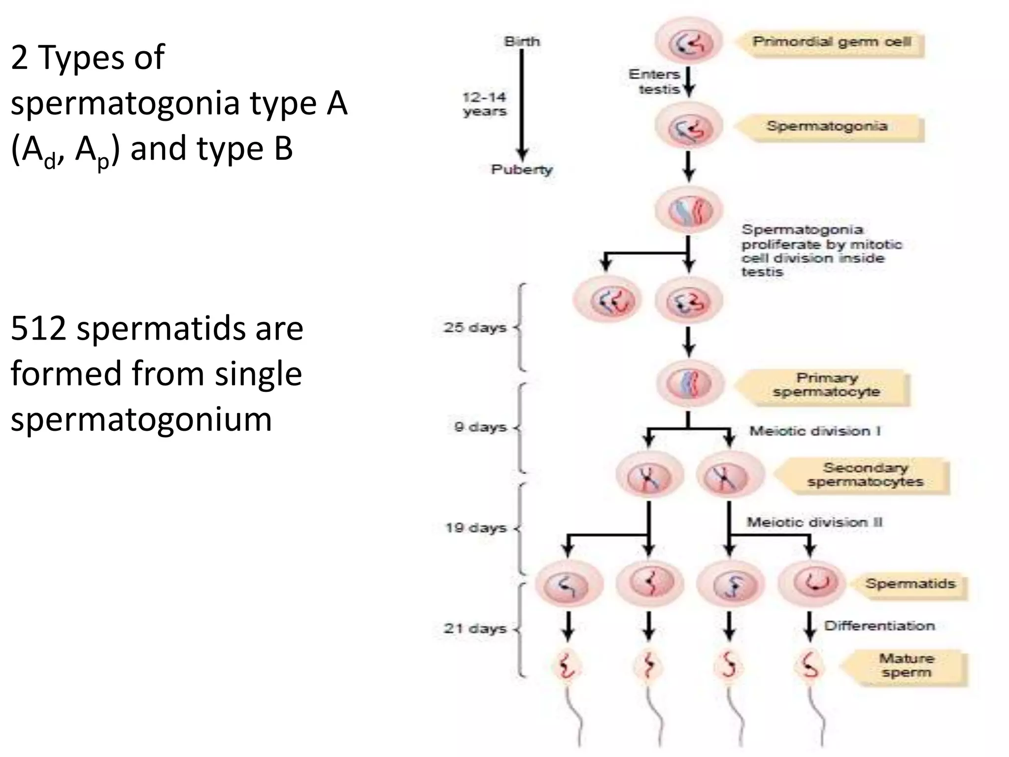
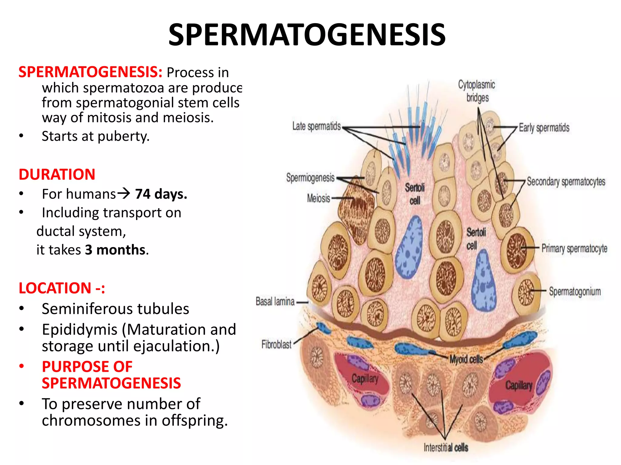
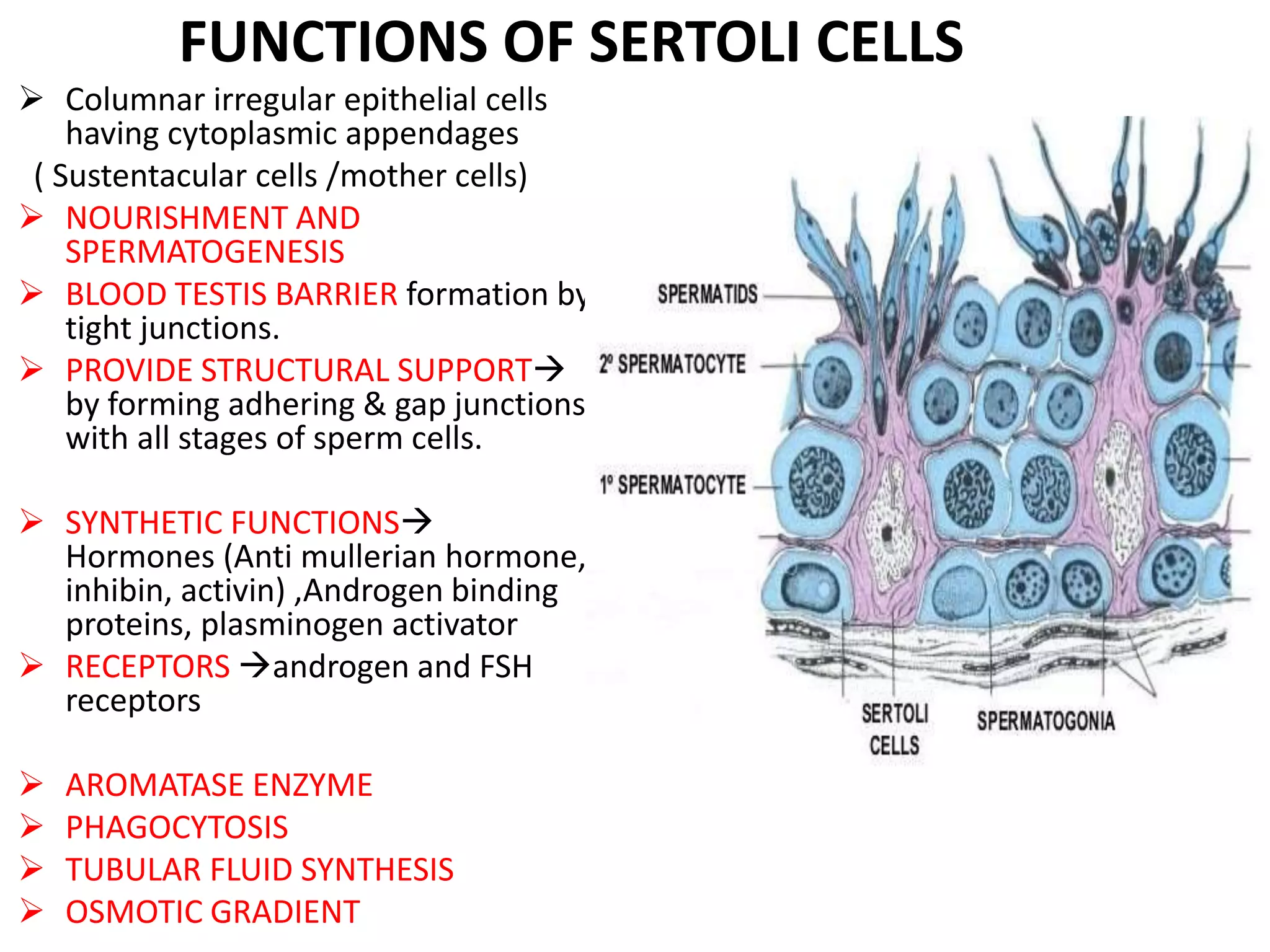
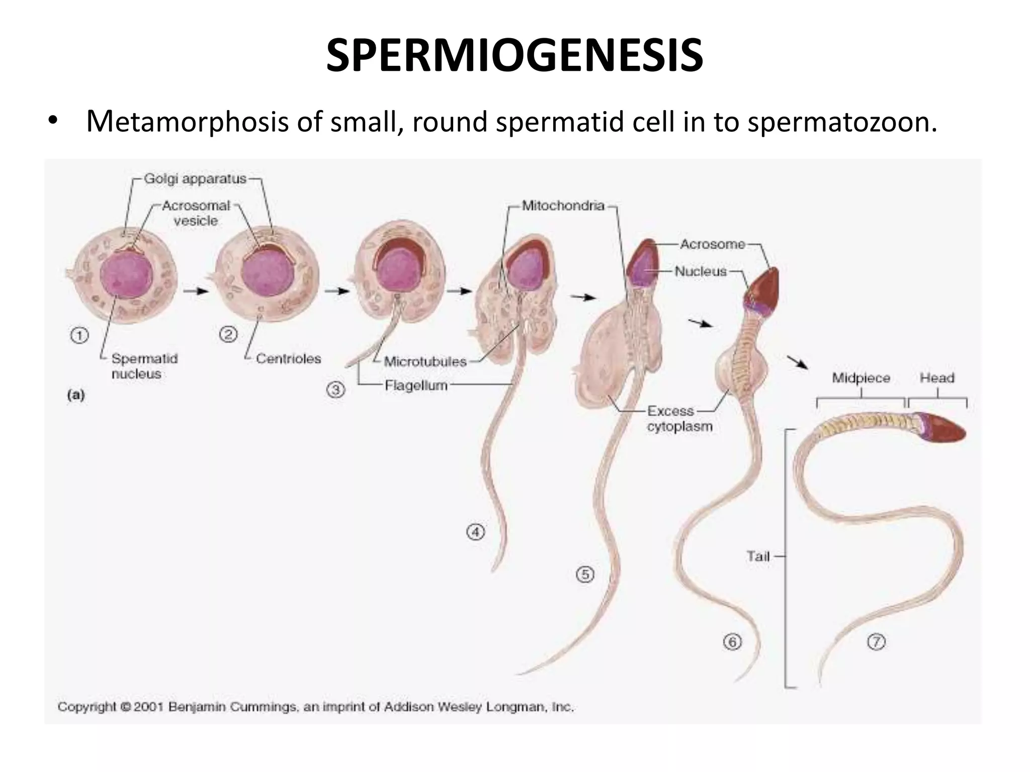
SPERMATOGENESIS
• Moderator -: Dr. Raj Kapoor
• Presented By-: Dr. Omprakash
• Physio-anatomy of male reproductive system
• Introduction
• Stages of spermatogenesis
• Further development of sperm
• Role of sertoli cells
• Sperm structure & count
• Semen composition
• Hormonal control & other affecting factors
• Role of testosterone
• Applied
PHYSIO-ANATOMY OF MALE REPRODUCTIVE TRACT
MALE GONAD / TESTIS
• 450-500
seminiferous
tubules per testis
each >0.5 m
• Total tubular
length in testis
approx. 400m
• Epididymis 6m
SPERMATOGENESIS
SPERMATOGENESIS: Process in
which spermatozoa are produced
from spermatogonial stem cells by
way of mitosis and meiosis.
• Starts at puberty.
DURATION
• For humans 74 days.
• Including transport on
ductal system,
it takes 3 months.
LOCATION -:
• Seminiferous tubules
• Epididymis (Maturation and
storage until ejaculation.)
• PURPOSE OF
SPERMATOGENESIS
• To preserve number of
chromosomes in offspring.
FUNCTIONS OF SERTOLI CELLS
 Columnar irregular epithelial cells
having cytoplasmic appendages
( Sustentacular cells /mother cells)
 NOURISHMENT AND
SPERMATOGENESIS
 BLOOD TESTIS BARRIER formation by
tight junctions.
 PROVIDE STRUCTURAL SUPPORT
by forming adhering & gap junctions
with all stages of sperm cells.
 SYNTHETIC FUNCTIONS
Hormones (Anti mullerian hormone,
inhibin, activin) ,Androgen binding
proteins, plasminogen activator
 RECEPTORS androgen and FSH
receptors
 AROMATASE ENZYME
 PHAGOCYTOSIS
 TUBULAR FLUID SYNTHESIS
 OSMOTIC GRADIENT
STAGES OF SPERMATOGENESIS
• 2 Types of spermatogonia type A (Ad, Ap) and type B
• 512 spermatids are formed from single spermatogonium.
• During division, cells bound to each other by cytoplasmic bridges for
synchronised development (incomplete cytogenesis)
1-spermatocytogenesis
2- spermatidogenesis
3-spermiogenesis
4- spermiation
5-maturation
SPERMATOGENESIS
SPERMIOGENESIS
• Metamorphosis of small, round spermatid cell in to spermatozoon.
SPERMATOZOON
• Haploid motile gamete cell Head, Neck, Body and tail.
• Axoneme microtubular structures in tail that propels sperm. (
9+2 structure)
• Acrosome membrane bound sac derived from golgi
apparatus. Also have hydrolytic enzymes for fertilization.
• Spiral arrangement of mitochondria Nebenkern
• Speed 1-4 mm/min. Maximum life span of ejaculated sperm 1-2
days
SEMEN
• Semen -: white, opalescent fluid , ejaculated at time of orgasm
• Average amount 3.5 ml (1 ml =120 million)
• >50% motile and >60% normal shape
• Specific gravity 1.028 pH7.35-7.50
• Components -:
• Vas deferens 10% fluid & sperm
• Seminal vesicle fluid 60% fructose, prostaglandins ,ascorbic acid,
phosphorylcholine, flavins, seminogelins
• Prostatic fluid 30% spermine, citric acid, cholesterol, phospholipids,
fibrogenase, fibrinolysin, zinc,acid phophatase
• Buffers phosphates & bicarbonates
• Hyaluronidase & PSA (Prostate specific antigen)
FURTHER DEVELOPMENT OF SPERMATOZOA
• Achieve progressive motility in epididymis.
• Activation of CatSper protein ( Ca++ ion channel) that permits
cAMP generalised Ca++ influx.
• Expression of olfactory receptors ( chemotaxis)
• Capacitation (only facilitatory)
• Acrosomal reaction
CAPACITATION
CAPACITATION -:
when sperm comes to contact of female genital tract, multiple changes
occurs that activate sperm for final process of fertilization
• Takes 1-10 hrs
CHANGES -:
• Washing away of inhibitory factors from uterine &tubal secretions
• Losing of cholesterol from head of sperm
• Increased permeability of Ca++ that
(a) increase whiplash activity &
(b) help in release of enzymes from head membrane
ACROSOMAL REACTION
• Hyaluronidase -: depolymerise hyaluronic acid polymers in the intercellular
cement that hold ovarian granulosa cells
• Proteolytic enzymes -: digest proteins in structural element of tissue cells
that still adhere to the ovum.
• Reason for entry of only one sperm in to the oocyte ??
HORMONAL CONTROL OF SPERMATOGENESIS
ABP – ANDROGEN BINDING
PROTEIN
SHBP – SEX HORMONE BINDING
PROTEIN
ROLE OF HORMONES IN SPERMATOGENESIS
TESTOSTERONE
• Growth & division of germ cells
LUTEINIZING HORMONE
• Stimulate leydig cells to secrete testosterone
FOLLICLE STIMULATING HORMONE
• Stimulate sertoli cells
ESTROGENS
• probably essential for spermiogenesis
GROWTH HORMONE
• Controls metabolic functions of testes
• Promote early division of spermatogonia
Pituitary dwarfs (spermatogenesis deficient or absent )
OTHER FACTORS AFFECTING SPERMATOGENESIS
Temperature:
• Seminiferous epithelium is sensitive to elevated temperature as high as
normal body temperature. Consequently, in scrotum. The optimal
temperature is maintained at 2 °C below body temperature.[ Achieved by
regulation of blood flow ]
• Scrotal reflex: Positioning towards and away from the heat of the body by
cremasteric muscle and Dartos smooth muscle in scrotum.
Others-:
• Dietary deficiencies (such as vitamins B, E and A), infectious diseases.
• Anabolic steroids , metals (cadmium and lead), x-ray exposure, dioxin,
alcohol, pesticides .
• DNA damage caused by oxidative stress.
BIOSYNTHESIS OF TESTOSTERONE
• ANDROGENS( Testosterone,Dihydrotestosterone & Androstenedione)
• C19 steroid with OH group in the 17 position
• LH hormone ↑ formation of cAMP  cholesterol synthesis
TRANSPORT & METABOLISM OF ANDROGENS
• Circulating testosterone -: sex hormone binding (SHBG)
globulin 60%, Albumin 30%, free state 2%.
• In target tissues, it converts to dihydrotestosterone, must
needed for certain characters.
• Converts into estrogens (liver & adipose tissues)
• Excreted in form of urinary 17-ketosteroid & conjugated
steroid
MECHANISM OF ACTION OF
TESTOSTERONE
• Congenital 5 alpha reductase deficiency male pseudohermaphroditism
• Penis @ 12 syndrome
TESTOSTERONE SECRETION
During fetal life, testis stimulated by chorionic gonadotropins from
placenta to produce testosterone up to 10 weeks or more after birth.
FUNCTIONS OF TESTOSTERONE
In fetal period -:
• Genital ridges secretes testosterone [& estrogens]
• fetal testes starts secretion @ 7 week
• Causes genital differentiation (wolffian duct)
• Differentiation of external genitalia
• Suppress formation of female genital organs (mullerian duct)
What happens if ?
• Injection of male sex hormones in large quantities in pregnant
animals having female fetus ?
• Removal of male testis in early male fetus ?
Effect of testosterone on descent of testis:
• Usually descend in last 2-3 months of gestation in to the scrotum
EFFECTS OF TESTOSTERONE
BASIS OF STERILITY IN STEROID ABUSE ?
MALE INFERTILITY
• Androgen dysfunction with normal count
1-hypothalamic pituitary defects
2-leydig cell defects
• Isolated dysfunction of sperm cell production with normal androgen
production.
Trauma, infection, abnormal sperms.
• Combined androgen &sperm cell production defects
1-development defects-: Klinefelter's syndrome, undescended testis,
2-acquired defects-: orchitis
• Failure of deposition of sperm in female genital tract
1-vas deferens obstruction
2-failure of erection, ejection
SPERM COUNT ABNORMALITIES
Normal count 120 million/ml [< 20 million/ml  sterile]
Aspermia: Absence of semen
Azoospermia: absence of sperm
Hypospermia: low semen volume
Hyperspermia: high semen volume
Oligozoospermia: Very low sperm count
Asthenozoospermia: poor sperm motility
Teratozoospermia: sperm carry more morphological defects than
usual
Necrozoospermia: all sperm in the ejaculate are dead
Leucospermia: a high level of white blood cells in semens
(>1 million/cc)
EFFECT OF SPERM MORPHOLOGY & MOTILITY
. ON FERTILITY
>50% should be motile
>60% should be of normal shape
CRYPTORCHIDISM
• Incomplete descent of testes:
• Normal descent 3 weeks to 1 month before birth.
• Spontaneous descent is the rule.
10% newborn males
2% at one year
Falls to 0.3% at puberty
• High temperature in undescended testis causes sterility
• Leydig cells are not affected so secondary characters develop
normally.
• Early treatment ( gonadotropic hormone) & surgical correction is
recommended because of high incidence of malignancies.
MALE HYPOGONADISM
• When testes of male fetus are non-functional during fetal life, none of the
male sexual characteristics develop in the fetus. Instead, female organs are
formed.
• But in the presence of testosterone, formation of female sexual organs is
suppressed, and instead, male organs are induced.
• When a boy loses his testes before puberty,
• A state of eunuchism having infantile sex organs and other infantile sexual
characteristic throughout life.
• Height  Slightly greater
• Bones thin
• Muscles weaker than those of a normal man.
• voice ---childlike.
• No loss of hair on the head.
• Loss of Normal adult masculine hair distribution.
WHEN A MAN IS CASTRATED AFTER PUBERTY
• Some secondary sexual characteristics revert to those of child and others remain
of adult masculine character.
• Sexual organs Regress slightly in size but not to a child like state
• Voice Regresses from the bass quality only slightly.
• Loss of masculine hair production.
• Loss of the thick masculine bones,
• Loss of the musculature of the virile male.
• Sexual desires  Are decreased but not lost.
• Erection  Can still occur as before, although with less ease, rarely ejaculation
can take place because the semen forming organs degenerate
• Loss of the testosterone-driven psychic desire.
ADIPOSOGENITAL SYNDROME, FRÖHLICH’S
SYNDROME, OR HYPOTHALAMIC EUNUCHISM
• Some instances of hypogonadism are caused by a genetic
inability of the hypothalamus to secrete normal amounts of
GnRH.
Often associated with a simultaneous abnormality of the feeding
center of the hypothalamus, causing the person to greatly
overeat.
• Consequently, obesity occurs along with eunuchism. A patient
with this condition is called Adiposogenital syndrome, Fröhlich’s
syndrome, or Hypothalamic eunuchism.
TESTICULAR TUMORS
INTERSTITIAL LEYDIG CELL TUMORS rare
• Produce testosterone 100 times. (Precocious pseudopuberty)
• In young children, cause rapid growth of the musculature and bones, early
uniting of epiphyses, adult height is less.
• Cause excessive development of male sexual organs, all skeletal muscles, and
other male sexual characteristics.
• In the adult male, difficult to diagnose because masculine features are
already present.
TUMORS OF THE GERMINAL EPITHELIUM common
• Causes teratoma because germinal cells are capable of differentiating into
almost any type of cell.
• May cause gynaecomastia ,if a significant quantity of placental tissue
develops in tumor.
Thank you
2 Types of
spermatogonia type A
(Ad, Ap) and type B
512 spermatids are
formed from single
spermatogonium
Spermatogenesis

Spermatogenesis

  • 1.
    SPERMATOGENESIS • Moderator -:Dr. Raj Kapoor • Presented By-: Dr. Omprakash
  • 2.
    • Physio-anatomy ofmale reproductive system • Introduction • Stages of spermatogenesis • Further development of sperm • Role of sertoli cells • Sperm structure & count • Semen composition • Hormonal control & other affecting factors • Role of testosterone • Applied
  • 3.
    PHYSIO-ANATOMY OF MALEREPRODUCTIVE TRACT
  • 4.
    MALE GONAD /TESTIS • 450-500 seminiferous tubules per testis each >0.5 m • Total tubular length in testis approx. 400m • Epididymis 6m
  • 5.
    SPERMATOGENESIS SPERMATOGENESIS: Process in whichspermatozoa are produced from spermatogonial stem cells by way of mitosis and meiosis. • Starts at puberty. DURATION • For humans 74 days. • Including transport on ductal system, it takes 3 months. LOCATION -: • Seminiferous tubules • Epididymis (Maturation and storage until ejaculation.) • PURPOSE OF SPERMATOGENESIS • To preserve number of chromosomes in offspring.
  • 6.
    FUNCTIONS OF SERTOLICELLS  Columnar irregular epithelial cells having cytoplasmic appendages ( Sustentacular cells /mother cells)  NOURISHMENT AND SPERMATOGENESIS  BLOOD TESTIS BARRIER formation by tight junctions.  PROVIDE STRUCTURAL SUPPORT by forming adhering & gap junctions with all stages of sperm cells.  SYNTHETIC FUNCTIONS Hormones (Anti mullerian hormone, inhibin, activin) ,Androgen binding proteins, plasminogen activator  RECEPTORS androgen and FSH receptors  AROMATASE ENZYME  PHAGOCYTOSIS  TUBULAR FLUID SYNTHESIS  OSMOTIC GRADIENT
  • 7.
    STAGES OF SPERMATOGENESIS •2 Types of spermatogonia type A (Ad, Ap) and type B • 512 spermatids are formed from single spermatogonium. • During division, cells bound to each other by cytoplasmic bridges for synchronised development (incomplete cytogenesis) 1-spermatocytogenesis 2- spermatidogenesis 3-spermiogenesis 4- spermiation 5-maturation
  • 8.
  • 9.
    SPERMIOGENESIS • Metamorphosis ofsmall, round spermatid cell in to spermatozoon.
  • 10.
    SPERMATOZOON • Haploid motilegamete cell Head, Neck, Body and tail. • Axoneme microtubular structures in tail that propels sperm. ( 9+2 structure) • Acrosome membrane bound sac derived from golgi apparatus. Also have hydrolytic enzymes for fertilization. • Spiral arrangement of mitochondria Nebenkern • Speed 1-4 mm/min. Maximum life span of ejaculated sperm 1-2 days
  • 11.
    SEMEN • Semen -:white, opalescent fluid , ejaculated at time of orgasm • Average amount 3.5 ml (1 ml =120 million) • >50% motile and >60% normal shape • Specific gravity 1.028 pH7.35-7.50 • Components -: • Vas deferens 10% fluid & sperm • Seminal vesicle fluid 60% fructose, prostaglandins ,ascorbic acid, phosphorylcholine, flavins, seminogelins • Prostatic fluid 30% spermine, citric acid, cholesterol, phospholipids, fibrogenase, fibrinolysin, zinc,acid phophatase • Buffers phosphates & bicarbonates • Hyaluronidase & PSA (Prostate specific antigen)
  • 12.
    FURTHER DEVELOPMENT OFSPERMATOZOA • Achieve progressive motility in epididymis. • Activation of CatSper protein ( Ca++ ion channel) that permits cAMP generalised Ca++ influx. • Expression of olfactory receptors ( chemotaxis) • Capacitation (only facilitatory) • Acrosomal reaction
  • 13.
    CAPACITATION CAPACITATION -: when spermcomes to contact of female genital tract, multiple changes occurs that activate sperm for final process of fertilization • Takes 1-10 hrs CHANGES -: • Washing away of inhibitory factors from uterine &tubal secretions • Losing of cholesterol from head of sperm • Increased permeability of Ca++ that (a) increase whiplash activity & (b) help in release of enzymes from head membrane
  • 14.
    ACROSOMAL REACTION • Hyaluronidase-: depolymerise hyaluronic acid polymers in the intercellular cement that hold ovarian granulosa cells • Proteolytic enzymes -: digest proteins in structural element of tissue cells that still adhere to the ovum. • Reason for entry of only one sperm in to the oocyte ??
  • 15.
    HORMONAL CONTROL OFSPERMATOGENESIS ABP – ANDROGEN BINDING PROTEIN SHBP – SEX HORMONE BINDING PROTEIN
  • 16.
    ROLE OF HORMONESIN SPERMATOGENESIS TESTOSTERONE • Growth & division of germ cells LUTEINIZING HORMONE • Stimulate leydig cells to secrete testosterone FOLLICLE STIMULATING HORMONE • Stimulate sertoli cells ESTROGENS • probably essential for spermiogenesis GROWTH HORMONE • Controls metabolic functions of testes • Promote early division of spermatogonia Pituitary dwarfs (spermatogenesis deficient or absent )
  • 17.
    OTHER FACTORS AFFECTINGSPERMATOGENESIS Temperature: • Seminiferous epithelium is sensitive to elevated temperature as high as normal body temperature. Consequently, in scrotum. The optimal temperature is maintained at 2 °C below body temperature.[ Achieved by regulation of blood flow ] • Scrotal reflex: Positioning towards and away from the heat of the body by cremasteric muscle and Dartos smooth muscle in scrotum. Others-: • Dietary deficiencies (such as vitamins B, E and A), infectious diseases. • Anabolic steroids , metals (cadmium and lead), x-ray exposure, dioxin, alcohol, pesticides . • DNA damage caused by oxidative stress.
  • 18.
    BIOSYNTHESIS OF TESTOSTERONE •ANDROGENS( Testosterone,Dihydrotestosterone & Androstenedione) • C19 steroid with OH group in the 17 position • LH hormone ↑ formation of cAMP  cholesterol synthesis
  • 19.
    TRANSPORT & METABOLISMOF ANDROGENS • Circulating testosterone -: sex hormone binding (SHBG) globulin 60%, Albumin 30%, free state 2%. • In target tissues, it converts to dihydrotestosterone, must needed for certain characters. • Converts into estrogens (liver & adipose tissues) • Excreted in form of urinary 17-ketosteroid & conjugated steroid
  • 20.
    MECHANISM OF ACTIONOF TESTOSTERONE • Congenital 5 alpha reductase deficiency male pseudohermaphroditism • Penis @ 12 syndrome
  • 21.
    TESTOSTERONE SECRETION During fetallife, testis stimulated by chorionic gonadotropins from placenta to produce testosterone up to 10 weeks or more after birth.
  • 22.
    FUNCTIONS OF TESTOSTERONE Infetal period -: • Genital ridges secretes testosterone [& estrogens] • fetal testes starts secretion @ 7 week • Causes genital differentiation (wolffian duct) • Differentiation of external genitalia • Suppress formation of female genital organs (mullerian duct) What happens if ? • Injection of male sex hormones in large quantities in pregnant animals having female fetus ? • Removal of male testis in early male fetus ? Effect of testosterone on descent of testis: • Usually descend in last 2-3 months of gestation in to the scrotum
  • 23.
  • 24.
    BASIS OF STERILITYIN STEROID ABUSE ?
  • 25.
    MALE INFERTILITY • Androgendysfunction with normal count 1-hypothalamic pituitary defects 2-leydig cell defects • Isolated dysfunction of sperm cell production with normal androgen production. Trauma, infection, abnormal sperms. • Combined androgen &sperm cell production defects 1-development defects-: Klinefelter's syndrome, undescended testis, 2-acquired defects-: orchitis • Failure of deposition of sperm in female genital tract 1-vas deferens obstruction 2-failure of erection, ejection
  • 26.
    SPERM COUNT ABNORMALITIES Normalcount 120 million/ml [< 20 million/ml  sterile] Aspermia: Absence of semen Azoospermia: absence of sperm Hypospermia: low semen volume Hyperspermia: high semen volume Oligozoospermia: Very low sperm count Asthenozoospermia: poor sperm motility Teratozoospermia: sperm carry more morphological defects than usual Necrozoospermia: all sperm in the ejaculate are dead Leucospermia: a high level of white blood cells in semens (>1 million/cc)
  • 27.
    EFFECT OF SPERMMORPHOLOGY & MOTILITY . ON FERTILITY >50% should be motile >60% should be of normal shape
  • 28.
    CRYPTORCHIDISM • Incomplete descentof testes: • Normal descent 3 weeks to 1 month before birth. • Spontaneous descent is the rule. 10% newborn males 2% at one year Falls to 0.3% at puberty • High temperature in undescended testis causes sterility • Leydig cells are not affected so secondary characters develop normally. • Early treatment ( gonadotropic hormone) & surgical correction is recommended because of high incidence of malignancies.
  • 29.
    MALE HYPOGONADISM • Whentestes of male fetus are non-functional during fetal life, none of the male sexual characteristics develop in the fetus. Instead, female organs are formed. • But in the presence of testosterone, formation of female sexual organs is suppressed, and instead, male organs are induced. • When a boy loses his testes before puberty, • A state of eunuchism having infantile sex organs and other infantile sexual characteristic throughout life. • Height  Slightly greater • Bones thin • Muscles weaker than those of a normal man. • voice ---childlike. • No loss of hair on the head. • Loss of Normal adult masculine hair distribution.
  • 30.
    WHEN A MANIS CASTRATED AFTER PUBERTY • Some secondary sexual characteristics revert to those of child and others remain of adult masculine character. • Sexual organs Regress slightly in size but not to a child like state • Voice Regresses from the bass quality only slightly. • Loss of masculine hair production. • Loss of the thick masculine bones, • Loss of the musculature of the virile male. • Sexual desires  Are decreased but not lost. • Erection  Can still occur as before, although with less ease, rarely ejaculation can take place because the semen forming organs degenerate • Loss of the testosterone-driven psychic desire.
  • 31.
    ADIPOSOGENITAL SYNDROME, FRÖHLICH’S SYNDROME,OR HYPOTHALAMIC EUNUCHISM • Some instances of hypogonadism are caused by a genetic inability of the hypothalamus to secrete normal amounts of GnRH. Often associated with a simultaneous abnormality of the feeding center of the hypothalamus, causing the person to greatly overeat. • Consequently, obesity occurs along with eunuchism. A patient with this condition is called Adiposogenital syndrome, Fröhlich’s syndrome, or Hypothalamic eunuchism.
  • 32.
    TESTICULAR TUMORS INTERSTITIAL LEYDIGCELL TUMORS rare • Produce testosterone 100 times. (Precocious pseudopuberty) • In young children, cause rapid growth of the musculature and bones, early uniting of epiphyses, adult height is less. • Cause excessive development of male sexual organs, all skeletal muscles, and other male sexual characteristics. • In the adult male, difficult to diagnose because masculine features are already present. TUMORS OF THE GERMINAL EPITHELIUM common • Causes teratoma because germinal cells are capable of differentiating into almost any type of cell. • May cause gynaecomastia ,if a significant quantity of placental tissue develops in tumor.
  • 33.
  • 35.
    2 Types of spermatogoniatype A (Ad, Ap) and type B 512 spermatids are formed from single spermatogonium

Editor's Notes

  • #4 Functions: Spermatogenesis Sperm maturation Sperm storage and emission Production and mixing of sperm with seminal contents Erection and ejaculation
  • #10 Major changes-: Massive reduction in cytoplasm Elongation of nucleus Acquirement of acrosomal cap Formation of middle piece and tail.
  • #14 Further development
  • #19 Leydig hormone act via G protein coupled LH receptor that increases formation of cAMP leads to cholesterol synthesis
  • #24 on distribution of body hair Baldness Skin changes Basal metabolism on red blood cells on electrolyte and water retention
  • #30 When testes of male fetus are nonfunctional during fetal life, none of the male sexual characteristics develop in the fetus. Instead, female organs are formed. Because basic genetic characteristic of the fetus, whether male or female, is to form female sexual organs if there are no sex hormones. But in the presence of testosterone, formation of female sexual organs is suppressed, and instead, male organs are induced. When a boy loses his testes before puberty, a state of eunuchism having infantile sex organs and other infantile sexual characteristic throughout life. The height is slightly greater than that of a normal man because the bone epiphyses are slow to unite, Bones are quite thin and the muscles are considerably weaker than those of a normal man. The voice is childlike, there is no loss of hair on the head, and the normal adult masculine hair distribution on the face and elsewhere does not occur.
  • #32  Some instances of hypogonadism are caused by a genetic inability of the hypothalamus to secrete normal amounts of GnRH. This often is associated with a simultaneous abnormality of the feeding center of the hypothalamus, causing the person to greatly overeat. Consequently, obesity occurs along with eunuchism. A patient with this condition is called adiposogenital syndrome, Fröhlich’s syndrome, or hypothalamic eunuchism.
  • #33  produce testosterone 100 times. In young children, cause rapid growth of the musculature and bones, early uniting of epiphyses, adult height is less. cause excessive development of male sexual organs, all skeletal muscles, and other male sexual characteristics. In the adult male, difficult to diagnose because masculine features are already present. TUMORS OF THE GERMINAL EPITHELIUM common causes teratoma because germinal cells are capable of differentiating into almost any type of cell. often secrete few hormones, may cause gynaecomastia if a significant quantity of placental tissue develops in tumor, it may secrete large quantities of hCG with functions similar to those of LH. Also, estrogenic hormones are sometimes secreted