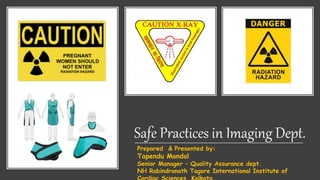
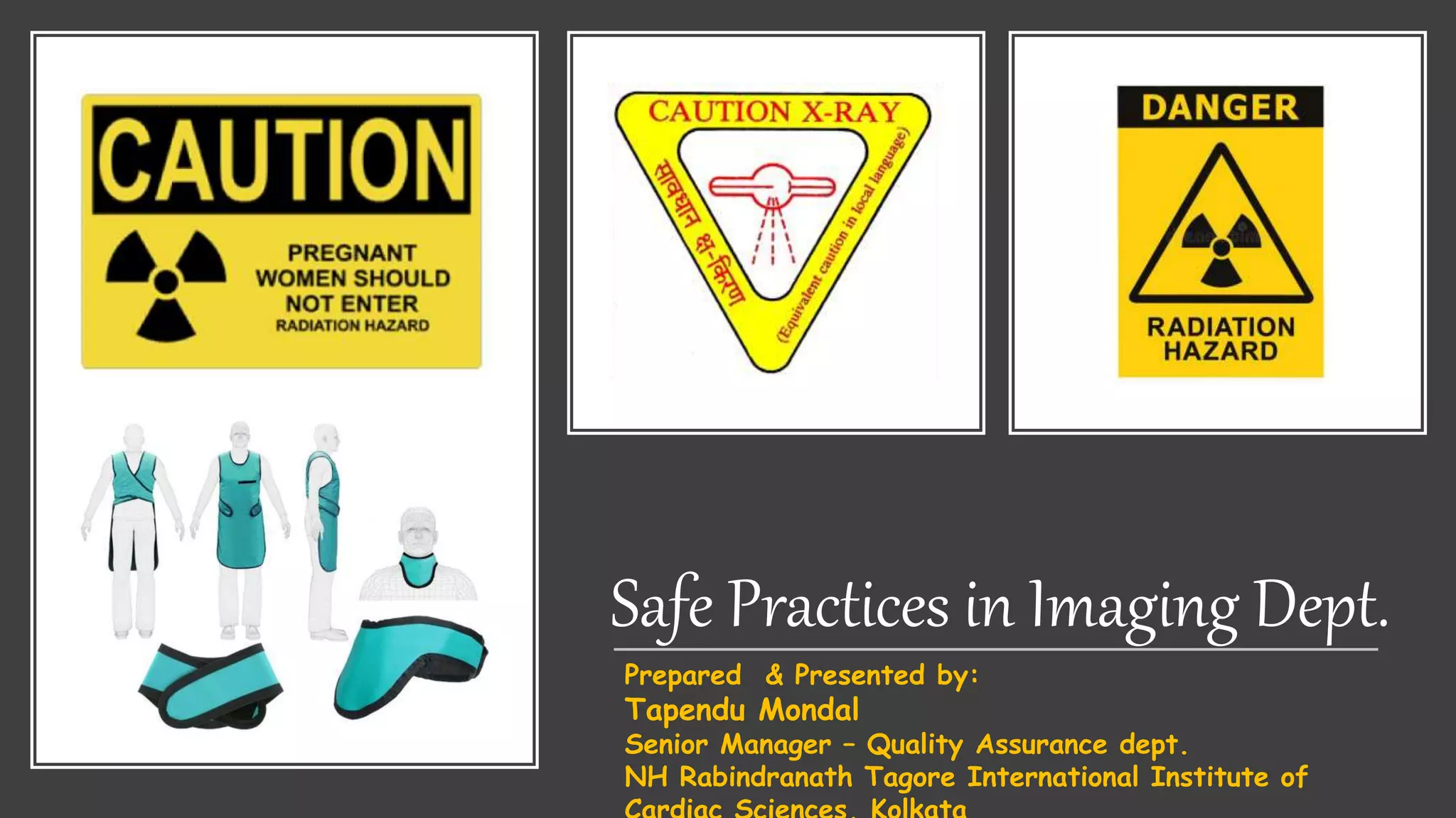
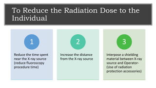
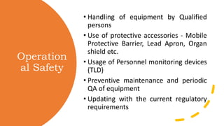
Safe Practices in Imaging Dept.
Prepared & Presented by:
Tapendu Mondal
Senior Manager – Quality Assurance dept.
NH Rabindranath Tagore International Institute of
To Reduce the Radiation Dose to the
Individual
Reduce the time spent
near the X-ray source
(reduce fluoroscopy
procedure time)
1
Increase the distance
from the X-ray source
2
Interpose a shielding
material between X-ray
source and Operator-
(Use of radiation
protection accessories)
3
Operation
al Safety
• Handling of equipment by Qualified
persons
• Use of protective accessories - Mobile
Protective Barrier, Lead Apron, Organ
shield etc.
• Usage of Personnel monitoring devices
(TLD)
• Preventive maintenance and periodic
QA of equipment
• Updating with the current regulatory
requirements
Ways to
minimize
radiation
exposure
• Always collimate to the area of interest, the larger the amount
of tissue the beam is allowed to irradiate the more scatter
radiation is produced.
• Avoid holding of infirm patient by staff, provide protective
apron to the attendant while holding such patients.
• Always operate the equipment from behind the protective
barrier or using lead equivalent apron.
• During fluoroscopy exams always make sure that built in safety
devices such as flaps /ceiling suspended glass/ couch hanging
flaps as applicable are available and use it on routine basis.
• During use of mobile x-ray equipment stand at least 6 feet
away from the patient and wear lead apron.
• During c-arm procedures standing on the side of the image
intensifier is safest because there is more scatter produced at
the entrance surface side of the patient.
• During use of Portable X-ray equipment, ensure that X-ray
tube is immobilized on the stand and film cassette shall not be
held in hand by any person.
Proper Storage of
Radiation
Protection
Devices-Lead
Apron
Personal Monitoring
Badge (TLD)
• Wear TLD badge inside the
Lead apron at chest level –
Radiography For R/F or in Cath
Lab
• After work, store TLD badges
in radiation free area
• Control TLD Card should be
kept away from radiation area
Radiation
Protection
Accessories
• Mobile Protective Barrier (MPB)- 1.5
mm Lead Eqv
• Lead Aprons - 0.25 mm Lead Eqv
• Thyroid Shield -0.25 mm Lead Eqv
• Gonads Shield- 0.25 mm Lead Eqv
• Eye Wear (Shield)- 0.25 mm Lead
Eqv
• Rubber hanging Flaps (In IR )-0.5 mm
Lead Eqv
• Hand Gloves -0.25 mm Lead Eqv
• Lead Glass window- 1.5 mm Lead
Eqv
• Door (Lead Lined)-1.7 mm Lead Eqv
Protectiv
e
Devices
–
Quality
Control
• All lead equivalent vinyl material (aprons, gloves,
etc) should comply with relevant international
standards. They should be tested at purchase
and regularly thereafter, at least every one years.
• Incorrect storage may lead to cracks in the
shielding but this may not be detectable by
visual inspection alone
• A simple test is to examine the devices using
fluoroscopy (at about 60 kVp). The use of
automatic dose rate or automatic brightness
controls should be avoided.
• This test will only detect flaws in the shielding.
Faulty devices should be discarded immediately
Radiatio
n
Safety…
• Carry out Quality Assurance testing of each
& every X-ray equipment once in TWO
YEARS.
• Improves imaging standard
• Increase the Life of the X-ray tube/
equipment by avoiding retakes
• Useful in reduction of unnecessary patient’s
Dose & hence dose to the staff
• A good knowledge of equipment
specification and characteristics is essential
for an effective optimization of radiation
protection
• Occupational doses can be reduced by
reducing patient dose. The proper use of
equipment (including shielding devices) is
essential.
Patient
dose
manageme
nt
• Quality control protocols –
• Justify the procedure
• Ask for records of previous diagnostic
procedures
• Plan the procedure (right patient, right
contrast, purpose of scan/investigation)
• Know well about your equipment settings
• Ensure only qualified and trained operator
should operate the equipment
• Inform patient about his/her procedure
• Handover procedure records (CD/disc and
reports) including radiation dose records
to patient.
• Ensure that daily and periodic QA are
performed and equipment performance is
satisfactory.
Patient
Protectio
n
Radiation safety during x-ray examination of
patient is ensured by:
• Limiting the total “beam–on” time
• Avoiding oblique lateral projections
• Prior to exposure, collimate the x-ray beam to the area
of interest , AVOID post exposure cropping of the image
• Selecting low dose rate protocol (ALARA)
• Use of correct exposure protocols for patient
examinations including paediatric protocols
• Try to avoid radiation exposed on pregnant women. On
pregnant patients, always use additional shields, if it is
felt that the abdomen is likely to get unnecessarily
exposed.
• record- keeping of patient’s doses for CT and IR
procedures.
Main
Responsibil
ities as a
Technologi
st/
Radiograph
er
Inform inform the radiologist and radiation safety officer (RSO) in the case of an accident
or incident.
Record take record of patient doses;
QA Perform regular quality control of radiological equipment;
Perform Perform the radiological procedure following an optimized protocol and ensuring
patient protection;
Selection Participate in optimization of imaging protocols;
Preparation Contribute to the preparation of specifications for new equipment;
Verify Verify that the female patient is non-pregnant;
Inform Inform accompanying persons;
Inform Inform the patient;
Identify Identify the patient;
CONCLU
SIONS
• The majority of the radiation dose received by the
operator (provided the primary beam is avoided) is
due to scattered radiation from the patient.
• Know your equipment
• Use protective shields (lead apron, mounted
shields/flaps, ceiling suspended screens as
applicable)
• Avoid holding of patient without adequate
protection.
• Keep hands out of the primary beam
• Stand in the correct place: opposite the X ray tube
rather than near the X ray tube.(knowledge of iso-
dose curve)
• Always wear your personal radiation monitoring
badge and use them in the right manner
• Make sure that x ray equipment is properly
functioning and periodically tested and maintained
• All actions to reduce patient dose will also reduce
staff dose.
• Follow Time, Distance, Shielding golden rules
Safe practices in imaging dept.

Safe practices in imaging dept.

  • 1.
    Safe Practices inImaging Dept. Prepared & Presented by: Tapendu Mondal Senior Manager – Quality Assurance dept. NH Rabindranath Tagore International Institute of
  • 2.
    To Reduce theRadiation Dose to the Individual Reduce the time spent near the X-ray source (reduce fluoroscopy procedure time) 1 Increase the distance from the X-ray source 2 Interpose a shielding material between X-ray source and Operator- (Use of radiation protection accessories) 3
  • 3.
    Operation al Safety • Handlingof equipment by Qualified persons • Use of protective accessories - Mobile Protective Barrier, Lead Apron, Organ shield etc. • Usage of Personnel monitoring devices (TLD) • Preventive maintenance and periodic QA of equipment • Updating with the current regulatory requirements
  • 4.
    Ways to minimize radiation exposure • Alwayscollimate to the area of interest, the larger the amount of tissue the beam is allowed to irradiate the more scatter radiation is produced. • Avoid holding of infirm patient by staff, provide protective apron to the attendant while holding such patients. • Always operate the equipment from behind the protective barrier or using lead equivalent apron. • During fluoroscopy exams always make sure that built in safety devices such as flaps /ceiling suspended glass/ couch hanging flaps as applicable are available and use it on routine basis. • During use of mobile x-ray equipment stand at least 6 feet away from the patient and wear lead apron. • During c-arm procedures standing on the side of the image intensifier is safest because there is more scatter produced at the entrance surface side of the patient. • During use of Portable X-ray equipment, ensure that X-ray tube is immobilized on the stand and film cassette shall not be held in hand by any person.
  • 5.
  • 6.
    Personal Monitoring Badge (TLD) •Wear TLD badge inside the Lead apron at chest level – Radiography For R/F or in Cath Lab • After work, store TLD badges in radiation free area • Control TLD Card should be kept away from radiation area
  • 7.
    Radiation Protection Accessories • Mobile ProtectiveBarrier (MPB)- 1.5 mm Lead Eqv • Lead Aprons - 0.25 mm Lead Eqv • Thyroid Shield -0.25 mm Lead Eqv • Gonads Shield- 0.25 mm Lead Eqv • Eye Wear (Shield)- 0.25 mm Lead Eqv • Rubber hanging Flaps (In IR )-0.5 mm Lead Eqv • Hand Gloves -0.25 mm Lead Eqv • Lead Glass window- 1.5 mm Lead Eqv • Door (Lead Lined)-1.7 mm Lead Eqv
  • 8.
    Protectiv e Devices – Quality Control • All leadequivalent vinyl material (aprons, gloves, etc) should comply with relevant international standards. They should be tested at purchase and regularly thereafter, at least every one years. • Incorrect storage may lead to cracks in the shielding but this may not be detectable by visual inspection alone • A simple test is to examine the devices using fluoroscopy (at about 60 kVp). The use of automatic dose rate or automatic brightness controls should be avoided. • This test will only detect flaws in the shielding. Faulty devices should be discarded immediately
  • 9.
    Radiatio n Safety… • Carry outQuality Assurance testing of each & every X-ray equipment once in TWO YEARS. • Improves imaging standard • Increase the Life of the X-ray tube/ equipment by avoiding retakes • Useful in reduction of unnecessary patient’s Dose & hence dose to the staff • A good knowledge of equipment specification and characteristics is essential for an effective optimization of radiation protection • Occupational doses can be reduced by reducing patient dose. The proper use of equipment (including shielding devices) is essential.
  • 10.
    Patient dose manageme nt • Quality controlprotocols – • Justify the procedure • Ask for records of previous diagnostic procedures • Plan the procedure (right patient, right contrast, purpose of scan/investigation) • Know well about your equipment settings • Ensure only qualified and trained operator should operate the equipment • Inform patient about his/her procedure • Handover procedure records (CD/disc and reports) including radiation dose records to patient. • Ensure that daily and periodic QA are performed and equipment performance is satisfactory.
  • 11.
    Patient Protectio n Radiation safety duringx-ray examination of patient is ensured by: • Limiting the total “beam–on” time • Avoiding oblique lateral projections • Prior to exposure, collimate the x-ray beam to the area of interest , AVOID post exposure cropping of the image • Selecting low dose rate protocol (ALARA) • Use of correct exposure protocols for patient examinations including paediatric protocols • Try to avoid radiation exposed on pregnant women. On pregnant patients, always use additional shields, if it is felt that the abdomen is likely to get unnecessarily exposed. • record- keeping of patient’s doses for CT and IR procedures.
  • 12.
    Main Responsibil ities as a Technologi st/ Radiograph er Informinform the radiologist and radiation safety officer (RSO) in the case of an accident or incident. Record take record of patient doses; QA Perform regular quality control of radiological equipment; Perform Perform the radiological procedure following an optimized protocol and ensuring patient protection; Selection Participate in optimization of imaging protocols; Preparation Contribute to the preparation of specifications for new equipment; Verify Verify that the female patient is non-pregnant; Inform Inform accompanying persons; Inform Inform the patient; Identify Identify the patient;
  • 13.
    CONCLU SIONS • The majorityof the radiation dose received by the operator (provided the primary beam is avoided) is due to scattered radiation from the patient. • Know your equipment • Use protective shields (lead apron, mounted shields/flaps, ceiling suspended screens as applicable) • Avoid holding of patient without adequate protection. • Keep hands out of the primary beam • Stand in the correct place: opposite the X ray tube rather than near the X ray tube.(knowledge of iso- dose curve) • Always wear your personal radiation monitoring badge and use them in the right manner • Make sure that x ray equipment is properly functioning and periodically tested and maintained • All actions to reduce patient dose will also reduce staff dose. • Follow Time, Distance, Shielding golden rules