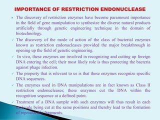
Dr. Nandadulal Sannigrahi,
Associate Professor,
Department of Botany,
Nistarini college, Purulia
D.B. Road, Purulia,
INDIA (W.B)
 Restriction mapping is a method used to map an unknown segment of
DNA by breaking it into pieces and then identifying the locations of the
breakpoints.
 This method relies upon the use of proteins called restriction enzymes,
which can cut, or digest, DNA molecules at short, specific sequences called
restriction sites.
 After a DNA segment has been digested using a restriction enzyme, the
resulting fragments can be examined using a laboratory method called gel
electrophoresis, which is used to separate pieces of DNA according to their
size.
 One common method for constructing a restriction map involves digesting
the unknown DNA sample in three ways.
 Here, two portions of the DNA sample are individually digested with
different restriction enzymes, and a third portion of the DNA sample is
double-digested with both restriction enzymes at the same time.
 Next, each digestion sample is separated using gel electrophoresis, and the
sizes of the DNA fragments are recorded.
 The total length of the fragments in each digestion will be equal.
 However, because the length of each individual DNA fragment depends
upon the positions of its restriction sites, each restriction site can be
mapped according to the lengths of the fragments.
 The information from the double-digestion is particularly useful for
correctly mapping the sites.
 The final drawing of the DNA segment that shows the positions of the
restriction sites is called a restriction map.
 So, the restriction mapping either circular or linear DNA is mostly done by
the use of multiple Restriction endonucleases extracted from diverse
bacterial sources to determine the relative position of the restriction sites.
 It is very indispensible tool in this regard for mapping of the Dna
sequences of the unknown one.
 Restriction mapping is a quite complex process that follows a number of
steps following single and double digestion by the restricted endonuclease,
 After the digestion by the enzymes, the relative length of the DNA
molecule is calculated and the measurements are done to find the relative
position of the DNA fragments intended for the same.
 The steps are as followed:
1. Isolation of sample DNA from the desired organism,
2. Digestion by Restricted Endonuclease (single),
3. Digestion by Restricted endonuclease (Double),
4. Agarose gel electrophoresis,
5. DNA pattern & Band visualization,
6. Band analysis,
7. Restriction map generation using the obtained data to find out the
restriction site of the sample DNA.
 ISOLATION OF SAMPLE DNA FROM THE ORGANISM:
 The investigation of environmentally and clinically significant
microorganisms is often a key component in student microbiology
laboratory projects.
 Many of these organisms, particularly bacteria, can be pathogenic and are
often the causative agent of disease in both humans and other animals,
 The purification of genomic DNA from bacterial cultures provides the
basis for downstream molecular analysis, and this process is often achieved
using commercially available kits.
 Kits are packaged (and often combined) reagent sets that are designed to
reduce the tedium behind multifaceted, complex procedures.
 They are user-friendly and efficient, albeit often at an increased cost, and
are ideal for researchers who are already familiar with the methods.
 DNA from the sample is isolated using the protocols as designed fro the
same.
 Digestion by Restricted Endonuclease (single)
 The DNA may be linear or circular and single digestion by the restricted
endonuclease is done to get the number of segments by single digestion,
 Restriction endonucleases recognize short DNA sequences and cleave
double-stranded DNA at specific sites within or adjacent to the recognition
sequences.
 Restriction endonuclease cleavage of DNA into discrete fragments is one
of the most basic procedures in molecular biology.
 The first method presented in this unit is the cleavage of a single DNA
sample with a single restriction endonuclease.
 For linear DNA, it will produce 2 fragments by following n+1 rule,
 For circular DNA, it will produce the single chain but having a option of
multiple fragments,
 The data of the digestion of a DNA molecule by more than one
endonucleases can be utilized to arrange the sites of breakage in a definite
order.
 The process involves double digestion ( enzyme A and B) to determine the
cleavage positions of DNA due to one enzyme with respect to another
enzyme,
 The result of reciprocal digests ( A followed by B and B followed by A)
show fragments,
 The original DNA sample is also digested by a mixture of both the
enzymes to confirm the results of individual successive digests,
 Overlapping region of different digests (A and B) can be detected that
allow to prep[are the restriction map.
 Three samples of a particular DNA species are taken,
 Each two of these samples is treated with separate restriction enzymes and
the other DNA sample is treated with both two separate restriction
enzymes,
 Then the three sets of fragments are compared following the Agarose gel
electrophoresis,
 The terminal end of the DNA molecule is also labeled with radioactive p32
to the restriction cleavage.
 Let 5000 bp length linear DNA fragments whose base sequences are unknown,
 Another circular plasmid having 5000 bp DNA length,
 In each case one restriction enzyme is present in each scenario we are looking
for,
 Now , the circular DNA is cut a single point and observe in an Agarose gel,
 If the 5000 bp long linear fragment is cut with restricted endonuclease, it will
cut into two pieces of 3000 bp and 2000 bp in length in each case cut by Eco
RI
 Now , if we cut them with restriction enzymes Bam, in each case, the
following data will be obtained,
 In case of circular, it will cut into 3000 bp, 1500 bp and 5 00 bp in each case,
 But if we cut the linear DNA fragments of both the EcoRI and Bam, it will cut
into three linear fragments-2000 bp. 1500 bp, 1000 bp & 500 bp in each,
 ,Through Agarose gel electrophoresis technique, the following restriction
cutting site will be obtained,
 Thus, the use of two restricted endonucleases enable to find out the exact
location of cutting site of the sequences of the entre length of DNA.
 The discovery of restriction enzymes have become paramount importance
in the field of gene manipulation to synthesize the diverse natural products
artificially through genetic engineering technique in the domain of
biotechnology.
 The discovery of the mode of action of the class of bacterial enzymes
known as restriction endonucleases provided the major breakthrough in
opening up the field of genetic engineering.
 In vivo, these enzymes are involved in recognizing and cutting up foreign
DNA entering the cell; their most likely role is thus protecting the bacteria
against phage infection.
 The property that is relevant to us is that these enzymes recognize specific
DNA sequences.
 The enzymes used in DNA manipulations are in fact known as Class II
restriction endonucleases; these enzymes cut the DNA within the
recognition sequence at a defined point.
 Treatment of a DNA sample with such enzymes will thus result in each
molecule being cut at the same positions and thereby lead to the formation
of reproducible fragments.
 ACKNOWLEDGEMENTS:
a. Google for images,
b. Different websites for enriching the course content,
c. Science Direct pages,
d. Cell and Molecular Biology-De Robertis & De Robertis, Jr.
e. Cell Biology- Verma & Agarwal
f. A textbook of Botany- Vol. III – Hait, Bhattacharya & Ghosh.
g. A Text Book of Cell and Molecular Biology- Ajay Paul,
h. Cell and Molecular Biology- Kar and Halder,
i. Concept of Genetics- Klug. Cummings, Spencer, Palladino,
DISCLAIMER:
 This presentation has been designed to address the academic fraternity
without any financial interest. This is absolutely free to use . The author
does not claim any kind of financial benefits from this content

Restriction Mapping: Linear and Circular.pdf

  • 1.
    Dr. Nandadulal Sannigrahi, AssociateProfessor, Department of Botany, Nistarini college, Purulia D.B. Road, Purulia, INDIA (W.B)
  • 2.
     Restriction mappingis a method used to map an unknown segment of DNA by breaking it into pieces and then identifying the locations of the breakpoints.  This method relies upon the use of proteins called restriction enzymes, which can cut, or digest, DNA molecules at short, specific sequences called restriction sites.  After a DNA segment has been digested using a restriction enzyme, the resulting fragments can be examined using a laboratory method called gel electrophoresis, which is used to separate pieces of DNA according to their size.  One common method for constructing a restriction map involves digesting the unknown DNA sample in three ways.  Here, two portions of the DNA sample are individually digested with different restriction enzymes, and a third portion of the DNA sample is double-digested with both restriction enzymes at the same time.
  • 3.
     Next, eachdigestion sample is separated using gel electrophoresis, and the sizes of the DNA fragments are recorded.  The total length of the fragments in each digestion will be equal.  However, because the length of each individual DNA fragment depends upon the positions of its restriction sites, each restriction site can be mapped according to the lengths of the fragments.  The information from the double-digestion is particularly useful for correctly mapping the sites.  The final drawing of the DNA segment that shows the positions of the restriction sites is called a restriction map.  So, the restriction mapping either circular or linear DNA is mostly done by the use of multiple Restriction endonucleases extracted from diverse bacterial sources to determine the relative position of the restriction sites.  It is very indispensible tool in this regard for mapping of the Dna sequences of the unknown one.
  • 5.
     Restriction mappingis a quite complex process that follows a number of steps following single and double digestion by the restricted endonuclease,  After the digestion by the enzymes, the relative length of the DNA molecule is calculated and the measurements are done to find the relative position of the DNA fragments intended for the same.  The steps are as followed: 1. Isolation of sample DNA from the desired organism, 2. Digestion by Restricted Endonuclease (single), 3. Digestion by Restricted endonuclease (Double), 4. Agarose gel electrophoresis, 5. DNA pattern & Band visualization, 6. Band analysis, 7. Restriction map generation using the obtained data to find out the restriction site of the sample DNA.
  • 6.
     ISOLATION OFSAMPLE DNA FROM THE ORGANISM:  The investigation of environmentally and clinically significant microorganisms is often a key component in student microbiology laboratory projects.  Many of these organisms, particularly bacteria, can be pathogenic and are often the causative agent of disease in both humans and other animals,  The purification of genomic DNA from bacterial cultures provides the basis for downstream molecular analysis, and this process is often achieved using commercially available kits.  Kits are packaged (and often combined) reagent sets that are designed to reduce the tedium behind multifaceted, complex procedures.  They are user-friendly and efficient, albeit often at an increased cost, and are ideal for researchers who are already familiar with the methods.  DNA from the sample is isolated using the protocols as designed fro the same.
  • 7.
     Digestion byRestricted Endonuclease (single)  The DNA may be linear or circular and single digestion by the restricted endonuclease is done to get the number of segments by single digestion,  Restriction endonucleases recognize short DNA sequences and cleave double-stranded DNA at specific sites within or adjacent to the recognition sequences.  Restriction endonuclease cleavage of DNA into discrete fragments is one of the most basic procedures in molecular biology.  The first method presented in this unit is the cleavage of a single DNA sample with a single restriction endonuclease.  For linear DNA, it will produce 2 fragments by following n+1 rule,  For circular DNA, it will produce the single chain but having a option of multiple fragments,  The data of the digestion of a DNA molecule by more than one endonucleases can be utilized to arrange the sites of breakage in a definite order.
  • 8.
     The processinvolves double digestion ( enzyme A and B) to determine the cleavage positions of DNA due to one enzyme with respect to another enzyme,  The result of reciprocal digests ( A followed by B and B followed by A) show fragments,  The original DNA sample is also digested by a mixture of both the enzymes to confirm the results of individual successive digests,  Overlapping region of different digests (A and B) can be detected that allow to prep[are the restriction map.  Three samples of a particular DNA species are taken,  Each two of these samples is treated with separate restriction enzymes and the other DNA sample is treated with both two separate restriction enzymes,  Then the three sets of fragments are compared following the Agarose gel electrophoresis,  The terminal end of the DNA molecule is also labeled with radioactive p32 to the restriction cleavage.
  • 9.
     Let 5000bp length linear DNA fragments whose base sequences are unknown,  Another circular plasmid having 5000 bp DNA length,  In each case one restriction enzyme is present in each scenario we are looking for,  Now , the circular DNA is cut a single point and observe in an Agarose gel,  If the 5000 bp long linear fragment is cut with restricted endonuclease, it will cut into two pieces of 3000 bp and 2000 bp in length in each case cut by Eco RI  Now , if we cut them with restriction enzymes Bam, in each case, the following data will be obtained,  In case of circular, it will cut into 3000 bp, 1500 bp and 5 00 bp in each case,  But if we cut the linear DNA fragments of both the EcoRI and Bam, it will cut into three linear fragments-2000 bp. 1500 bp, 1000 bp & 500 bp in each,  ,Through Agarose gel electrophoresis technique, the following restriction cutting site will be obtained,  Thus, the use of two restricted endonucleases enable to find out the exact location of cutting site of the sequences of the entre length of DNA.
  • 11.
     The discoveryof restriction enzymes have become paramount importance in the field of gene manipulation to synthesize the diverse natural products artificially through genetic engineering technique in the domain of biotechnology.  The discovery of the mode of action of the class of bacterial enzymes known as restriction endonucleases provided the major breakthrough in opening up the field of genetic engineering.  In vivo, these enzymes are involved in recognizing and cutting up foreign DNA entering the cell; their most likely role is thus protecting the bacteria against phage infection.  The property that is relevant to us is that these enzymes recognize specific DNA sequences.  The enzymes used in DNA manipulations are in fact known as Class II restriction endonucleases; these enzymes cut the DNA within the recognition sequence at a defined point.  Treatment of a DNA sample with such enzymes will thus result in each molecule being cut at the same positions and thereby lead to the formation of reproducible fragments.
  • 12.
     ACKNOWLEDGEMENTS: a. Googlefor images, b. Different websites for enriching the course content, c. Science Direct pages, d. Cell and Molecular Biology-De Robertis & De Robertis, Jr. e. Cell Biology- Verma & Agarwal f. A textbook of Botany- Vol. III – Hait, Bhattacharya & Ghosh. g. A Text Book of Cell and Molecular Biology- Ajay Paul, h. Cell and Molecular Biology- Kar and Halder, i. Concept of Genetics- Klug. Cummings, Spencer, Palladino, DISCLAIMER:  This presentation has been designed to address the academic fraternity without any financial interest. This is absolutely free to use . The author does not claim any kind of financial benefits from this content