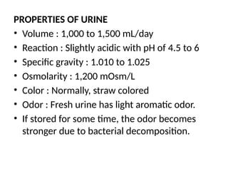
PROPERTIES OF URINE
•Volume : 1,000 to 1,500 mL/day
• Reaction : Slightly acidic with pH of 4.5 to 6
• Specific gravity : 1.010 to 1.025
• Osmolarity : 1,200 mOsm/L
• Color : Normally, straw colored
• Odor : Fresh urine has light aromatic odor.
• If stored for some time, the odor becomes
stronger due to bacterial decomposition.
4.
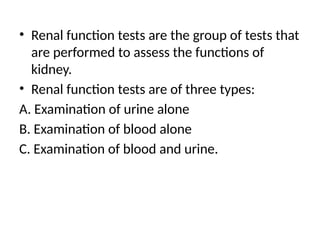
• Renal functiontests are the group of tests that
are performed to assess the functions of
kidney.
• Renal function tests are of three types:
A. Examination of urine alone
B. Examination of blood alone
C. Examination of blood and urine.
5.
EXAMINATION OF URINE– URINALYSIS
• Urinalysis:
i. Physical examination
ii. Microscopic examination
iii. Chemical analysis.
6.
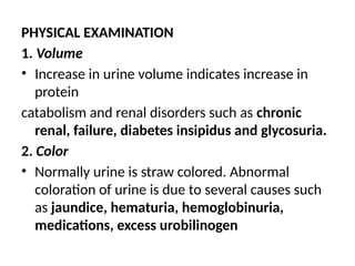
PHYSICAL EXAMINATION
1. Volume
•Increase in urine volume indicates increase in
protein
catabolism and renal disorders such as chronic
renal, failure, diabetes insipidus and glycosuria.
2. Color
• Normally urine is straw colored. Abnormal
coloration of urine is due to several causes such
as jaundice, hematuria, hemoglobinuria,
medications, excess urobilinogen
7.
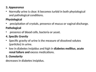
3. Appearance
• Normallyurine is clear. It becomes turbid in both physiological
and pathological conditions.
Physiological
• precipitation of crystals, presence of mucus or vaginal discharge.
Pathological
• presence of blood cells, bacteria or yeast.
4. Specific Gravity
• Specific gravity of urine is the measure of dissolved solutes
(particles) in urine.
• low in diabetes insipidus and high in diabetes mellitus, acute
renal failure and excess medications.
5. Osmolarity
decreases in diabetes insipidus.
8.
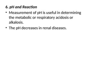
6. pH andReaction
• Measurement of pH is useful in determining
the metabolic or respiratory acidosis or
alkalosis.
• The pH decreases in renal diseases.
9.
MICROSCOPIC EXAMINATION
• Microscopicexamination of centrifuged sediment
of urine is useful in determining the renal diseases.
1. Red Blood Cells
• Presence of red blood cells in urine indicates
glomerular disease such as glomerulonephritis.
2. White Blood Cells
• Normally few white blood cells appear in high
power field.
• The number increases in acute glomerulonephritis,
• infection of urinary tract, vagina or cervix.
10.
3. Epithelial Cells
Normallyfew tubular epithelial cells slough into urine.
• Presence of many epithelial cells suggests nephrotic
syndrome and tubular necrosis.
4. Casts
Casts may be hyaline, granular or cellular in nature.
The number increases in proteinuria due to
glomerulonephritis.
• Red blood cell casts appear in urine during
glomerulonephritis and tubular necrosis.
• White blood cell casts appear in pyelonephritis.
• Epithelial casts are formed during acute tubular necrosis.
11.
5. Crystals
• Commoncrystals are the crystals of calcium oxalate,,
calcium phosphate, uric acid and triple phosphate,
(calcium, ammonium and magnesium).
• Abnormal crystals such as crystals of cystine and
tyrosine appear in liver diseases.
6. Bacteria
• Bacteria are common in urine specimens because of
normal microbial flora of urinary tract, urethra and
vagina and because of their ability to multiply rapidly
in urine.
• Culture to determine the presence of bacteria in urine.
12.
CHEMICAL ANALYSIS
1. Glucose
•Glucose appears in urine when the blood glucose level
increases above 180 mg/dL. Glycosuria (presence of
glucose in urine) may be the first indicator of diabetes
mellitus.
2. Protein
• Presence of excess protein (proteinuria) particularly
albumin (albuminuria) in urine indicates renal diseases.
• Urinary excretion of albumin in a normal healthy adultis
about 30 mg/day. It exceeds this level in glome
rulonephritis.
13.
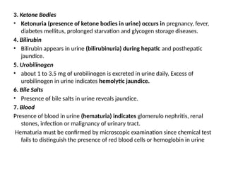
3. Ketone Bodies
•Ketonuria (presence of ketone bodies in urine) occurs in pregnancy, fever,
diabetes mellitus, prolonged starvation and glycogen storage diseases.
4. Bilirubin
• Bilirubin appears in urine (bilirubinuria) during hepatic and posthepatic
jaundice.
5. Urobilinogen
• about 1 to 3.5 mg of urobilinogen is excreted in urine daily. Excess of
urobilinogen in urine indicates hemolytic jaundice.
6. Bile Salts
• Presence of bile salts in urine reveals jaundice.
7. Blood
Presence of blood in urine (hematuria) indicates glomerulo nephritis, renal
stones, infection or malignancy of urinary tract.
Hematuria must be confirmed by microscopic examination since chemical test
fails to distinguish the presence of red blood cells or hemoglobin in urine
14.
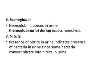
8. Hemoglobin
• Hemoglobinappears in urine
(hemoglobinuria) during excess hemolysis.
9. Nitrite
• Presence of nitrite in urine indicates presence
of bacteria in urine since some bacteria
convert nitrate into nitrite in urine.
15.
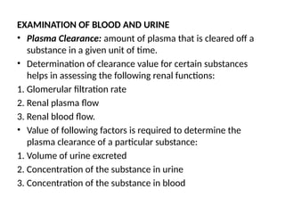
EXAMINATION OF BLOODAND URINE
• Plasma Clearance: amount of plasma that is cleared off a
substance in a given unit of time.
• Determination of clearance value for certain substances
helps in assessing the following renal functions:
1. Glomerular filtration rate
2. Renal plasma flow
3. Renal blood flow.
• Value of following factors is required to determine the
plasma clearance of a particular substance:
1. Volume of urine excreted
2. Concentration of the substance in urine
3. Concentration of the substance in blood
16.
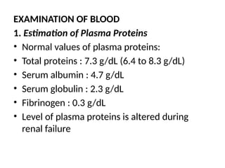
EXAMINATION OF BLOOD
1.Estimation of Plasma Proteins
• Normal values of plasma proteins:
• Total proteins : 7.3 g/dL (6.4 to 8.3 g/dL)
• Serum albumin : 4.7 g/dL
• Serum globulin : 2.3 g/dL
• Fibrinogen : 0.3 g/dL
• Level of plasma proteins is altered during
renal failure
17.
• Renal failure:failure of excretory functions of
kidney. It is usually, characterized by decrease
in glomerular filtration rate (GFR).
• GFR is considered as the best index of renal
failure.
• Decrease in GFR is not affected much during
the initial stages of renal failure.
• If 50% of the nephrons are affected, GFR
decreases only by 20% to 30%. It is because of
the compensatory mechanism by the
unaffected nephrons
18.
ACUTE RENAL FAILURE
•Acute renal failure: abrupt or sudden stoppage
of renal functions.
• reversible within few days to few weeks.
• result in sudden life-threatening reactions in
the body with the need for emergency
treatment.
19.
CAUSES
1. Acute nephritis(inflammation of kidneys)
2. Damage of renal tissues by poisons
3. Renal ischemia, which develops during circulatory shock
4. Acute tubular necrosis (necrosis of tubular cells in kidney)
caused by burns, hemorrhage, snake bite, toxins (like
insecticides, heavy metals and carbon tetrachloride) and drugs
(like diuretics, aminoglycosides and platinum derivatives)
5. Severe transfusion reactions
6. Sudden fall in blood pressure during hemorrhage, diarrhea,
severe burns and cholera
7. Blockage of ureter due to the formation of calculi (renal stone)
or tumor.
20.
FEATURES
1. Oliguria (decreasedurinary output)
2. Anuria (cessation of urine formation) in severe cases
3. Proteinuria (appearance of proteins in urine) including
albuminuria (excretion of albumin in urine)
4. Hematuria (presence of blood in urine)
5. Edema due to increased volume of extracellular fluid (ECF)
caused by retention of sodium and water
6. Hypertension within few days because of increased ECF
volume
7. Acidosis due to the retention of metabolic end products
8. Coma due to severe acidosis (if the patient is not treated in
time) resulting in death within 10 to 14 days.
21.
• Chronic renalfailure
• progressive, long standing and irreversible impairment of renal
functions.
• when more and more nephrons start losing the function over the
months or years, the compensatory mechanism fails and chronic
renal failure develops.
„ CAUSES
1. Chronic nephritis
2. Polycystic kidney disease
3. Renal calculi (kidney stones)
4. Urethral constriction
5. Hypertension
6. Atherosclerosis
7. Tuberculosis
8. Slow poisoning by drugs or metals.
22.
FEATURES
• 1. Uremia;excess accumulation of end products of protein
metabolism such as urea, nitrogen and creatinine in blood.
• Uremia occurs because of the failure of kidney to excrete the
metabolic end products and toxic substances.
• Common features of uremia
i. Anorexia (loss of appetite)
ii. Lethargy
iii. Drowsiness
iv. Nausea and vomiting
v. Pigmentation of skin
vi. Muscular twitching, tetany and convulsion
vii. Confusion and mental deterioration
viii. Coma.
23.
2. Acidosis
• Uremiaresults in acidosis, which leads to coma and death.
3. Edema
• Failure of kidney to excrete sodium and electrolytes causes increase in
extracellular fluid volume resulting in development of edema.
4. Blood Loss
• Gastrointestinal bleeding accompanied by platelet dysfunction leads to
heavy loss of blood.
5. Anemia
• Since, erythropoietin is not secreted in the kidney during renal failure, the
production of RBC decreases resulting in normocytic normochromic
anemia.
6. Hyperparathyroidism
• Secondary hyperparathyroidism is developed due to the deficiency of
calcitriol (1,25dihydroxycholecalciferol).
• It increases the removal of calcium from bones resulting in osteomalacia.