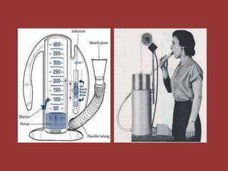
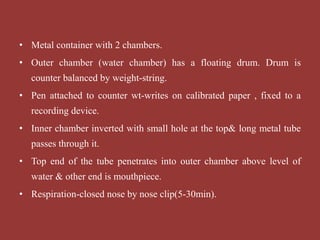
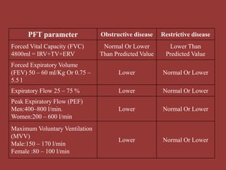
Pulmonary Function
Tests (PFT)
KRVS Chaitanya
Aspects of pulmonary function:
– Perfusion: Blood flow.
– Diffusion: Movement of O2 & CO2.
– Ventilation: Air exchange btwn alveolar spaces &
the atmosphere.
Introduction
Parameter in PFT
• Tidal Volume(TV)
• Inspiratory Reserve Volume(IRV)
• Inspiratory Capacity(IC)
• Functional Residual Capacity(FRC)
• Expiratory Reserve Volume(ERV)
• Residual Volume(RV)
• Vital Capacity(VC)
• Alveolar Ventilation
• Assessment of various aspects of pulmonary physiology.
• Detect & quantify the respiratory disease.
• Evaluation of the disease & its response to therapy.
• Provide valuable clinical information
Indication of PFT:
Pulmonary Function Test (Techniques)
1. Spirometry
2. Bronchial provocation test
3. Static lung volumes
4. Carbon monoxide diffusing capacity
5. Alveolar arterial oxygen gradient (AAOG)
6. Exercise testing (CPET)
• Measures the volume of air inspired or expired &
records the time –the volume change occurs.
• Mechanical signal → electrical signal records the amt
of gas breathed in & out→ SPIROGRAM.
• 2 major categories:-
– Mechanical Or Volume-displacement
– Electronic Or Flow Sensing.
1. SPIROMETRY
• Determines the effectiveness of various mechanical forces
involved in lung & chest wall movement.
• The values obtained provide quantitative information about the
degree of obstruction to expiratory airflow or the degree of
restriction of inspired air
• Metal container with 2 chambers.
• Outer chamber (water chamber) has a floating drum. Drum is
counter balanced by weight-string.
• Pen attached to counter wt-writes on calibrated paper , fixed to a
recording device.
• Inner chamber inverted with small hole at the top& long metal tube
passes through it.
• Top end of the tube penetrates into outer chamber above level of
water & other end is mouthpiece.
• Respiration-closed nose by nose clip(5-30min).
• The forced expiratory volumes exhaled within 1,2,3 seconds –
timed vital capacities (FEV1,FEV2 & FEV3) Respectively.
• FEF 25-75 –forced expiratory flow btwn 25% &75%.
• Measurements are useful for evaluating a patient’s response to
bronchodilators.
• If FEV1 <80% of predicted or FEF25-75% is <60% of
predicted, bronchodilators are administered –nebulizer;
Spirometry is repeated.
• Measured (actual) Spirometry values are compared with predicted
values- age , height, weight & gender; expressed as a % of predicted
value.(>80% normal limits).
• During expiration floating drum moves up , counter weight comes
down downward deflection on the graph.
• Inspiration the drum moves down, counter weight comes up, upward
deflection on the graph.
• Spirometry test –measured twice both before and after given a
bronchodilator.
• Improvement in measurements means patient will respond well.
Disadvantage :
• Only for single use due to CO2 accumulation & O2 cannot be
supplied.
• Residual Volume, Functional Residual Capacity and Total
Lung Capacity cannot be measured.
Indications of test
– To establish baseline ventilator function.
– To detect disease.
– To follow course of disease.
– Monitoring treatment.
– Evaluation of impairment.
– Pre-operative evaluation.
– Occupational surveys.
• Provocative material is given by inhalation & bronchospasm
provoked in the laboratory.
• 3 Types of provocative materials used:
1. Nonspecific pharmacologic- Histamine, Methacholine.
2. Nonspecific irritant- Sulphur dioxide , smoke, citric acid.
3. Specific-Individual allergen.
• 20% fall in FEV1 from basal values – positive response.
• Histamine solution by nebulisation - 8mg/ml.
2. BRONCHIAL PROVOCATION TEST
Indications For Testing:-
• To identify patients with hypersensitive or hyper reactive
airways- diagnosing asthma.
• To identify specific provocative factors.
• Research tool to study pathophysiology of acute reversible
bronchospasm.
Basic techniques for measurement of lung volumes:-
• Gas dilution methods.
• Body plethysmography.
• Radiographic techniques
3. STATIC LUNG VOLUMES
Indications For Static Lung Volumes:-
• Restrictive lung diseases –
– Confirming diagnosis.
– Establishing baseline & quantitating the level of
impairment.
– Evaluating response to therapy & following the course of
the disease.
• Obstructive lung diseases –
– To assess severity of disease.
– Assess clinical course and response to therapy.
4. CARBON MONOXIDE DIFFUSING
CAPACITY
• Diffusing capacity –
– The rate at which gas enters the blood divided by the driving
pressure of the gas.
• Factors Affecting Diffusing Capacity-
– Changes in alveolar capillary membrane.
– Ventilation to perfusion distribution.
– Haemoglobin concentration.
– Pulmonary circulation.
Reduced diffusing capacity is seen in:-
– Anaemia.
– Multiple Pulmonary Emboli.
– Emphysema.
– Pulmonary Resection Interstitial Lung Disease.
– Severe Bronchospasm.
Indications For CO Diffusing Capacity:-
– Unexplained dyspnoea due to early interstitial lung disease.
– Differentiating emphysema from chronic bronchitis
– Diagnosing asthma.
– Diagnosis & follow up of patients with interstitial lung
disease.
– Diagnosis of recurrent multiple pulmonary emboli.
• Alveolar oxygen tension is calculated & arterial oxygen tension
measured by blood gas estimation.
• Difference between the two gives a measurement of alveolar to
arterial oxygen gradient.
• In normal →5-15 mm Hg.
• Increased AAOG is due to 3 mechanisms:-
– Ventilation perfusion mismatch.
– Increased right to left shunt.
– Diffusion block.
5. ALVEOLAR ARTERIAL OXYGEN
GRADIENT (AAOG)
• Alveolar arterial oxygen gradient are measured after exercise.
• Gradient reduces but if it increases it indicates ventilation
perfusion mismatch.
• Absolute shunt fraction is also calculated when AAG are
measured 10 min after 100% oxygen.
• Increased gradient after 100% oxygen with an increased shunt,
indicate presence of right to left shunt.
6. CARDIOPULMONARY EXERCISE TEST
(CPET)
• Cardiopulmonary stress test ,with the addition pulmonary
factors are also evaluated during exercise.
• Evaluate the response of the cardiovascular & respiratory
systems to exercise, allows measurement of gas exchange.
• It categorizes disorders that limit exercise tolerance by
documenting their pathophysiology.
• Allows for an objective assessment of the patients symptoms ,
accurate prescription.
PFT parameter Obstructive disease Restrictive disease
Forced Vital Capacity (FVC)
4800ml = IRV+TV+ERV
Normal Or Lower
Than Predicted Value
Lower Than
Predicted Value
Forced Expiratory Volume
(FEV) 50 – 60 ml/Kg Or 0.75 –
5.5 l
Lower Normal Or Lower
Expiratory Flow 25 – 75 % Lower Normal Or Lower
Peak Expiratory Flow (PEF)
Men:400–800 l/min.
Women:200 – 600 l/min
Lower Normal Or Lower
Maximum Voluntary Ventilation
(MVV)
Male:150 – 170 l/min
Female :80 – 100 l/min
Lower Normal Or Lower
PFT parameter Obstructive disease Restrictive disease
Slow Vital Capacity(SVC) Normal Or Lower Lower
Total Lung Capacity (TLC)
6000ml= IRV+TV+ERV+RV
Normal Or Higher Lower
Functional Residual Capacity
(FRC) 2200ml = ERV + RV
Higher Normal or Lower
Residual Volume (RV)1200ml
Male:1.2 L
Female:1.1L
Higher Normal
Expiratory Reserve Volume
(ERV) 1000ml
Men : 1.0 L
Women:0.7 L
Normal Or Lower Normal Or Lower
BRONCHOSPASM:-
– Aspirin ACE inhibitors:- Ramipril , Captopril, Enalapril
etc.
– Beta adrenergic receptor blockers:- Acebutolol, Atenolol,
Metaprolol, Celiprolol.
– Inhalational agents/Aerosols:- Beclomethasone,
Fluticasone, Flunisolide.
– Nonsteroidal Anti-inflammatory drugs. Penicillin.
• PULMONARY EDEMA:-
• Dextran
• Heroin
• Hydrochlorothiazide
• Methadone
• Tricyclic Antidepressants.
• INTERSTITIAL PNEUMONITIS:-
• Etoposide
• Phenytoin
• Procarbazine
• Sulphonamides.
PULMONARY FIBROSIS:-
–Amiodarone
–Bleomycin
–Bromocriptine
–Cyclophosphamide
–6-Mercaptopurine
–Methotrexate
–Nitrofurantoin
Thank You

PULMONARY FUNCTION TEST

  • 1.
  • 2.
    Aspects of pulmonaryfunction: – Perfusion: Blood flow. – Diffusion: Movement of O2 & CO2. – Ventilation: Air exchange btwn alveolar spaces & the atmosphere. Introduction
  • 3.
    Parameter in PFT •Tidal Volume(TV) • Inspiratory Reserve Volume(IRV) • Inspiratory Capacity(IC) • Functional Residual Capacity(FRC) • Expiratory Reserve Volume(ERV) • Residual Volume(RV) • Vital Capacity(VC) • Alveolar Ventilation
  • 4.
    • Assessment ofvarious aspects of pulmonary physiology. • Detect & quantify the respiratory disease. • Evaluation of the disease & its response to therapy. • Provide valuable clinical information Indication of PFT:
  • 5.
    Pulmonary Function Test(Techniques) 1. Spirometry 2. Bronchial provocation test 3. Static lung volumes 4. Carbon monoxide diffusing capacity 5. Alveolar arterial oxygen gradient (AAOG) 6. Exercise testing (CPET)
  • 6.
    • Measures thevolume of air inspired or expired & records the time –the volume change occurs. • Mechanical signal → electrical signal records the amt of gas breathed in & out→ SPIROGRAM. • 2 major categories:- – Mechanical Or Volume-displacement – Electronic Or Flow Sensing. 1. SPIROMETRY
  • 7.
    • Determines theeffectiveness of various mechanical forces involved in lung & chest wall movement. • The values obtained provide quantitative information about the degree of obstruction to expiratory airflow or the degree of restriction of inspired air
  • 9.
    • Metal containerwith 2 chambers. • Outer chamber (water chamber) has a floating drum. Drum is counter balanced by weight-string. • Pen attached to counter wt-writes on calibrated paper , fixed to a recording device. • Inner chamber inverted with small hole at the top& long metal tube passes through it. • Top end of the tube penetrates into outer chamber above level of water & other end is mouthpiece. • Respiration-closed nose by nose clip(5-30min).
  • 10.
    • The forcedexpiratory volumes exhaled within 1,2,3 seconds – timed vital capacities (FEV1,FEV2 & FEV3) Respectively. • FEF 25-75 –forced expiratory flow btwn 25% &75%. • Measurements are useful for evaluating a patient’s response to bronchodilators. • If FEV1 <80% of predicted or FEF25-75% is <60% of predicted, bronchodilators are administered –nebulizer; Spirometry is repeated.
  • 11.
    • Measured (actual)Spirometry values are compared with predicted values- age , height, weight & gender; expressed as a % of predicted value.(>80% normal limits). • During expiration floating drum moves up , counter weight comes down downward deflection on the graph. • Inspiration the drum moves down, counter weight comes up, upward deflection on the graph. • Spirometry test –measured twice both before and after given a bronchodilator. • Improvement in measurements means patient will respond well.
  • 12.
    Disadvantage : • Onlyfor single use due to CO2 accumulation & O2 cannot be supplied. • Residual Volume, Functional Residual Capacity and Total Lung Capacity cannot be measured.
  • 13.
    Indications of test –To establish baseline ventilator function. – To detect disease. – To follow course of disease. – Monitoring treatment. – Evaluation of impairment. – Pre-operative evaluation. – Occupational surveys.
  • 14.
    • Provocative materialis given by inhalation & bronchospasm provoked in the laboratory. • 3 Types of provocative materials used: 1. Nonspecific pharmacologic- Histamine, Methacholine. 2. Nonspecific irritant- Sulphur dioxide , smoke, citric acid. 3. Specific-Individual allergen. • 20% fall in FEV1 from basal values – positive response. • Histamine solution by nebulisation - 8mg/ml. 2. BRONCHIAL PROVOCATION TEST
  • 15.
    Indications For Testing:- •To identify patients with hypersensitive or hyper reactive airways- diagnosing asthma. • To identify specific provocative factors. • Research tool to study pathophysiology of acute reversible bronchospasm.
  • 16.
    Basic techniques formeasurement of lung volumes:- • Gas dilution methods. • Body plethysmography. • Radiographic techniques 3. STATIC LUNG VOLUMES
  • 18.
    Indications For StaticLung Volumes:- • Restrictive lung diseases – – Confirming diagnosis. – Establishing baseline & quantitating the level of impairment. – Evaluating response to therapy & following the course of the disease. • Obstructive lung diseases – – To assess severity of disease. – Assess clinical course and response to therapy.
  • 19.
    4. CARBON MONOXIDEDIFFUSING CAPACITY • Diffusing capacity – – The rate at which gas enters the blood divided by the driving pressure of the gas. • Factors Affecting Diffusing Capacity- – Changes in alveolar capillary membrane. – Ventilation to perfusion distribution. – Haemoglobin concentration. – Pulmonary circulation.
  • 20.
    Reduced diffusing capacityis seen in:- – Anaemia. – Multiple Pulmonary Emboli. – Emphysema. – Pulmonary Resection Interstitial Lung Disease. – Severe Bronchospasm.
  • 21.
    Indications For CODiffusing Capacity:- – Unexplained dyspnoea due to early interstitial lung disease. – Differentiating emphysema from chronic bronchitis – Diagnosing asthma. – Diagnosis & follow up of patients with interstitial lung disease. – Diagnosis of recurrent multiple pulmonary emboli.
  • 22.
    • Alveolar oxygentension is calculated & arterial oxygen tension measured by blood gas estimation. • Difference between the two gives a measurement of alveolar to arterial oxygen gradient. • In normal →5-15 mm Hg. • Increased AAOG is due to 3 mechanisms:- – Ventilation perfusion mismatch. – Increased right to left shunt. – Diffusion block. 5. ALVEOLAR ARTERIAL OXYGEN GRADIENT (AAOG)
  • 23.
    • Alveolar arterialoxygen gradient are measured after exercise. • Gradient reduces but if it increases it indicates ventilation perfusion mismatch. • Absolute shunt fraction is also calculated when AAG are measured 10 min after 100% oxygen. • Increased gradient after 100% oxygen with an increased shunt, indicate presence of right to left shunt.
  • 24.
    6. CARDIOPULMONARY EXERCISETEST (CPET) • Cardiopulmonary stress test ,with the addition pulmonary factors are also evaluated during exercise. • Evaluate the response of the cardiovascular & respiratory systems to exercise, allows measurement of gas exchange. • It categorizes disorders that limit exercise tolerance by documenting their pathophysiology. • Allows for an objective assessment of the patients symptoms , accurate prescription.
  • 25.
    PFT parameter Obstructivedisease Restrictive disease Forced Vital Capacity (FVC) 4800ml = IRV+TV+ERV Normal Or Lower Than Predicted Value Lower Than Predicted Value Forced Expiratory Volume (FEV) 50 – 60 ml/Kg Or 0.75 – 5.5 l Lower Normal Or Lower Expiratory Flow 25 – 75 % Lower Normal Or Lower Peak Expiratory Flow (PEF) Men:400–800 l/min. Women:200 – 600 l/min Lower Normal Or Lower Maximum Voluntary Ventilation (MVV) Male:150 – 170 l/min Female :80 – 100 l/min Lower Normal Or Lower
  • 26.
    PFT parameter Obstructivedisease Restrictive disease Slow Vital Capacity(SVC) Normal Or Lower Lower Total Lung Capacity (TLC) 6000ml= IRV+TV+ERV+RV Normal Or Higher Lower Functional Residual Capacity (FRC) 2200ml = ERV + RV Higher Normal or Lower Residual Volume (RV)1200ml Male:1.2 L Female:1.1L Higher Normal Expiratory Reserve Volume (ERV) 1000ml Men : 1.0 L Women:0.7 L Normal Or Lower Normal Or Lower
  • 27.
    BRONCHOSPASM:- – Aspirin ACEinhibitors:- Ramipril , Captopril, Enalapril etc. – Beta adrenergic receptor blockers:- Acebutolol, Atenolol, Metaprolol, Celiprolol. – Inhalational agents/Aerosols:- Beclomethasone, Fluticasone, Flunisolide. – Nonsteroidal Anti-inflammatory drugs. Penicillin.
  • 28.
    • PULMONARY EDEMA:- •Dextran • Heroin • Hydrochlorothiazide • Methadone • Tricyclic Antidepressants. • INTERSTITIAL PNEUMONITIS:- • Etoposide • Phenytoin • Procarbazine • Sulphonamides.
  • 29.
  • 30.