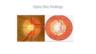
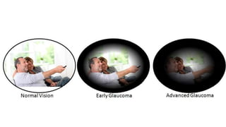
POAG
Dr.Syeda Fahmida Farzana Aziz
Introduction
Definition:
Primary open-angle glaucoma (POAG) is a commonly disease of adult
onset.
It is characterized by:
• IOP >21 mmHg at some stage.
• Glaucomatous optic nerve damage.
• An open anterior chamber angle.
Introduction Cont..
• Characteristic visual field loss as damage progresses.
• Absence of signs of secondary glaucoma or a non-
glaucomatous cause for the optic neuropathy.
Risk factors
• The higher the IOP, the greater the likelihood of
glaucoma.
• POAG is more common in older individuals.
• More common in black individuals.
• Family history of POAG
• Diabetes mellitus
• Myopia
Risk factors Cont..
• Contraceptive pill.
• Vascular disease.
• Translaminar pressure gradient.
• Large discs may be more vulnerable to damage
• Ocular perfusion pressure
Pathogenesis of glaucomatous
optic neuropathy
• Direct mechanical damage
damage to retinal nerve fibres at the
optic nerve head, perhaps as they pass through the lamina
Cribrosa.
• Ischaemic damage
due to compression of blood
vessels supplying the optic nerve head.
Changes in glaucoma
(a) the optic nerve head
(b) the peripapillary area and
(c) the retinal nerve fibre layer
Subtypes of glaucomatous damage
• Focal ischaemic discs
• Myopic disc with glaucoma
• Sclerotic discs
• Concentrically enlarging discs
Non-specific signs of glaucomatous damage
• Disc haemorrhages
• Baring of circumlinear blood vessels
• Bayoneting
• Collaterals between two veins
• Loss of nasal NRR
• The laminar dot sign
• ‘Sharpened edge’ or ‘sharpened rim’
Optic Disc findings
Steroid responsiveness
Elevation of IOP in response to a course of steroid is more
marked in patients with POAG and their close relatives.
Screening
Routine screening eye examinations
• Visual field assessment
• Tonometry
• Ophthalmoscopy
Investigation
• Pachymetry for CCT.
• Perimetry
• Imaging of the optic disc, peripapillary RNFL and/or
ganglion cell complex, e.g.
–Red-free photography,
–Stereo disc photography,
Investigation Cont..
–OCT,
–Confocal scanning laser ophthalmoscopy
–Scanning laser polarimetry.
Treatment goals
1. Target pressure: IOP level is identified below which further damage
is considered unlikely.
2. Proportional reduction in IOP by a certain percentage – often 30%
and then monitor, aiming for a further reduction if progression
occurs.
3.Response to progression.
Treatment goals
Target IOP depend upon
• IOP at which damage has occurred.
• Severity of visual field damage.
• Rate of progression of damage.
• Age and Life expectancy.
Management
The primary aim of treatment is to prevent functional
impairment of vision within the patient’s lifetime by slowing
the rate of ganglion cell loss.
 Currently the only proven method of achieving this is the
lowering of IOP.
Management
Medical - Antiglaucoma drugs
LASER – SLT, ALT, MLT
Surgical
Types of medications
Beta blockers Alpha-2 agonists Prostaglandin
analogues
CAIs Miotics Hyperosmotic agents
Medical therapy
• 1. Commencing medical therapy
Initial treatment is usually with one drug in its lowest
concentration with the desired therapeutic effect & fewest
potential side effects .
2.Review:
• Response to the drug is assessed against the target IOP.
• If the response is satisfactory subsequent assessment is
generally done for a further 3-4 months.
Medical therapy(contd)
• If there has been little or no response the initial drug is
withdrawn and another substituted.
• If there has been an apparently incomplete response another
drug may be added or a fixed combination substituted.
• When two separate drugs are used the patient should be
instructed to wait five minutes before instilling the second
drug to prevent washout of the first.
Medical therapy(contd)
3. If IOP control is good and glaucomatous damage mild or
moderate with no substantial threat to central vision, annual
perimetry is generally sufficient.
4. Gonioscopy should be performed annually because anterior
chamber angle tends to narrow with age.
5. Optic disc examination
Surgical treatment
• Trabeculectomy is the surgical procedure most commonly
performed for POAG.
• Phacoemulsification alone is frequently associated with a
significant fall in IOP, but is generally only offered to patients in
whom significant lens opacity is present.
Surgical treatment cont..
Indications Of Surgery
1. Progressive deterioration despite maximum medical therapy
2. Avoidance of excessive polypharmacy and drug intolerance
3. Primary therapy- advanced disease requiring very low target
pressure
4. Patient preference
Laser Treatment
Indication of Laser trabeculoplasty
• Intolerance to topical medication
• Failure of medical therapy
• Avoidance of polypharmacy
• Avoidance of Surgery
• Primary therapy
Patient counselling
 Full informations regarding the disease
 Informations regarding the medications.
 Mode of administering the drug.
 Informations regarding expected side effects
Prognosis
• The great majority of patients diagnosed with POAG will not be
blind in their lifetime but the incidence of blindness varies
considerably.
• Bad prognostic factors are:
 Advance damage at diagnosis
 Non-compliance with treatment
 Ethnic origin-black patients.
Take home message
Glaucoma is the second leading cause of blindness .
Glaucoma is a treatable disease, if detected earlier & treated
properly we can stop the progression of glaucomatous
damage.
By making good awareness about glaucoma
we can minimize family, socio-economic & as a whole
national burden.

Primary Open Angle Glaucoma

  • 1.
  • 2.
    Introduction Definition: Primary open-angle glaucoma(POAG) is a commonly disease of adult onset. It is characterized by: • IOP >21 mmHg at some stage. • Glaucomatous optic nerve damage. • An open anterior chamber angle.
  • 3.
    Introduction Cont.. • Characteristicvisual field loss as damage progresses. • Absence of signs of secondary glaucoma or a non- glaucomatous cause for the optic neuropathy.
  • 4.
    Risk factors • Thehigher the IOP, the greater the likelihood of glaucoma. • POAG is more common in older individuals. • More common in black individuals. • Family history of POAG • Diabetes mellitus • Myopia
  • 5.
    Risk factors Cont.. •Contraceptive pill. • Vascular disease. • Translaminar pressure gradient. • Large discs may be more vulnerable to damage • Ocular perfusion pressure
  • 6.
    Pathogenesis of glaucomatous opticneuropathy • Direct mechanical damage damage to retinal nerve fibres at the optic nerve head, perhaps as they pass through the lamina Cribrosa. • Ischaemic damage due to compression of blood vessels supplying the optic nerve head.
  • 7.
    Changes in glaucoma (a)the optic nerve head (b) the peripapillary area and (c) the retinal nerve fibre layer
  • 8.
    Subtypes of glaucomatousdamage • Focal ischaemic discs • Myopic disc with glaucoma • Sclerotic discs • Concentrically enlarging discs
  • 9.
    Non-specific signs ofglaucomatous damage • Disc haemorrhages • Baring of circumlinear blood vessels • Bayoneting • Collaterals between two veins • Loss of nasal NRR • The laminar dot sign • ‘Sharpened edge’ or ‘sharpened rim’
  • 10.
  • 12.
    Steroid responsiveness Elevation ofIOP in response to a course of steroid is more marked in patients with POAG and their close relatives.
  • 13.
    Screening Routine screening eyeexaminations • Visual field assessment • Tonometry • Ophthalmoscopy
  • 14.
    Investigation • Pachymetry forCCT. • Perimetry • Imaging of the optic disc, peripapillary RNFL and/or ganglion cell complex, e.g. –Red-free photography, –Stereo disc photography,
  • 15.
    Investigation Cont.. –OCT, –Confocal scanninglaser ophthalmoscopy –Scanning laser polarimetry.
  • 16.
    Treatment goals 1. Targetpressure: IOP level is identified below which further damage is considered unlikely. 2. Proportional reduction in IOP by a certain percentage – often 30% and then monitor, aiming for a further reduction if progression occurs. 3.Response to progression.
  • 17.
    Treatment goals Target IOPdepend upon • IOP at which damage has occurred. • Severity of visual field damage. • Rate of progression of damage. • Age and Life expectancy.
  • 18.
    Management The primary aimof treatment is to prevent functional impairment of vision within the patient’s lifetime by slowing the rate of ganglion cell loss.  Currently the only proven method of achieving this is the lowering of IOP.
  • 19.
    Management Medical - Antiglaucomadrugs LASER – SLT, ALT, MLT Surgical
  • 21.
    Types of medications Betablockers Alpha-2 agonists Prostaglandin analogues CAIs Miotics Hyperosmotic agents
  • 22.
    Medical therapy • 1.Commencing medical therapy Initial treatment is usually with one drug in its lowest concentration with the desired therapeutic effect & fewest potential side effects . 2.Review: • Response to the drug is assessed against the target IOP. • If the response is satisfactory subsequent assessment is generally done for a further 3-4 months.
  • 23.
    Medical therapy(contd) • Ifthere has been little or no response the initial drug is withdrawn and another substituted. • If there has been an apparently incomplete response another drug may be added or a fixed combination substituted. • When two separate drugs are used the patient should be instructed to wait five minutes before instilling the second drug to prevent washout of the first.
  • 24.
    Medical therapy(contd) 3. IfIOP control is good and glaucomatous damage mild or moderate with no substantial threat to central vision, annual perimetry is generally sufficient. 4. Gonioscopy should be performed annually because anterior chamber angle tends to narrow with age. 5. Optic disc examination
  • 25.
    Surgical treatment • Trabeculectomyis the surgical procedure most commonly performed for POAG. • Phacoemulsification alone is frequently associated with a significant fall in IOP, but is generally only offered to patients in whom significant lens opacity is present.
  • 26.
    Surgical treatment cont.. IndicationsOf Surgery 1. Progressive deterioration despite maximum medical therapy 2. Avoidance of excessive polypharmacy and drug intolerance 3. Primary therapy- advanced disease requiring very low target pressure 4. Patient preference
  • 27.
    Laser Treatment Indication ofLaser trabeculoplasty • Intolerance to topical medication • Failure of medical therapy • Avoidance of polypharmacy • Avoidance of Surgery • Primary therapy
  • 28.
    Patient counselling  Fullinformations regarding the disease  Informations regarding the medications.  Mode of administering the drug.  Informations regarding expected side effects
  • 29.
    Prognosis • The greatmajority of patients diagnosed with POAG will not be blind in their lifetime but the incidence of blindness varies considerably. • Bad prognostic factors are:  Advance damage at diagnosis  Non-compliance with treatment  Ethnic origin-black patients.
  • 30.
    Take home message Glaucomais the second leading cause of blindness . Glaucoma is a treatable disease, if detected earlier & treated properly we can stop the progression of glaucomatous damage. By making good awareness about glaucoma we can minimize family, socio-economic & as a whole national burden.