![KENNETH C. FORTGANG, MD ,[object Object]](data:image/gif;base64,R0lGODlhAQABAIAAAAAAAP///yH5BAEAAAAALAAAAAABAAEAAAIBRAA7)
Recommended
Recommended
More Related Content
What's hot
What's hot (20)
Similar to Identifying Radial Head Fractures Online Lesson
Similar to Identifying Radial Head Fractures Online Lesson (20)
Recently uploaded
Recently uploaded (20)
Identifying Radial Head Fractures Online Lesson
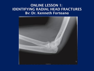
- 1. ONLINE LESSON 1: IDENTIFYING RADIAL HEAD FRACTURES By: Dr. Kenneth Fortgang
- 3. UNIV OF SOUTHERN CAL/LAC; SURGICAL INTERNSHIP
- 4. USC/LACOUNTY; RADIOLOGY RESIDENCY AND INTERVENTIONAL FELLOWSHIP
- 5. NORTH BROWARD HOSPITAL DISTRICT; LEVEL I AND LEVEL II TRAUMA CENTERS
- 6. RADIAL HEAD FRACTURES Kenneth C. Fortgang, MD Medical Director Premier Radiology Services
- 10. U L N A R A D I U S TROC CAP H U M E R U S Anatomy ©Ken L Schreibman, PhD/MD 2003
- 11. H U M E R U S U L N A TROC OLECRANON CORONOID Anatomy ©Ken L Schreibman, PhD/MD 2003 R A D I U S CAP
- 12. Fat Pads H U M E R U S CAP Anterior Fad Pad (Coronoid Fossa) Posterior Fad Pad (Olecranon Fossa) ©Ken L Schreibman, PhD/MD 2003 U L N A R A D I U S
- 14. CAN YOU SEE THE FRACTURE?
- 15. CAN YOU SEE THE FRACTURE?
- 17. Alignment
- 18. Bone Contour
- 19. Margins
- 20. Density
- 22. Soft tissues
- 24. Look for sail sign and posterior fat pad
- 25. If these signs are present but no fracture is identified, radial head fracture is likely.
- 26. Look for a fracture line and contour deformity
- 27. Radiographic Signs of Radial head fracture on Lateral view
- 29. Type II: angulated or >2 mm displaced
- 30. Type III: comminuted
- 31. RADIAL HEAD FRACTURE WITH FAT PADS
- 32. Anterior and Posterior Fat Pad
- 33. Figure 1. Lateral radiograph shows a positive fat pad sign in a patient with a nondisplaced fracture of the radial head. Goswami G K Radiology 2002;222:419-420 ©2002 by Radiological Society of North America
- 35. No FAT PAD
- 36. FAT PADS
- 38. CT FRACTURE AND EFFUSION
- 39. EFFUSION
- 41. RADIAL NECK FRACTURE AND MINIMAL EFFUSION
- 42. RADIAL HEAD FRACTURE WITH EFFUSION
- 43. MRI EFFUSION TAKE THIS HOME
- 46. If elbow can’t be extended, obtain AP/lat of both humerus and forearm
Editor's Notes
- Type I: less than 2 mm displacement Type II: Angulation or >2 mm displacement Type III: comminuted
- Figure 1. Lateral radiograph shows a positive fat pad sign in a patient with a nondisplaced fracture of the radial head. The anterior lucency (arrow) represents the elevated anterior fat pad, and the posterior lucency (arrowhead) represents the elevated posterior fat pad.