
Recommended
More Related Content
Similar to pneumonia and cholecystitis.pptx
Similar to pneumonia and cholecystitis.pptx (20)
Recently uploaded
Recently uploaded (20)
pneumonia and cholecystitis.pptx
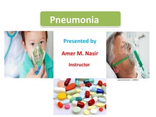
- 1. Pneumonia Presented by Amer M. Nasir Instructor
- 2. Pneumonia Pneumonia is an inflammation of the lung parenchyma caused by various microorganisms, including bacteria, mycobacteria, fungi, and viruses .Bacterial pneumonia is the most common type in adults.
- 3. • Pneumonias are classified as : 1.community acquired pneumonia (CAP). 2. hospital-acquired (nosocomial) pneumonia (HAP). 3. pneumonia in the immunocompromised host. 4. aspiration pneumonia. Pathophysiology An inflammatory reaction can occur in the alveoli, producing an exudate that interferes with the diffusion of oxygen and carbon dioxide; bronchospasm may also occur if the patient has reactive airway disease.
- 4. Risk factors of pneumonia 1. infants from birth to age 2 years, and individuals ages 65 years or older 2. people who have had a stroke, have problems swallowing, or are bedridden 3. people with weakened immune systems because of disease or use of medications such as steroids or certain cancer drugs 4. people who smoke, misuse certain types of illicit drugs, or drink excessive amounts of alcohol 5. people with certain chronic medical conditions such as asthma, cystic fibrosis, diabetes, or heart failure
- 5. • Clinical Manifestations Clinical features vary depending on the causative organism and the patient’s disease. 1. Sudden chills and rapidly rising fever (38.5C to 40.5C [101.F to 105.F]). 2. Pleuritic chest pain aggravated by respiration and coughing. 3. Severely ill patient has marked tachypnea (25 to 45 breaths/min) and dyspnea; orthopnea when not propped up. 4. Pulse rapid and bounding; may increase 10 beats/min per degree of temperature elevation (Celsius). 5. A relative bradycardia for the amount of fever suggests viral infection, mycoplasma infection, or infection with a Legionella organism.
- 6. 6. infection, headache, low-grade fever, pleuritic pain, myalgia, rash, and pharyngitis; after a few days, mucoid or mucopurulent sputum is expectorated. 7. Severe pneumonia: flushed cheeks; lips and nail beds demonstrating central cyanosis. 8. Sputum purulent, rusty, blood-tinged, viscous, or green depending on etiologic agent. 9. Appetite is poor, and the patient is diaphoretic and tires easily. 10 Signs and symptoms of pneumonia may also depend on a patient’s underlying condition (eg, different signs occur in patients with conditions such as cancer, and in those who are undergoing treatment with immunosuppressants, which decrease the resistance to infection).
- 8. Cause of pneumonia 1. Bacteria causes of pneumonia which including Streptococcus pneumoniae Haemophilus influenzae Chlamydophila pneumoniae Mycoplasma pneumonia Loginella pneumophlea 2. Viruses causes of pneumonia • Rhinoviruses • Coronaviruses • Influenza Virus • Respiratory Syncytial Virus (Rsv) • Adenovirus • Parainfluenza. • Herpes Simplex Virus
- 9. Causes of pneumonia 3- Fungi causing pneumonia Histoplasma Capsulatum Cryptococcus Neoformans Pneumocystis Jiroveci Coccidioides Immitis 4- Aspiration When aspiration gastric contain may be lead to aspiration pneumonia such as during surgery or G.E.R.D
- 10. Diagnosis test of pneumonia • • Primarily history, physical examination • Blood tests. Blood tests are used to confirm an infection and to try to identify the type of organism causing the infection. • Chest X-ray. determine the extent and location of the infection. a. • Pulse oximetry. This measures the oxygen level in blood. Pneumonia can prevent lungs from moving enough oxygen into bloodstream.
- 11. Diagnosis Sputum test. A sample of fluid from lungs (sputum) is taken after a deep cough and analyzed to help pinpoint the cause of the infection. CT scan Pleural fluid culture. A fluid sample is taken by putting a needle between ribs from the pleural area and analyzed to help determine the type of infection.
- 12. Prevention of pneumonia Vaccination against causative pneumonia Environmental Measures which includes avoid any stimulus of the pneumonia such as smoke, dust or any pollution Appropriate Treatment of other health problems. It is involved antibiotics and anti-emetics, control of the fever
- 13. • Medical Management • Antibiotics are prescribed on the basis of Gram stain results and antibiotic guidelines (resistance patterns, risk factors, etiology must be considered). Combination therapy may also be used. • Supportive treatment includes hydration, antipyretics, antitussive medications, antihistamines, or nasal decongestants. • Bed rest is recommended until infection shows signs of clearing. • Oxygen therapy is given for hypoxemia. • Respiratory support includes high inspiratory oxygen concentrations, endotracheal intubation, and mechanical ventilation. • Treatment of pleural effusion, shock, respiratory failure, or superinfection is instituted, if needed. • For groups at high risk for CAP, pneumococcal vaccination is advised.
- 14. Cholelithiasis (and Cholecystitis) Amer M. Nasir Master Degree in Medical_ Surgical Nursing
- 15. Cholelithiasis :calculi (gallstones) usually form in the gallbladder from solid constituents of bile and vary greatly in size, shape, and composition. • There are two major types of gallstones: 1. pigment stones, which contain an excess of unconjugated pigments in the bile. 2. cholesterol stones (the more common form), which result from bile supersaturated with cholesterol due to increased synthesis of cholesterol and decreased synthesis of bile acids that dissolve cholesterol. • Risk factors for pigment stones include: 1. cirrhosis, 2. hemolysis, 3. infections of the biliary tract. These stones cannot be dissolved and must be removed surgically.
- 16. Risk factors for cholesterol stones include: 1. gender (women are two to three times more likely to develop cholesterol stones). 2. use of oral contraceptives. 3. estrogens, and clofibrate; 4. age (usually older than 40 years); 5. multiparous status. 6. obesity. 7. There is also an increased risk related to diabetes, GI tract disease, T-tube fistula,and ileal resection or bypass.
- 17. Cholecystitis is refers to a painful inflammation of the gallbladder's wall. The disorder can occur a single time (acute), or can recur multiple times (chronic).
- 18. Clinical Manifestations of Cholelithiasis (and Cholecystitis) 1. May be silent, producing no pain and only mild GI symptoms 2. May be acute or chronic with epigastric distress (fullness, abdominal distention, and vague upper right quadrant pain); may follow a meal rich in fried or fatty foods. 3. If the cystic duct is obstructed, the gallbladder becomes distended, inflamed, and eventually infected; fever and palpable abdominal mass; biliary colic ,abdominal pain, radiating to back or right shoulder with nausea and vomiting several hours after a heavy meal; restlessness and constant or colicky pain. 4. Jaundice, accompanied by marked itching, with obstruction of the common bile duct, in a small percentage of patients 5. Very dark urine; grayish or clay-colored stool 6. Deficiencies of vitamins A, D, E, and K (fat-soluble vitamins)
- 19. Diagnosis of cholecystitis Cholecystogram, cholangiogram; celiac axis arteriography • Laparoscopy • Ultrasonography • Helical CT scans and MRI; ERCP • Serum alkaline phosphatase; gamma-glutamyl (GGT),gamma-glutamyl transpeptidase (GGTP), LDH • Cholesterol levels
- 20. Risk factors of cholecystis The following factors may increase the risk of developing gallstones: 1. A family history of gallstones on the mother's side of the family 2. Crohn's disease 3. Diabetes 4. End-stage kidney disease 5. Hyperlipidemia 6. Losing weight rapidly 7. Obesity 8. Older age 9. Pregnancy
- 21. Complications of cholecystitis 1. Gangrene (death tissue) 2. Preformation of gallbladder 3. Pancreatitis 4. Persistent bile duct blockage 5. Inflammation of bile common duct
- 22. • Medical Management •Achieve remission with rest, IV fluids, nasogastric suction, • analgesia, and antibiotics. • Diet immediately after an episode is usually low-fat liquids with high protein and carbohydrates followed by solid soft foods as tolerated, avoiding eggs, cream, pork.
- 23. Surgical Management • Goal of surgery is to relieve persistent symptoms, to remove the cause of biliary colic, and to treat acute cholecystitis. 1. Laparoscopic cholecystectomy: performed through a small incision or puncture made through the abdominal wall in the umbilicus. 2. Cholecystectomy: Gallbladder is removed through an abdominal incision (usually right subcostal) after ligation of the cystic duct and artery. 3. Minicholecystectomy: Gallbladder is removed through a small incision. 4. Choledochostomy: incision into the common duct for stone removal.