objectives
Structure of pituitarygland:
Anterior pituitary cell types and hormones.
Posterior pituitary cell types and hormones.
Hypothalamic control of pituitary gland:
1. Hypothalamo-hypophysial portal system.
2. Hypothalamo-hypophysial tract.
Feedback mechanisms:
➢ Positive feedback.
3.
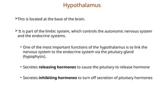
Hypothalamus
This is locatedat the base of the brain.
It is part of the limbic system, which controls the autonomic nervous system
and the endocrine systems.
• One of the most important functions of the hypothalamus is to link the
nervous system to the endocrine system via the pituitary gland
(hypophysis).
• Secretes releasing hormones to cause the pituitary to release hormone
• Secretes inhibiting hormones to turn off secretion of pituitary hormones
4.
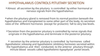
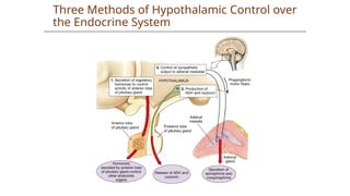
Almost all secretionby the pituitary is controlled by either hormonal or
nervous signals from the hypothalamus
when the pituitary gland is removed from its normal position beneath the
hypothalamus and transplanted to some other part of the body, its secretion
rates of the different hormones (except for prolactin) fall to very low levels.
Secretion from the posterior pituitary is controlled by nerve signals that
originate in the hypothalamus and terminate in the posterior pituitary.
secretion by the anterior pituitary is controlled by hormones called
hypothalamic releasing and hypothalamic inhibitory hormones secreted within
the hypothalamus and then conducted, to the anterior pituitary through
minute blood vessels called hypothalamic-hypophysial portal vessels.
HYPOTHALAMUS CONTROLS PITUITARY SECRETION
5.
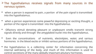
The hypothalamusreceives signals from many sources in the
nervous system.
when a person is exposed to pain, a portion of the pain signal is transmitted
into the hypothalamus.
when a person experiences some powerful depressing or exciting thought, a
portion of the signal is transmitted into the hypothalamus.
Olfactory stimuli denoting pleasant or unpleasant smells transmit strong
signals directly and through the amygdaloid nuclei into the hypothalamus.
Even the concentrations of nutrients, electrolytes, water, and various
hormones in the blood excite or inhibit various portions of the hypothalamus.
the hypothalamus is a collecting center for information concerning the
internal well-being of the body, and much of this information is used to
control secretions of the many globally important pituitary hormones.
PITUITARY GLAND ANDITS RELATION TO THE
HYPOTHALAMUS
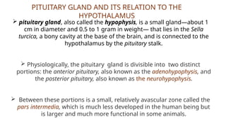
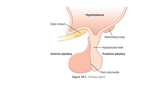
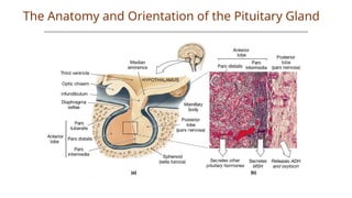
pituitary gland, also called the hypophysis, is a small gland—about 1
cm in diameter and 0.5 to 1 gram in weight— that lies in the Sella
turcica, a bony cavity at the base of the brain, and is connected to the
hypothalamus by the pituitary stalk.
Physiologically, the pituitary gland is divisible into two distinct
portions: the anterior pituitary, also known as the adenohypophysis, and
the posterior pituitary, also known as the neurohypophysis.
Between these portions is a small, relatively avascular zone called the
pars intermedia, which is much less developed in the human being but
is larger and much more functional in some animals.
9.
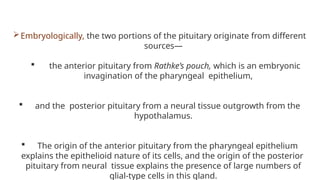
Embryologically, the twoportions of the pituitary originate from different
sources—
the anterior pituitary from Rathke’s pouch, which is an embryonic
invagination of the pharyngeal epithelium,
and the posterior pituitary from a neural tissue outgrowth from the
hypothalamus.
The origin of the anterior pituitary from the pharyngeal epithelium
explains the epithelioid nature of its cells, and the origin of the posterior
pituitary from neural tissue explains the presence of large numbers of
glial-type cells in this gland.
10.
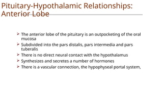
Pituitary-Hypothalamic Relationships:
Anterior Lobe
The anterior lobe of the pituitary is an outpocketing of the oral
mucosa
Subdivided into the pars distalis, pars intermedia and pars
tuberalis
There is no direct neural contact with the hypothalamus
Synthesizes and secretes a number of hormones
There is a vascular connection, the hypophyseal portal system,
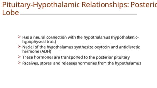
11.
Has aneural connection with the hypothalamus (hypothalamic-
hypophyseal tract)
Nuclei of the hypothalamus synthesize oxytocin and antidiuretic
hormone (ADH)
These hormones are transported to the posterior pituitary
Receives, stores, and releases hormones from the hypothalamus
Pituitary-Hypothalamic Relationships: Posterio
Lobe
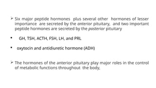
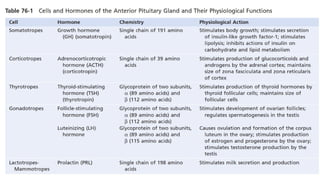
Six majorpeptide hormones plus several other hormones of lesser
importance are secreted by the anterior pituitary, and two important
peptide hormones are secreted by the posterior pituitary
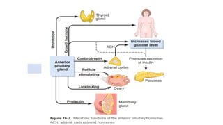
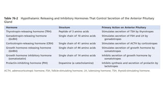
GH, TSH, ACTH, FSH, LH, and PRL
oxytocin and antidiuretic hormone (ADH)
The hormones of the anterior pituitary play major roles in the control
of metabolic functions throughout the body,
14.
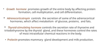
Growth hormonepromotes growth of the entire body by affecting protein
formation, cell multiplication, and cell differentiation.
Adrenocorticotropin controls the secretion of some of the adrenocortical
hormones, which affect metabolism of glucose, proteins, and fats.
Thyroid-stimulating hormone controls the secretion rate of thyroxine and
triiodothyronine by the thyroid gland, and these hormones control the rates
of most intracellular chemical reactions in the body.
• Prolactin promotes mammary gland development and milk production.
15.
.
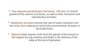
Two separategonadotropic hormones, FSH and LH control
growth of the ovaries and testes, as well as their hormonal and
reproductive activities.
Antidiuretic hormone controls the rate of water excretion into
the urine, thus helping to control the concentration of water in
the body fluids.
Oxytocin helps express milk from the glands of the breast to
the nipples during suckling and helps in the delivery of the
baby at the end of gestation
18.
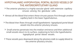
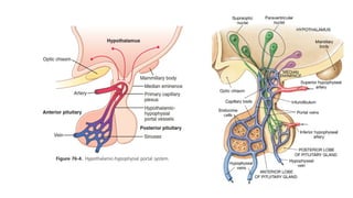
HYPOTHALAMIC-HYPOPHYSIAL PORTAL BLOODVESSELS OF
THE ANTERIORPITUITARY GLAND
The anterior pituitary is a highly vascular gland with extensive capillary
sinuses among the glandular cells.
Almost all the blood that enters these sinuses passes first through another
capillary bed in the lower hypothalamus.
The blood then flows through small hypothalamic- hypophysial portal blood
vessels into the anterior pituitary sinuses.
Small arteries penetrate into the median eminence and then additional
small vessels return to its surface, coalescing to form the hypothalamic-
hypophysial portal blood vessels.
These vessels pass downward along the pituitary stalk to supply blood to
the anterior pituitary sinuses.
20.
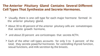
The Anterior PituitaryGland Contains Several Different
Cell Types That Synthesize and Secrete Hormones.
Usually, there is one cell type for each major hormone formed in
the anterior pituitary gland.
About 30 to 40 percent of the anterior pituitary cells are somatotropes
that secrete growth hormone,
and about 20 percent are corticotropes that secrete ACTH.
Each of the other cell types accounts for only 3 to 5 percent of the
total; they secrete powerful hormones for controlling thyroid function,
sexual functions, and milk secretion by the breasts.
21.
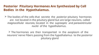
Posterior Pituitary HormonesAre Synthesized by Cell
Bodies in the Hypothalamus.
The bodies of the cells that secrete the posterior pituitary hormones
are not located in the pituitary gland but are large neurons, called
magnocellular neurons, located in the supraoptic and paraventricular
nuclei of the hypothalamus.
The hormones are then transported in the axoplasm of the
neurons’ nerve Fibers passing from the hypothalamus to the posterior
pituitary gland
22.
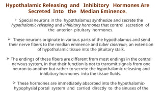
Hypothalamic Releasing andInhibitory Hormones Are
Secreted Into the Median Eminence.
Special neurons in the hypothalamus synthesize and secrete the
hypothalamic releasing and inhibitory hormones that control secretion of
the anterior pituitary hormones.
These neurons originate in various parts of the hypothalamus and send
their nerve fibers to the median eminence and tuber cinereum, an extension
of hypothalamic tissue into the pituitary stalk.
The endings of these fibers are different from most endings in the central
nervous system, in that their function is not to transmit signals from one
neuron to another but rather to secrete the hypothalamic releasing and
inhibitory hormones into the tissue fluids.
These hormones are immediately absorbed into the hypothalamic-
hypophysial portal system and carried directly to the sinuses of the
24.
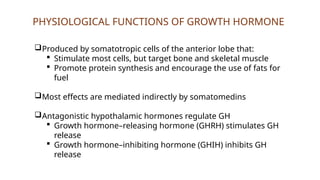
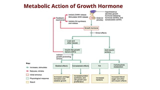
PHYSIOLOGICAL FUNCTIONS OFGROWTH HORMONE
Produced by somatotropic cells of the anterior lobe that:
Stimulate most cells, but target bone and skeletal muscle
Promote protein synthesis and encourage the use of fats for
fuel
Most effects are mediated indirectly by somatomedins
Antagonistic hypothalamic hormones regulate GH
Growth hormone–releasing hormone (GHRH) stimulates GH
release
Growth hormone–inhibiting hormone (GHIH) inhibits GH
release
25.
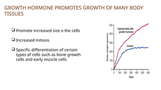
GROWTH HORMONE PROMOTESGROWTH OF MANY BODY
TISSUES
Promote increased size o the cells
Increased mitosis
Specific differentiation of certain
types of cells such as bone growth
cells and early muscle cells
26.
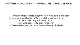
GROWTH HORMONE HASSEVERAL METABOLIC EFFECTS
1. Increased rate of proteins synthesis in most cells of the body
2- Increased mobilization of fatty acids from adipose tissue
Increased free fatty acid in the blood,
increased use of fatty acids for energy
3- Decreased rate of glucose utilization through the body
27.
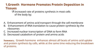
1.Growth Hormone PromotesProtein Deposition in
Tissues
A. Enhancement of amino acid transport through the cell membrane
B. Enhancement of RNA translation to cause protein synthesis by the
ribosomes
C. Increased nuclear transcription of DNA to form RNA
D. Decreased catabolism of protein and amino acids
Summary. Growth hormone enhances almost all facets of amino acid uptake
and protein synthesis by cells, while at the same time reducing the breakdown
of proteins.
Increased rate of proteins synthesis in most cells
of the body by;
28.
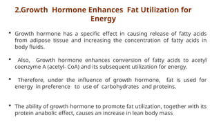
2.Growth Hormone EnhancesFat Utilization for
Energy
Growth hormone has a specific effect in causing release of fatty acids
from adipose tissue and increasing the concentration of fatty acids in
body fluids.
Also, Growth hormone enhances conversion of fatty acids to acetyl
coenzyme A (acetyl- CoA) and its subsequent utilization for energy.
Therefore, under the influence of growth hormone, fat is used for
energy in preference to use of carbohydrates and proteins.
The ability of growth hormone to promote fat utilization, together with its
protein anabolic effect, causes an increase in lean body mass.
29.
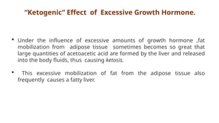
“Ketogenic” Effect ofExcessive Growth Hormone.
Under the influence of excessive amounts of growth hormone ,fat
mobilization from adipose tissue sometimes becomes so great that
large quantities of acetoacetic acid are formed by the liver and released
into the body fluids, thus causing ketosis.
This excessive mobilization of fat from the adipose tissue also
frequently causes a fatty liver.
30.
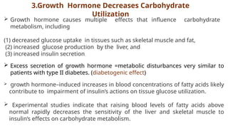
3.Growth Hormone DecreasesCarbohydrate
Utilization
Growth hormone causes multiple effects that influence carbohydrate
metabolism, including
(1) decreased glucose uptake in tissues such as skeletal muscle and fat,
(2) increased glucose production by the liver, and
(3) increased insulin secretion.
Excess secretion of growth hormone =metabolic disturbances very similar to
patients with type II diabetes. (diabetogenic effect)
growth hormone–induced increases in blood concentrations of fatty acids likely
contribute to impairment of insulin’s actions on tissue glucose utilization.
Experimental studies indicate that raising blood levels of fatty acids above
normal rapidly decreases the sensitivity of the liver and skeletal muscle to
insulin’s effects on carbohydrate metabolism.
31.
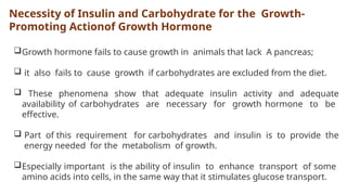
Necessity of Insulinand Carbohydrate for the Growth-
Promoting Actionof Growth Hormone
Growth hormone fails to cause growth in animals that lack A pancreas;
it also fails to cause growth if carbohydrates are excluded from the diet.
These phenomena show that adequate insulin activity and adequate
availability of carbohydrates are necessary for growth hormone to be
effective.
Part of this requirement for carbohydrates and insulin is to provide the
energy needed for the metabolism of growth.
Especially important is the ability of insulin to enhance transport of some
amino acids into cells, in the same way that it stimulates glucose transport.
32.
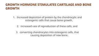
GROWTH HORMONE STIMULATESCARTILAGE AND BONE
GROWTH
1. Increased deposition of protein by the chondrocytic and
osteogenic cells that cause bone growth,
2. increased rate of reproduction of these cells, and
3. converting chondrocytes into osteogenic cells, that
causing deposition of new bone.
33.
GROWTH HORMONE EXERTSMUCH OF ITS EFFECT
THROUGH INTERMEDIATE SUBSTANCES CALLED
SOMATOMEDINS
growth hormone causes the liver to form several small proteins called
somatomedins that have the potent effect of increasing all aspects of bone
growth.
Many of the somatomedin effects on growth are similar to the effects of
insulin on growth. Therefore, the somatomedins are also called insulin-like
growth factors (IGFs).
At least four somatomedins have been isolated, but by far the most
important of these is somatomedin C (also called insulin-like growth factor-
1, or IGF-I)
The molecular weight of somatomedin C is about 7500, and its
concentration in the plasma closely follows the rate of growth hormone
34.
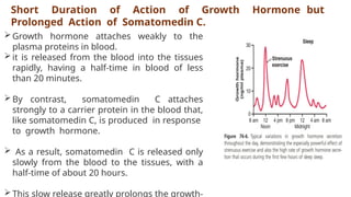
Short Duration ofAction of Growth Hormone but
Prolonged Action of Somatomedin C.
Growth hormone attaches weakly to the
plasma proteins in blood.
it is released from the blood into the tissues
rapidly, having a half-time in blood of less
than 20 minutes.
By contrast, somatomedin C attaches
strongly to a carrier protein in the blood that,
like somatomedin C, is produced in response
to growth hormone.
As a result, somatomedin C is released only
slowly from the blood to the tissues, with a
half-time of about 20 hours.
This slow release greatly prolongs the growth-
35.
REGULATION OF GROWTHHORMONE
SECRETION
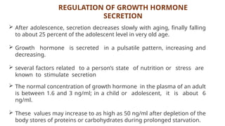
After adolescence, secretion decreases slowly with aging, finally falling
to about 25 percent of the adolescent level in very old age.
Growth hormone is secreted in a pulsatile pattern, increasing and
decreasing.
several factors related to a person’s state of nutrition or stress are
known to stimulate secretion
The normal concentration of growth hormone in the plasma of an adult
is between 1.6 and 3 ng/ml; in a child or adolescent, it is about 6
ng/ml.
These values may increase to as high as 50 ng/ml after depletion of the
body stores of proteins or carbohydrates during prolonged starvation.
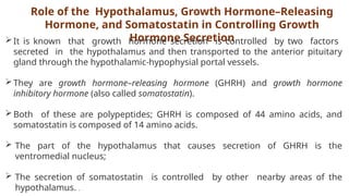
Role of theHypothalamus, Growth Hormone–Releasing
Hormone, and Somatostatin in Controlling Growth
Hormone Secretion
It is known that growth hormone secretion is controlled by two factors
secreted in the hypothalamus and then transported to the anterior pituitary
gland through the hypothalamic-hypophysial portal vessels.
They are growth hormone–releasing hormone (GHRH) and growth hormone
inhibitory hormone (also called somatostatin).
Both of these are polypeptides; GHRH is composed of 44 amino acids, and
somatostatin is composed of 14 amino acids.
The part of the hypothalamus that causes secretion of GHRH is the
ventromedial nucleus;
The secretion of somatostatin is controlled by other nearby areas of the
hypothalamus. .
40.
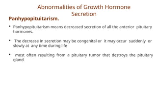
Abnormalities of GrowthHormone
Secretion
Panhypopituitarism.
Panhypopituitarism means decreased secretion of all the anterior pituitary
hormones.
The decrease in secretion may be congenital or it may occur suddenly or
slowly at any time during life
most often resulting from a pituitary tumor that destroys the pituitary
gland.
41.
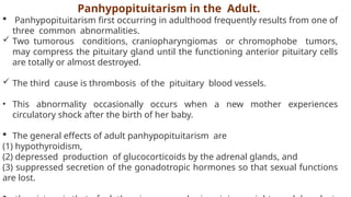
Panhypopituitarism in theAdult.
Panhypopituitarism first occurring in adulthood frequently results from one of
three common abnormalities.
Two tumorous conditions, craniopharyngiomas or chromophobe tumors,
may compress the pituitary gland until the functioning anterior pituitary cells
are totally or almost destroyed.
The third cause is thrombosis of the pituitary blood vessels.
• This abnormality occasionally occurs when a new mother experiences
circulatory shock after the birth of her baby.
The general effects of adult panhypopituitarism are
(1) hypothyroidism,
(2) depressed production of glucocorticoids by the adrenal glands, and
(3) suppressed secretion of the gonadotropic hormones so that sexual functions
are lost.
42.
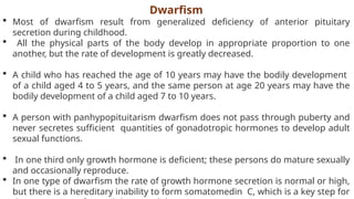
Dwarfism
Most ofdwarfism result from generalized deficiency of anterior pituitary
secretion during childhood.
All the physical parts of the body develop in appropriate proportion to one
another, but the rate of development is greatly decreased.
A child who has reached the age of 10 years may have the bodily development
of a child aged 4 to 5 years, and the same person at age 20 years may have the
bodily development of a child aged 7 to 10 years.
A person with panhypopituitarism dwarfism does not pass through puberty and
never secretes sufficient quantities of gonadotropic hormones to develop adult
sexual functions.
In one third only growth hormone is deficient; these persons do mature sexually
and occasionally reproduce.
In one type of dwarfism the rate of growth hormone secretion is normal or high,
but there is a hereditary inability to form somatomedin C, which is a key step for
43.
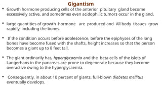
Gigantism
Growth hormoneproducing cells of the anterior pituitary gland become
excessively active, and sometimes even acidophilic tumors occur in the gland.
large quantities of growth hormone are produced and All body tissues grow
rapidly, including the bones.
If the condition occurs before adolescence, before the epiphyses of the long
bones have become fused with the shafts, height increases so that the person
becomes a giant up to 8 feet tall.
The giant ordinarily has, hyperglycaemia and the beta cells of the islets of
Langerhans in the pancreas are prone to degenerate because they become
overactive owing to the hyperglycaemia.
Consequently, in about 10 percent of giants, full-blown diabetes mellitus
eventually develops.
44.
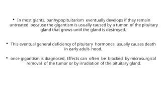
In mostgiants, panhypopituitarism eventually develops if they remain
untreated because the gigantism is usually caused by a tumor of the pituitary
gland that grows until the gland is destroyed.
This eventual general deficiency of pituitary hormones usually causes death
in early adult- hood.
once gigantism is diagnosed, Effects can often be blocked by microsurgical
removal of the tumor or by irradiation of the pituitary gland.
45.
If anacidophilic tumor occurs after adolescence—after the epiphyses of the
long bones have fused with the shafts the person cannot grow taller, but the
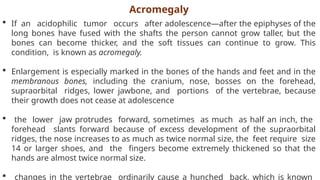
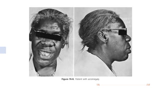
bones can become thicker, and the soft tissues can continue to grow. This
condition, is known as acromegaly.
Enlargement is especially marked in the bones of the hands and feet and in the
membranous bones, including the cranium, nose, bosses on the forehead,
supraorbital ridges, lower jawbone, and portions of the vertebrae, because
their growth does not cease at adolescence
the lower jaw protrudes forward, sometimes as much as half an inch, the
forehead slants forward because of excess development of the supraorbital
ridges, the nose increases to as much as twice normal size, the feet require size
14 or larger shoes, and the fingers become extremely thickened so that the
hands are almost twice normal size.
changes in the vertebrae ordinarily cause a hunched back, which is known
Acromegaly
47.
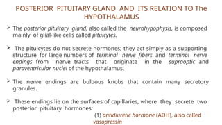
POSTERIOR PITUITARY GLANDAND ITS RELATION TO The
HYPOTHALAMUS
The posterior pituitary gland, also called the neurohypophysis, is composed
mainly of glial-like cells called pituicytes.
The pituicytes do not secrete hormones; they act simply as a supporting
structure for large numbers of terminal nerve fibers and terminal nerve
endings from nerve tracts that originate in the supraoptic and
paraventricular nuclei of the hypothalamus.
The nerve endings are bulbous knobs that contain many secretory
granules.
These endings lie on the surfaces of capillaries, where they secrete two
posterior pituitary hormones:
(1) antidiuretic hormone (ADH), also called
vasopressin
48.
If thepituitary stalk is cut above the pituitary gland but the entire
hypothalamus is left intact, the posterior pituitary hormones continue to
be secreted normally, after a transient decrease for a few days;
they are then secreted by the cut ends of the fibers within the
hypothalamus and not by the nerve endings in the posterior pituitary.
The reason for this is that the hormones are initially synthesized in the
cell bodies of the supraoptic and paraventricular nuclei and are then
transported in combination with “carrier” proteins called neurophysins
ADH is formed primarily in the supraoptic nuclei, whereas oxytocin is
formed primarily in the paraventricular nuclei.
49.
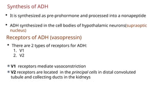
Synthesis of ADH
It is synthesized as pre-prohormone and processed into a nonapeptide
ADH synthesized in the cell bodies of hypothalamic neurons(supraoptic
nucleus)
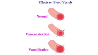
Receptors of ADH (vasopressin)
There are 2 types of receptors for ADH:
1. V1
2. V2
V1 receptors mediate vasoconstriction
V2 receptors are located in the principal cells in distal convoluted
tubule and collecting ducts in the kidneys
50.
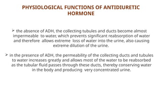
PHYSIOLOGICAL FUNCTIONS OFANTIDIURETIC
HORMONE
the absence of ADH, the collecting tubules and ducts become almost
impermeable to water, which prevents significant reabsorption of water
and therefore allows extreme loss of water into the urine, also causing
extreme dilution of the urine.
in the presence of ADH, the permeability of the collecting ducts and tubules
to water increases greatly and allows most of the water to be reabsorbed
as the tubular fluid passes through these ducts, thereby conserving water
in the body and producing very concentrated urine.
51.
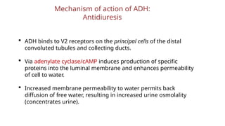
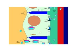
Mechanism of actionof ADH:
Antidiuresis
ADH binds to V2 receptors on the principal cells of the distal
convoluted tubules and collecting ducts.
Via adenylate cyclase/cAMP induces production of specific
proteins into the luminal membrane and enhances permeability
of cell to water.
Increased membrane permeability to water permits back
diffusion of free water, resulting in increased urine osmolality
(concentrates urine).
53.
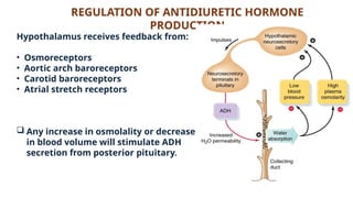
REGULATION OF ANTIDIURETICHORMONE
PRODUCTION
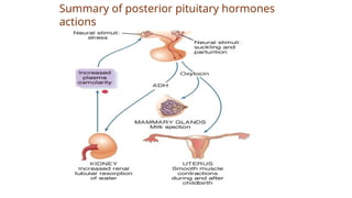
Hypothalamus receives feedback from:
• Osmoreceptors
• Aortic arch baroreceptors
• Carotid baroreceptors
• Atrial stretch receptors
Any increase in osmolality or decrease
in blood volume will stimulate ADH
secretion from posterior pituitary.
55.
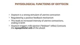
PHYSIOLOGICAL FUNCTIONS OFOXYTOCIN
• Oxytocin is a strong stimulant of uterine contraction
• Regulated by a positive feedback mechanism
• This leads to increased intensity of uterine contractions,
ending in birth
• Oxytocin triggers milk ejection (“letdown” reflex) Contracts
the myoepithelial cells of the alveoli