
Recommended
Recommended
More Related Content
Similar to physiology of muscle contraction and basics of exercise
Similar to physiology of muscle contraction and basics of exercise (20)
More from Joe Antony
More from Joe Antony (20)
Recently uploaded
Recently uploaded (20)
physiology of muscle contraction and basics of exercise
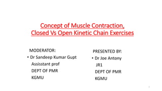
- 1. Concept of Muscle Contraction, Closed Vs Open Kinetic Chain Exercises MODERATOR: • Dr Sandeep Kumar Gupt Assisstant prof DEPT OF PMR KGMU PRESENTED BY: • Dr Joe Antony JR1 DEPT OF PMR KGMU 1
- 2. Contents • Muscle Microanatomy • Molecular characteristics of myofibril • Physiology of muscle contraction • Muscle fiber types • Factors affecting muscle strength and performance • Types of muscle contraction • Effects of exercise training • Progressive resistance exercise • Effects of ageing • Open chain exercises • Closed chain exercises 2
- 3. Muscle Microanatomy • Each skeletal muscle is made of many muscle fibers, which range in diameter between 10 and 80 μm. • Each muscle fiber contains hundreds to thousands of myofibrils. • Each myofibril comprises approximately 1500 myosin (thick) filaments and 3000 actin (thin) filaments, which are responsible for muscle contraction Braddom 6th edition 3
- 4. • Myosin and actin filaments partially interdigitate, causing myofibrils to have alternate light and dark bands. • The light bands contain only actin filaments and are called I bands (because they are isotropic to polarized light). • Dark bands contain myosin as well as ends of actin filaments where they overlap myosin, and are called A bands (anisotropic to polarized light) Braddom 6th edition 4
- 5. • The ends of actin filaments are attached to Z disks. • From Z disk, actin filaments extend in either direction, interdigitating with myosin filaments. • The Z disk passes from myofibril to myofibril, attaching myofibrils across muscle fiber. • Thus entire muscle fiber has light and dark bands, as do individual myofibrils, and thus striated appearance of muscle fib Braddom 6th edition 5
- 6. • The portion of a myofibril or whole muscle fiber between two Z disks is called a sarcomere. • The myofibrils within muscle fibers are suspended in a matrix called sarcoplasm. • The sarcoplasm contains potassium, magnesium, phosphate, enzymes, and mitochondria. • The sarcoplasm also contains sarcoplasmic reticulum, an extensive endoplasmic reticulum important in control of muscle contraction. Braddom 6th edition 6
- 7. Molecular Characteristics of Contractile Filaments • Each myosin filament is composed of 200 or more myosin molecules. • Each myosin molecule is composed of six polypeptide chains: two heavy chains and four light chains. • The two heavy chains are wrapped around each other to form a double helix, tail and arm of myosin molecule. • One end of each of chains is folded into a globular mass called myosin head. • Therefore two myosin heads are lying side by side. • The four light chains are also parts of myosin heads, two to each head Braddom 6th edition 7
- 8. • The tails of myosin molecules are bonded together, forming body of myosin filament. • Protruding from body, arm and heads of myosin molecules are called crossbridges, which are flexible at two points called hinges. • In addition to serving as a component of cross-bridge, myosin head also functions as adenosine triphosphatase (ATPase), allowing head to cleave ATP and energize contraction Braddom 6th edition,Guyton and Hall 12th edition 8
- 9. Physiology of muscle contraction • A motor unit is made up of a motor neuron and all of the skeletal muscle fibers innervated by the neuron's axon terminals, including the neuromuscular junctions between the neuron and the fibres. • muscle contraction is initiated by release of acetylcholine from motor nerve. • Acetylcholine opens protein channels in muscle fiber membrane, allowing sodium to flow into muscle fiber membrane and initiating a muscle action potential. • action potential depolarizes muscle fiber membrane, causing sarcoplasmic reticulum to release calcium. • Calcium, in turn, generates attraction between actin and myosin crossbridges, causing them to slide together. Braddom 6th edition 9
- 10. Sliding filament mechanism • In relaxed state ends of actin filaments, derived from two successive Z disks, barely overlap each other while at same time completely overlapping myosin filaments. • In contracted state actin filaments overlap each other to a great extent, and Z disks are pulled up to end of myosin filaments Braddom 6th edition 10
- 11. • Actin filaments are composed of three protein components: actin, tropomyosin, and troponin. • Several G-actin molecules form strands of F-actin. Two F-actin strands are then wound in a double helix. • One molecule of ADP is attached to each G-actin molecule. • These ADP molecules represent active sites of actin filaments with which myosin cross-bridges interact to cause muscle contraction Braddom 6th edition, Guyton and hall 12 th edition 11
- 12. • Tropomyosin molecules are wrapped around F-actin helix. In resting state, tropomyosin covers active sites of actin strands, preventing contraction. • Attached near one end of each tropomyosin molecule is a troponin molecule. • Each troponin molecule consists of three protein subunits. Troponin I has a strong affinity for actin, troponin T for tropomyosin, and troponin C for calcium Braddom 6th edition 12
- 13. • In resting state troponin-tropomyosin complex is thought to cover active sites of actin, inhibiting contraction. • In presence of calcium, this inhibitory effect is removed, allowing contraction to proceed. Braddom 6th edition 13
- 14. Muscle fibre types Type 1 fiber • slow oxidative • best suited for endurance activities requiring aerobic metabolism. Type 2 fiber • fast twitch • most active during activities requiring strength and speed. • 2A (fast, oxidative glycolytic) • a type of hybrid that retains some oxidative capacity. • 2B (fast glycolytic) Braddom 6th edition 14
- 15. Braddom 6th edition 15
- 16. During anaerobic activity, • slow oxidative fibers (type 1) are used almost exclusively at intensities below 70% of ˙VO2max. • Beyond this intensity, anaerobic pathways are stimulated. • At ˙VO2max, both type 1 and type 2 are relied on, and oxygen debt eventually occurs secondary to aerobic metabolism Braddom 6th edition 16
- 17. During isometric contractions, • type 2 fibers are generally recruited when force exceeds 20% of maximal voluntary contraction. • If sustained for long periods, however, type 2 can be recruited at thresholds below 20% of maximal voluntary contraction. Braddom 6th edition 17
- 18. Muscle fibre orientation • Parallel muscles have their fibers arranged parallel to length of muscle and produce a greater range of motion than similarsized muscles with pennate arrangement. • Pennate muscles have shorter fibers that are arranged obliquely to their tendons (similar to makeup of a feather), increasing cross-sectional area and power produced. Braddom 6th edition 18
- 19. Factors affecting muscle strength and perfomance • Cross sectional area • The ability of a muscle to produce force is directly proportional to its cross- sectional area. • For parallel muscles, this corresponds to cross-section at bulkiest part of muscle. • For pennate muscles, multiple cross-sections are taken at right angles to each of muscle fibers. • Pennate muscles are particularly adapted to force production because many more muscle fibers are contained in pennate muscles and these fibers are shorter Braddom 6th edition 19
- 20. • Length-Tension Relationship • Maximum force of contraction occurs when a muscle is at its normal resting muscle length. • For muscles, this corresponds to about midrange of joint motion or slightly longer, and is length at which tension just begins to exceed zero. • If a muscle is stretched beyond resting length before contraction, resting tension develops, and active tension (the increase in tension during contraction) decreases. • Efficiency (percentage of energy that is converted into work instead of heat) occurs at a velocity of contraction of approximately 30% of maximal. Braddom 6th edition 20
- 21. • Torque-Velocity Relationship • The greatest amount of force is generated by a muscle during fast eccentric (lengthening) contractions. • The least amount of force is produced during fast concentric (shortening) contractions. • The amount of force developed in order of most force to least force can be summarized as follows: fast eccentric, isometric, slow concentric, and fast concentric Braddom 6th edition 21
- 22. Types of muscle contraction • Isometric contractions - contractions in which there is no change in length of muscle. • No joint or limb motion occurs. • internal force does not overcome external force – Exertion against immovable objects or by holding joint in a static position • characteristics of isotonic contraction depend on the load against which muscle contracts, as well as inertia of load Braddom 6th edition 22
- 23. • Isotonic contractions occur when muscle changes length, producing limb motion • Concentric contractions occur when muscle shortens. • Eccentric contractions occur when muscle lengthens. More fast twitch fibers are recruited during eccentric contractions. Braddom 6th edition, Board review 3 rd edition 23
- 24. • Isokinetic contractions -occur when muscle contraction is performed at a constant velocity. • This can be done only with assistance of a preset rate-limiting device. • This type of exercise does not exist in nature. Braddom 6th edition 24
- 25. Effects of exercise training • The SAID principle (specific adaptations to imposed demands) states that a muscle will adapt to a specific demand imposed on it, making it better able to handle greater load. • Neural Adaptations • Observed strength gains within first few weeks of a weightlifting program are mostly because of neuromuscular adaptations. • The nervous system recruits larger motor units with higher frequencies of stimulation to provide force necessary to overcome imposed resistance. • Early strength gains and increased muscle tension production from training therefore result from a more efficient neural recruitment process. • This means that most of improvement in strength-related functional activities gained on inpatient rehabilitation units is attributable to neural recruitment rather than muscle hypertrophy, as a result of relatively short length of stays Braddom 6th edition 25
- 26. Muscle Hypertrophy • Enlargement of total muscle mass and cross-sectional area. • Muscle hypertrophy is more common in fast-twitch than in slow- twitch muscles. • Type 2A fibers exhibit greatest growth, more so than type 2B and type 1 fibers. • Muscle hypertrophy is typically experienced after 6 to 7 weeks of resistance training. • Conversely, muscle atrophy resulting from disuse occurs primarily in type 2 fibers Braddom 6th edition 26
- 27. • Virtually all muscle hypertrophy occurs from hypertrophy of individual muscle fibers. • During muscle hypertrophy rate of muscle contractile protein synthesis is greater than decay, leading to greater numbers of actin and myosin filaments in myofibrils. • The myofibrils within each muscle fiber split, resulting in more myofibrils in each muscle fiber. • Only under very rare conditions of extreme muscle force generation do numbers of muscle fibers increase (fiber hyperplasia), and even then by only a few percent. Braddom 6th edition 27
- 28. • When muscles are stretched to a greater-than-normal length, causing new sarcomeres to be added at ends of muscle fibers where they attach to tendons. • Conversely, when a muscle remains shortened at less than its resting length, sarcomeres at end of muscle fibers disappear Braddom 6th edition 28
- 29. Progressive resistance exercise • Delorme • three sets are performed for each exercise. • Ten repetitions are performed in each set. • The weight for the first set is 50% of the 10-RM; the second set, 75% of the 10-RM; and the third set, 100% of the 10-RM. • This is usually referred to clinically as progressive resistive exercise. Braddom 6th edition 29
- 30. • Oxford • individual starts with 10 repetitions at 100% of the 10-RM, • then 10 repetitions at 75% of the 10-RM, • then a third set of 10 repetitions at 50% of the 10-RM. • This is usually referred to in the clinical setting as regressive resistive exercise. 30
- 31. DAPRE (Daily Adjusted Progressive Resistence Exercise) • The 1st set in DAPRE involves 10 repetitions at 50% of the athlete’s predetermined 6-RM. • The 2nd set consists of six repetitions at 75% of the athlete’s 6-RM. • The 3rd set consists of as many repetitions as can be performed at the athlete’s 6-RM. • The number of repetitions performed in the third set determines the resistance for the fourth set. • If 5-7 repetitions were performed in the third set, resistance stays the same. • If fewer than 5 repetitions were performed in the third set, the weight is lowered by 5 lb. • If more than 7 repetitions are performed in the third set, the weight is raised by 5 lb. • 4th set consists of as many repetitions as can be performed to fatigue • working weight for the next day is established based on the athlete’s performance during the 4th set 31
- 32. Effects of Aging • Although it was previously believed that strength training in older adults was attributable only to learning or neural factors, reports have also demonstrated that muscles of older individuals can demonstrate hypertrophy after strength training 32
- 33. Open-Chain Exercises • Involve motions in which distal segment (hand or foot) is free to move in space, without necessarily causing simultaneous motions at adjacent joints • Limb movement only occurs distal to moving joint. Muscle activation occurs in muscles that cross moving joint. • For example, during knee flexion in an open-chain exercise action of hamstrings is independent of recruitment of other hip or ankle musculature. • Open-chain exercises also are typically performed in non-weight-bearing positions. • In addition, during resistance training, exercise load (resistance) is applied to moving distal segment. 33
- 34. 34
- 35. Closed-chain exercises • involve motions in which body moves on a distal segment that is fixed or stabilized on a support surface. • Movement at one joint causes simultaneous motions at distal as well as proximal joints in a relatively predictable manner. • For example, when a patient is performing a bilateral short-arc squatting motion (mini-squat) and then returning to an erect position, as knees flex and extend, hips and ankles move in a predictable pattern along with knees. • Closed-chain exercises are primarily performed in weight- bearing positions Bilateral closed-chain resisted hip and knee extension. 35
- 36. Examples in upper extremities include balance activities in quadruped, sitting press-ups, wall push-offs, or prone push- ups; examples in lower extremities include lunges, squats, step- up or step-down exercises, or heel rises 36
- 37. CHARACTERISTICS OF OPEN & CLOSED CHAIN EXERCISES 37
- 38. Rationale for Use of Open-Chain and Closed-Chain Exercises • Rationale is based on goals of an individualized rehabilitation program and a critical analysis of potential benefits and limitations inherent in either form of exercise. • Functional activities involve many combinations and considerable variations of open- and closed-chain motions • So, inclusion and integration of task-specific open-chain and closed- chain exercises into a rehabilitation or conditioning program is both appropriate and prudent. 38
- 39. Rationale of Open-Chain Training • Because open-chain training typically is performed in nonweight-bearing postures, it may be only option when weight bearing is contraindicated or must be significantly restricted or when unloading in a closed-chain position is not possible. • Soft tissue pain and swelling or restricted motion of any segment of chain may also necessitate use of open-chain exercises at adjacent joints. • Any activity that involves open-chain motions can easily be replicated with open-chain exercises, first by developing isolated control and strength of weak musculature and then by combining motions to simulate functional patterns. 39
- 40. • After a fracture of tibia, for example, lower extremity usually is immobilized in a long leg cast, and weight bearing is restricted for at least a few weeks. • During this period, hip strengthening exercises in an open chain manner can still be initiated and gradually progressed until partial weight bearing and closed-chain activities are permissible. 40
- 41. Closed-Chain Exercises and Weight-Bearing Restrictions • While partial weight bearing on involved extremity. This is simple to achieve in upper extremity; but in lower extremity, because patient is in an upright position during closed-chain exercises, axial loading in one or both lower extremities must be reduced 41
- 42. Parameters and Progression of Closed- Chain Exercises 42
- 43. Thank you • References • Braddoms 6th edition • Guyton and hall 12 th edition • ACSM’s exercise physiology • Board review of physical medicine and rehabilitation 43
Editor's Notes
- Parallel- abdominal uscles Multipennate- pectoralis major,deltoid
- Isometric has developed as per funvtion. Occular muscle contract immmediatly,
- Repetition maximum (RM): "maximal number of times a load can be lifted before fatigue using good form and technique (ACSM, 1998)." A "1RM" signifies the maximum resistance a person can move in one repetition of an exercise