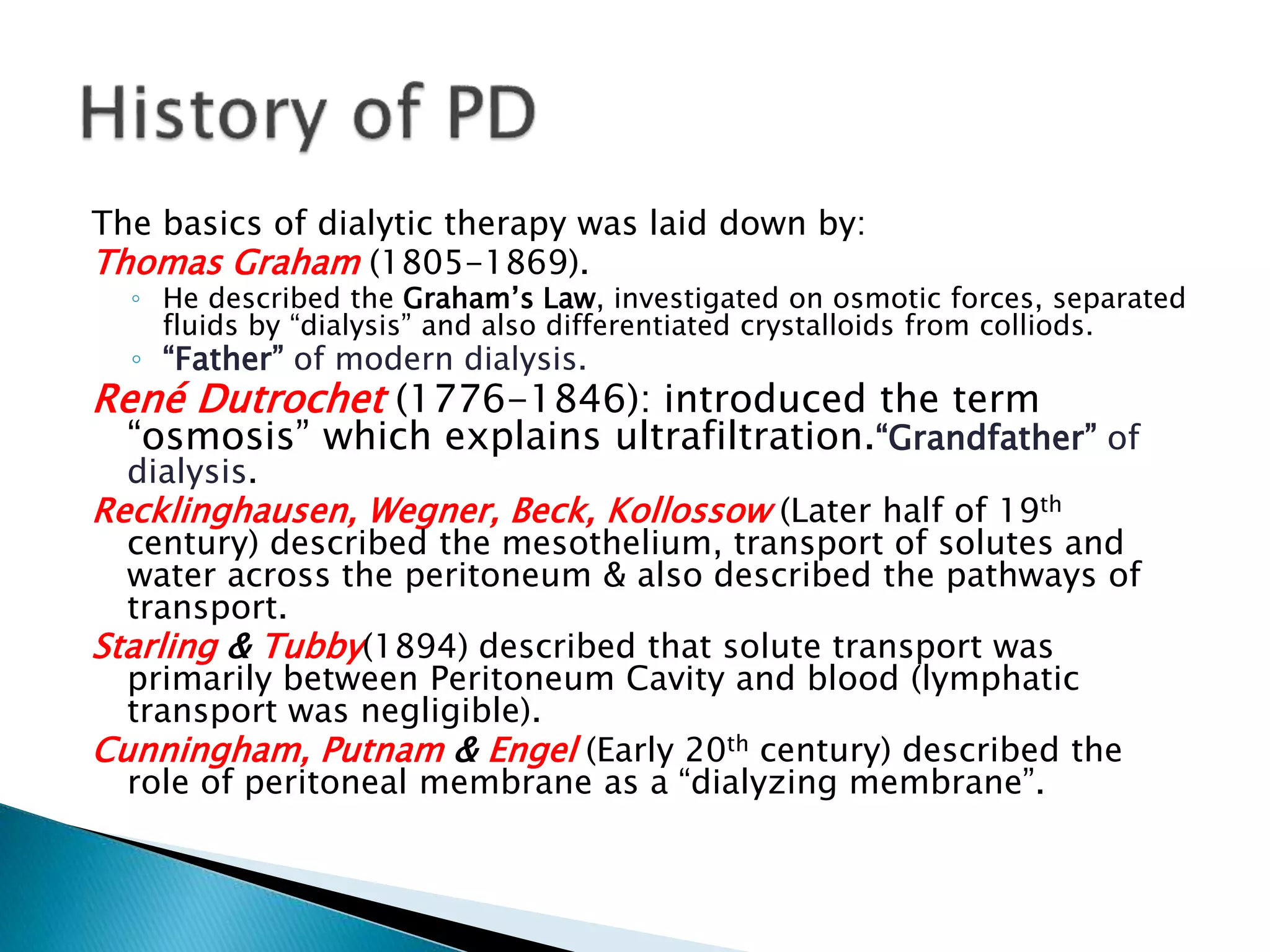
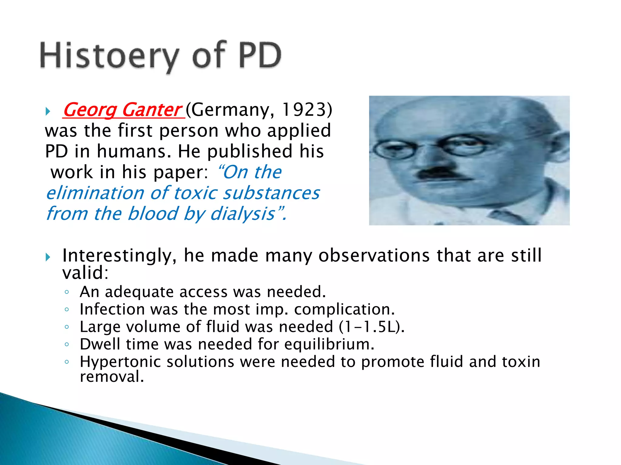
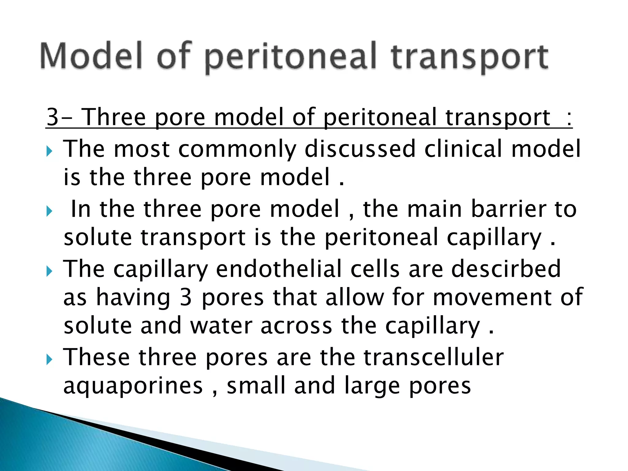
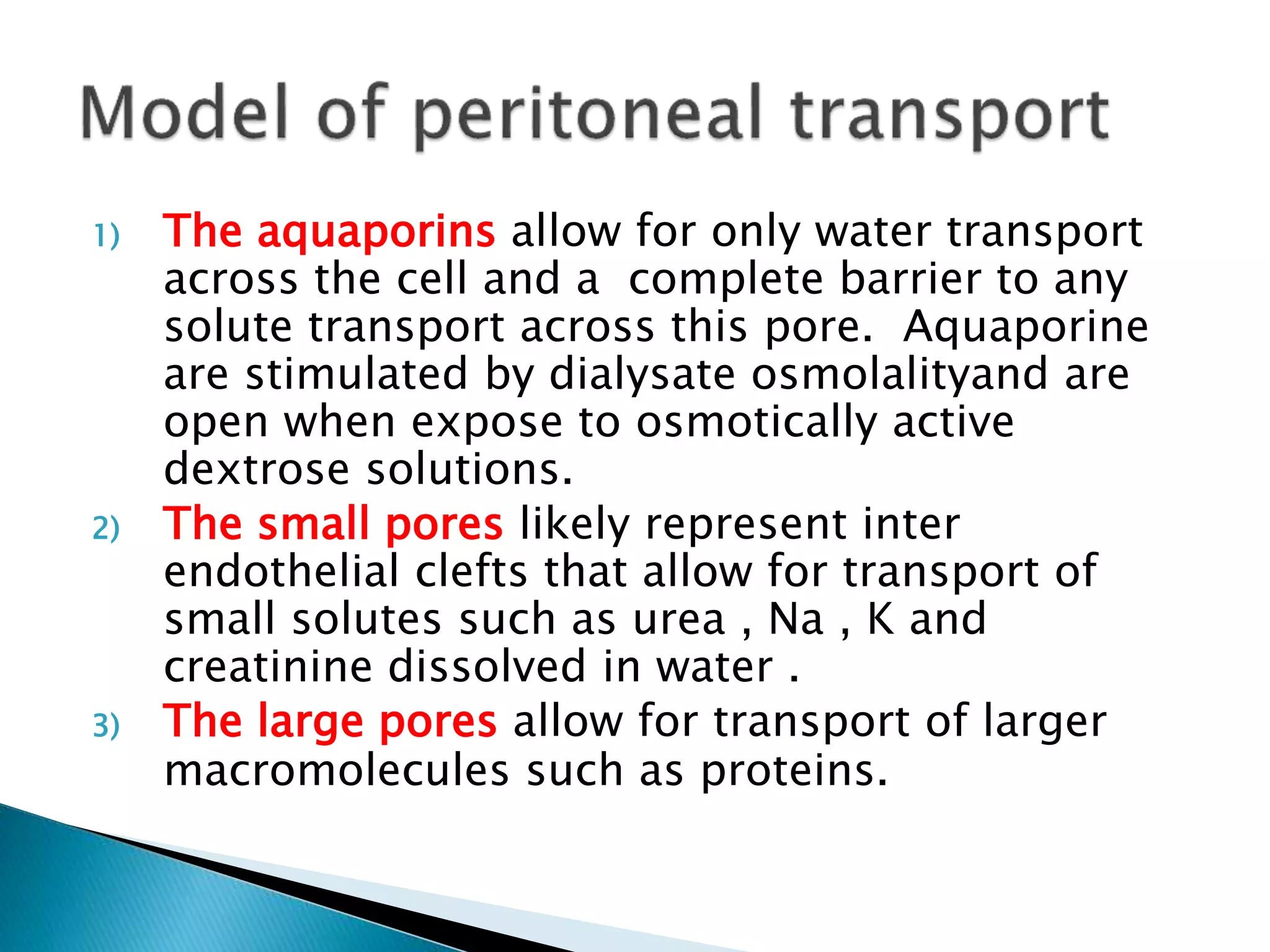
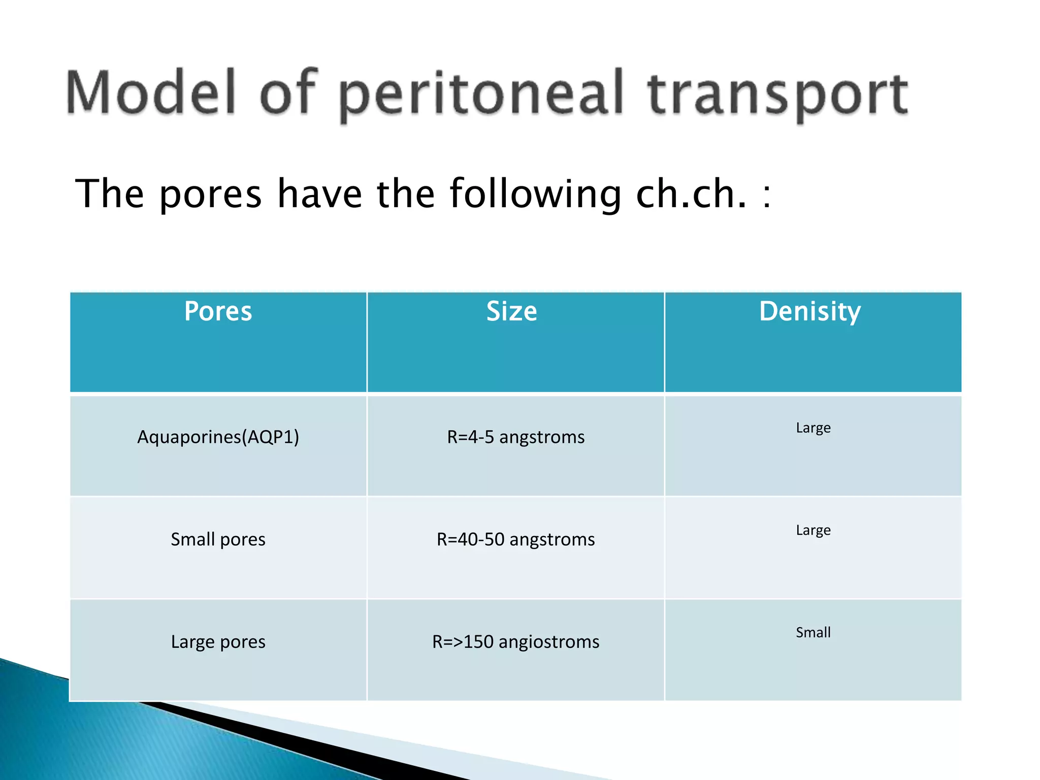
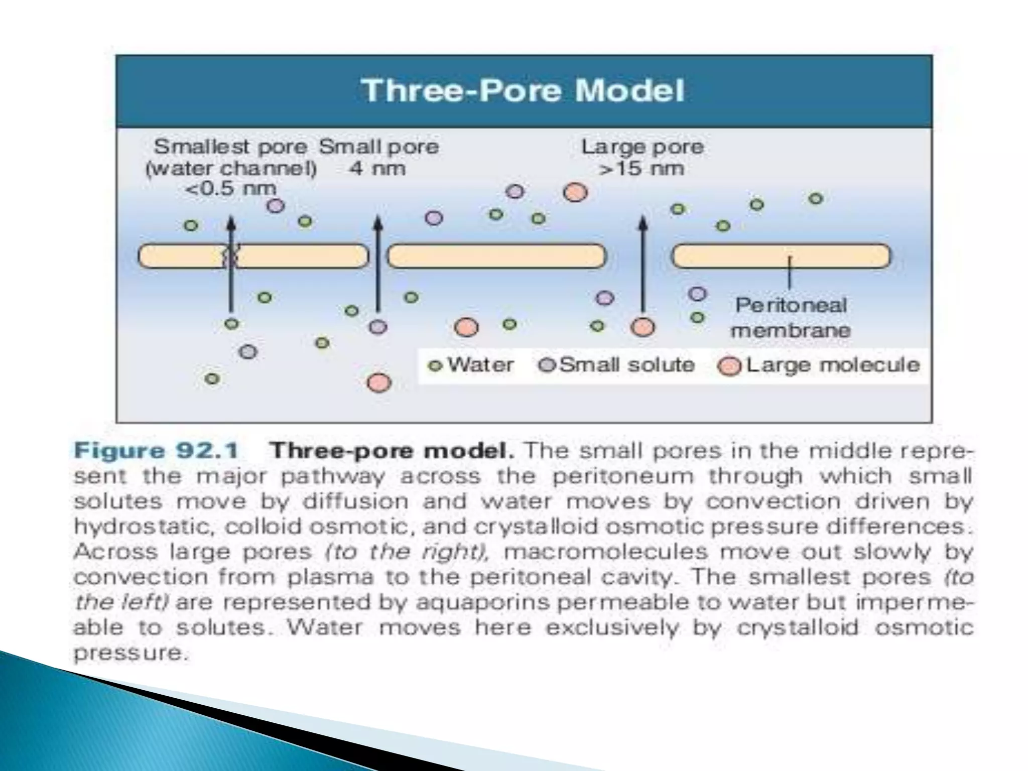
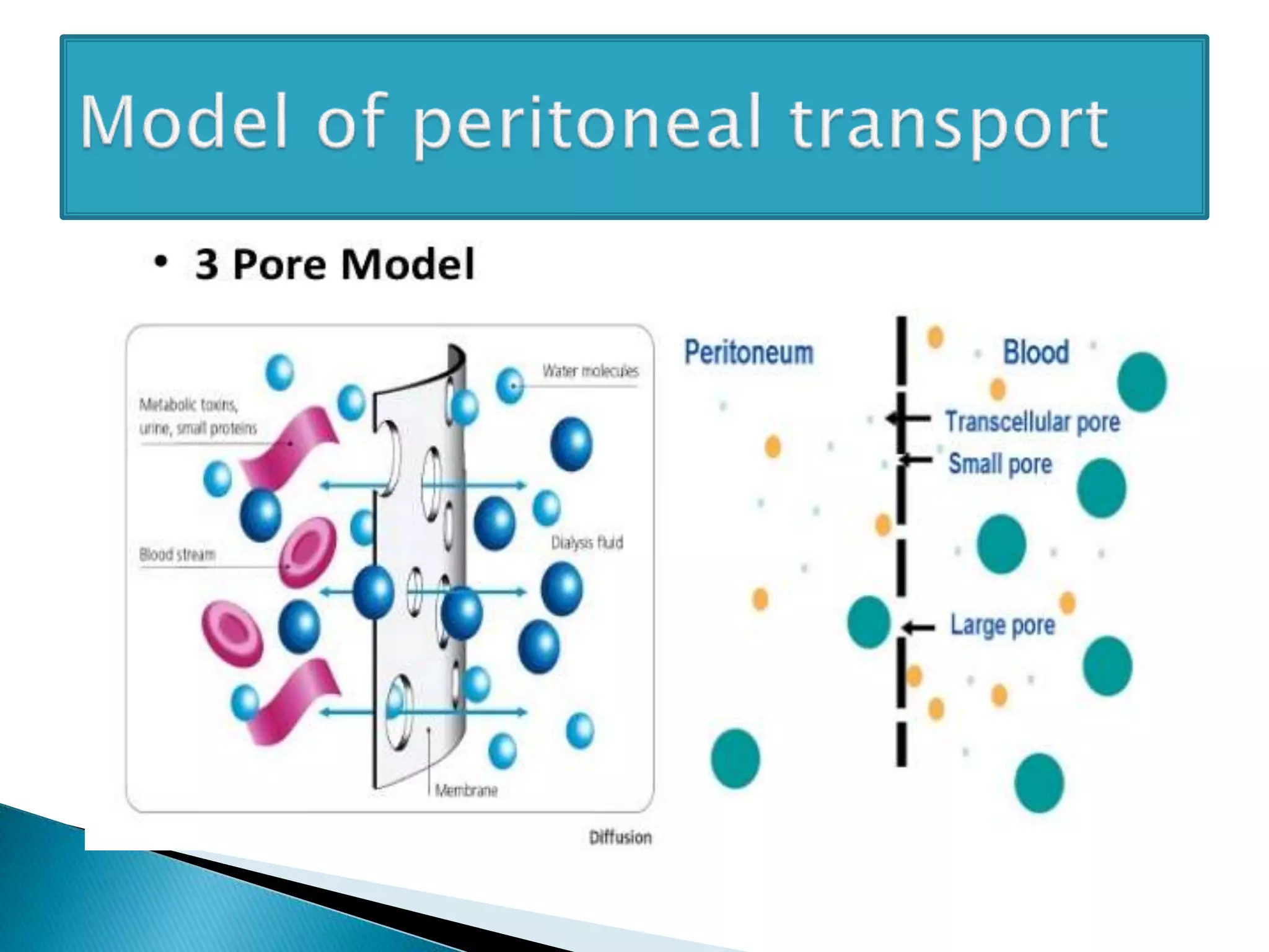
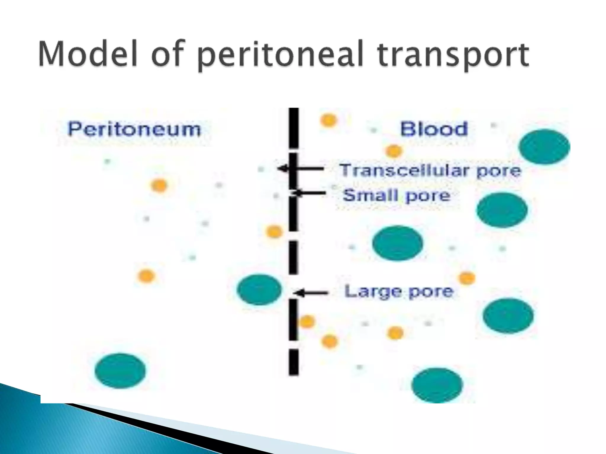
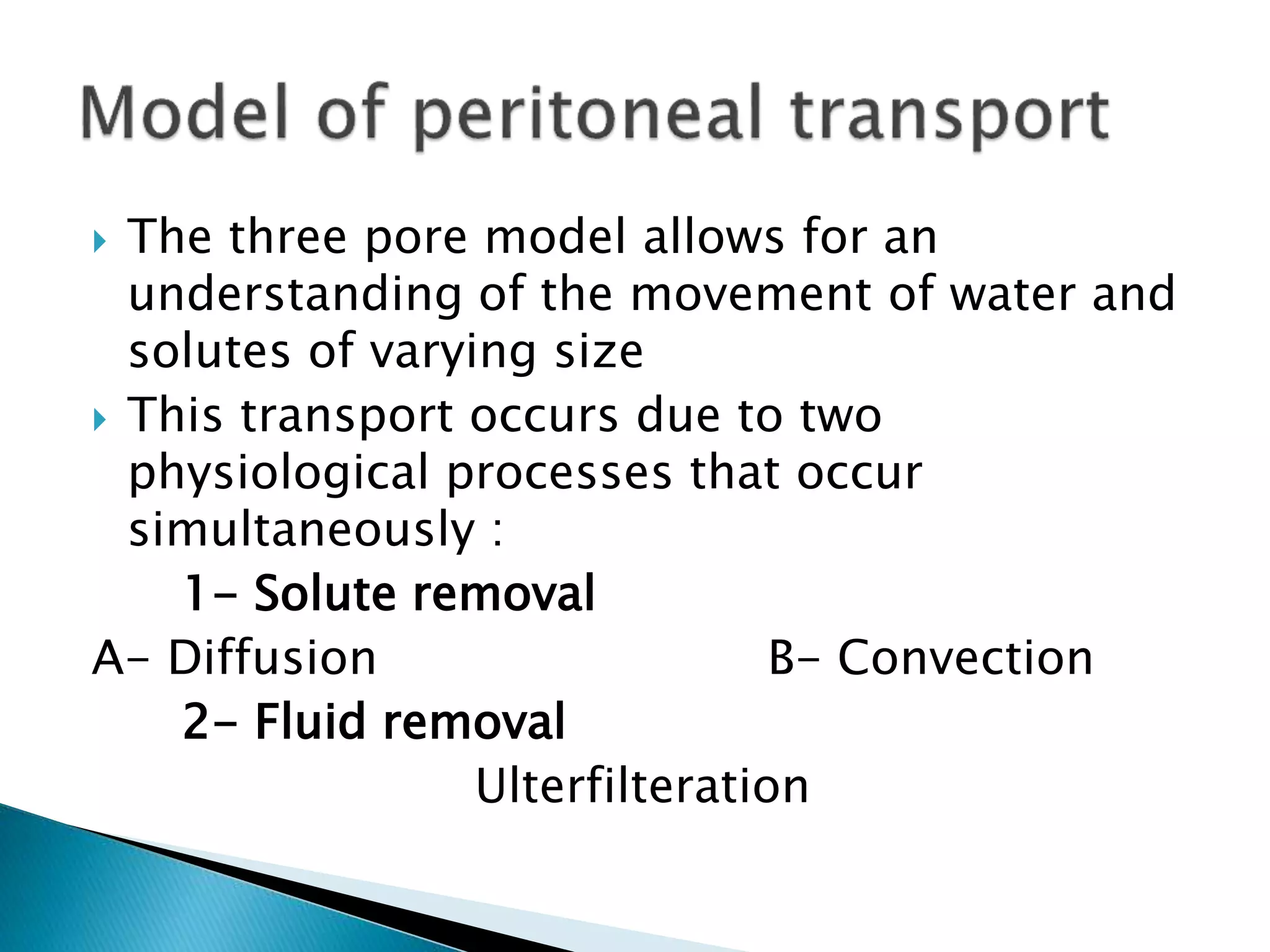
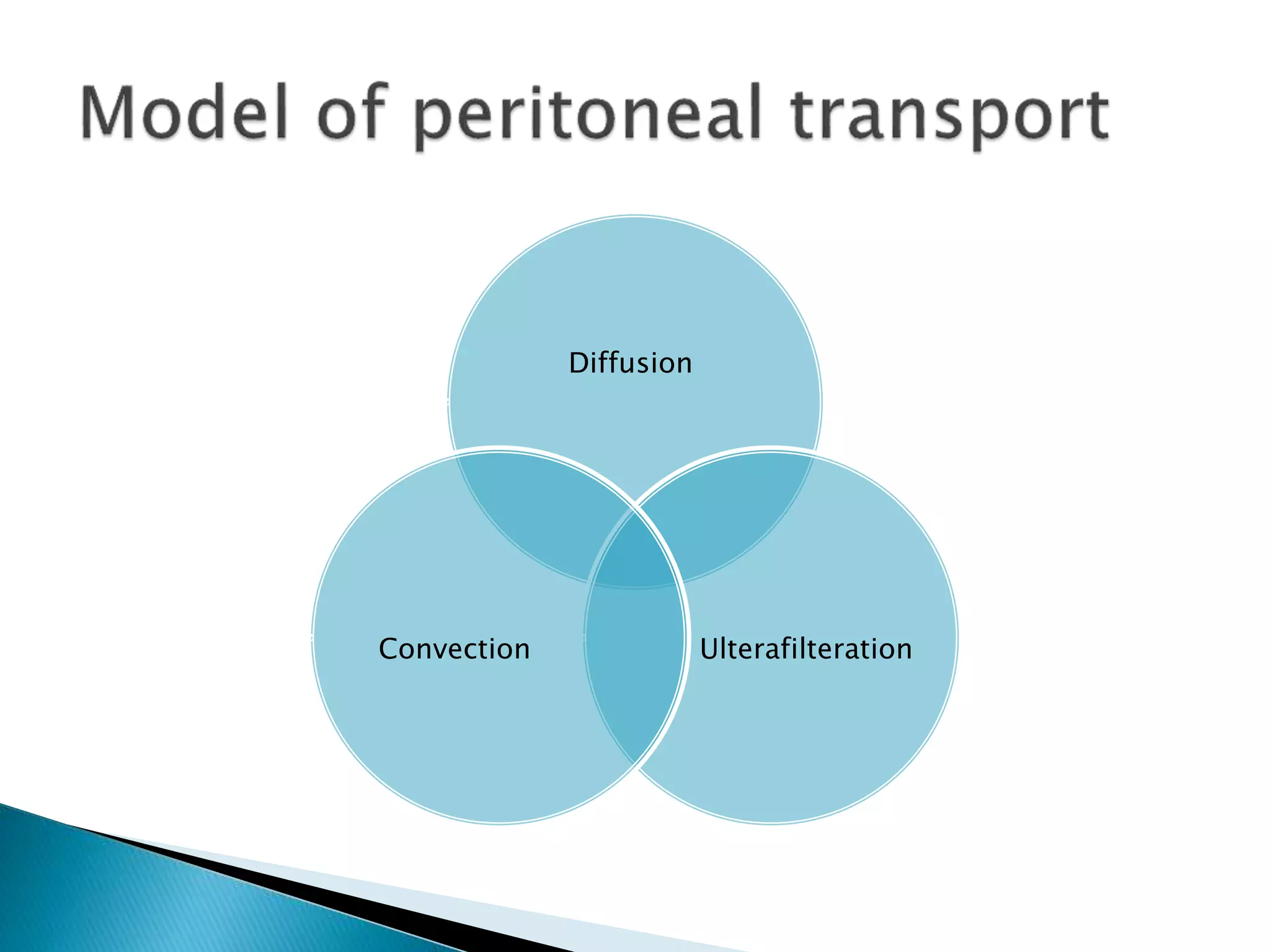
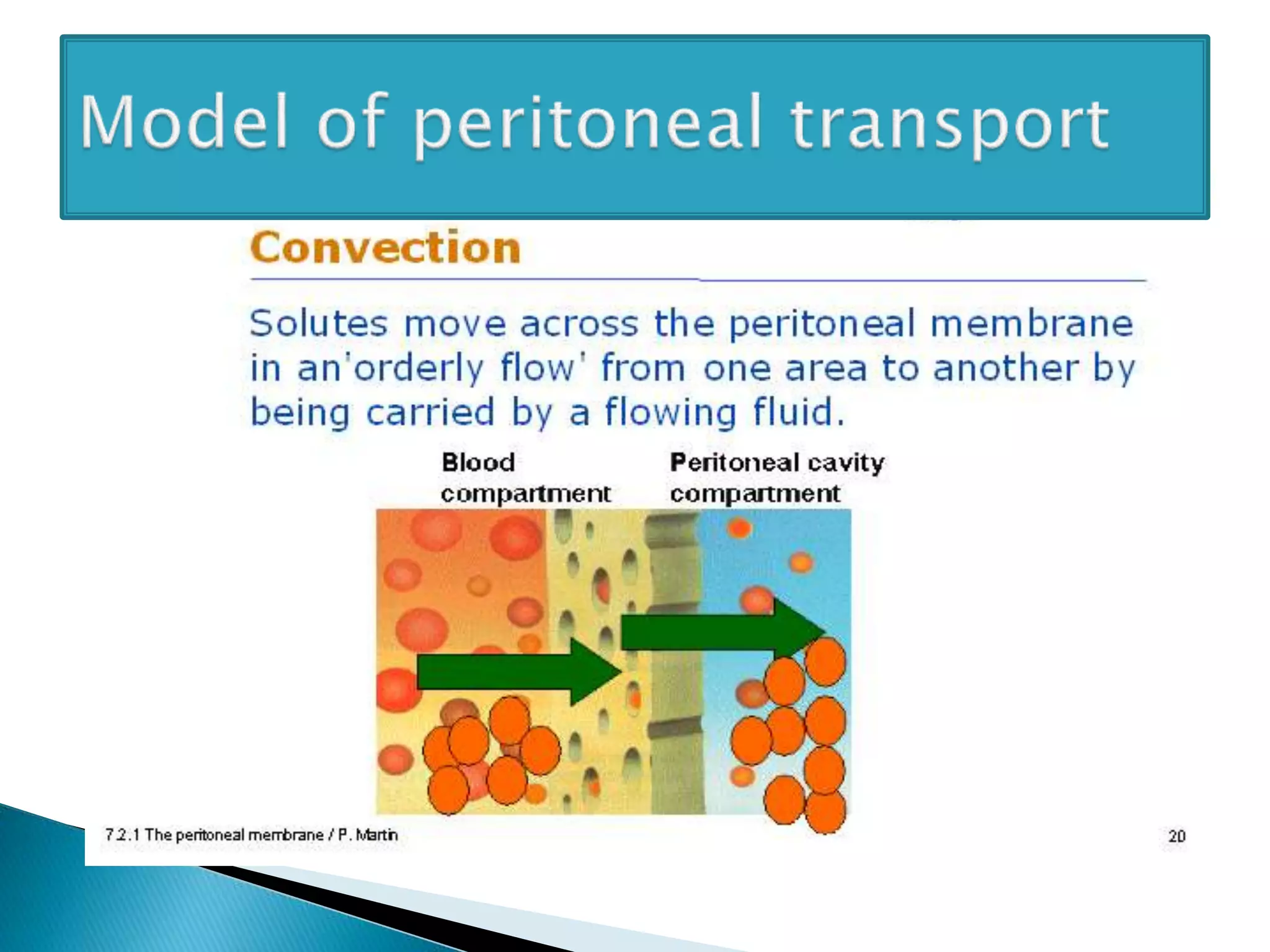
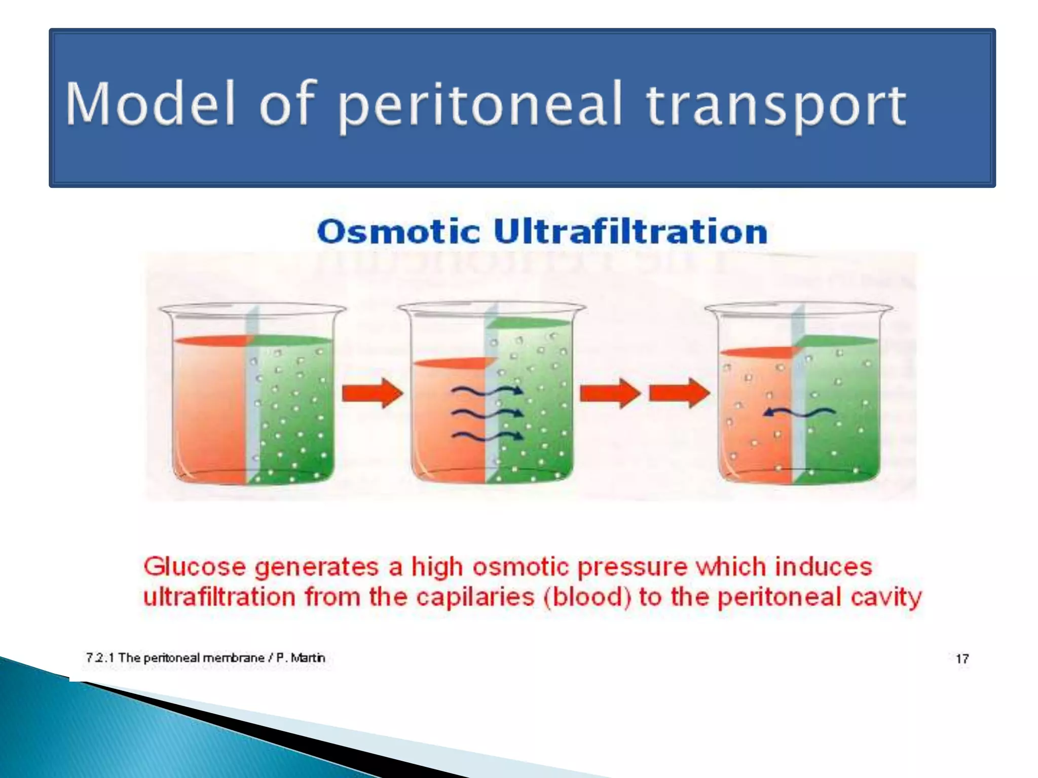
By
Dr Ahmed Salah Younes
Nephrologist at Rustaq Hospital
An understanding of membrane physiology is
required for proper determination of the PD
prescription .
Dr .steven Guest
The basics of dialytic therapy was laid down by:
Thomas Graham (1805-1869).
◦ He described the Graham’s Law, investigated on osmotic forces, separated
fluids by “dialysis” and also differentiated crystalloids from colliods.
◦ “Father” of modern dialysis.
René Dutrochet (1776-1846): introduced the term
“osmosis” which explains ultrafiltration.“Grandfather” of
dialysis.
Recklinghausen, Wegner, Beck, Kollossow (Later half of 19th
century) described the mesothelium, transport of solutes and
water across the peritoneum & also described the pathways of
transport.
Starling & Tubby(1894) described that solute transport was
primarily between Peritoneum Cavity and blood (lymphatic
transport was negligible).
Cunningham, Putnam & Engel (Early 20th century) described the
role of peritoneal membrane as a “dialyzing membrane”.
 Georg Ganter (Germany, 1923)
was the first person who applied
PD in humans. He published his
work in his paper: “On the
elimination of toxic substances
from the blood by dialysis”.
 Interestingly, he made many observations that are still
valid:
◦ An adequate access was needed.
◦ Infection was the most imp. complication.
◦ Large volume of fluid was needed (1-1.5L).
◦ Dwell time was needed for equilibrium.
◦ Hypertonic solutions were needed to promote fluid and toxin
removal.
Richard Ruben (San Fransisco, 1959): he was
the first to initiate long term IPD in CRF. The
first patient was Mae Stewart, 33/F.
 Boen (Seattle, 1962):
◦ First long term PD programmed.
◦ First automated PD machine.
◦ Repeated puncture method using “Boen’s button.”
◦ develop fluid factory ,
◦ explain diffusion curve ,
◦ peritoneal clearance ,
◦ described the influnce of glucose on ulterafilteration
◦ modeled the correction of metabolic acidosis with add of
bicarbonate to fliud ,
◦ He develop completely closed system with limit the risk of
infection
◦ He designed larger infusion bottles to allow for repeated
infusion , monitor fluid removed he automated the entire
PD system at home
 The PD take place between the blood in the capillary
located in intersteium of the peritonium and infused
dialysis solution across peritoneal membrane
 The peritoneal membrane serves as dialyzing membrane
that can allow solutes to move from the capillary blood
compartment to the dialysate in the peritoneal cavity
 The peritoneal membrane act as semipermeable
membrane had a surface area about 1 to 2 m2
 The total membrane area includes the visceral peritoneum
(60%) , peritoneal covering the mesentery and omental
surfaces (30%) , and the parital peritoneum (10%)
 The peritoneal membrane has an extensive
blood supply
1. The visceral peritoneum supplied by
mesenteric arteries that drain ultimately
into the portal circulation
2. The parietal peritoneium supplied by
smaller epigasteric , intercostals and lumber
arteries that drain directely into inferior
vena cava the estimated total peritoneal
blood flow is 50 to 100 ml/min
 Three major peritoneal components could filter
toxins from the blood compartment into
peritoneal space :
1- Mesothelial cell layer
2- Interstitial space
3- Capillary endothelium and basement membrane
(most important to determined solute transport )
 The parietal peritoneum is more important in
transport than the visceral as only 25-30 % of
the viscarl peritoneum it is in contact with the
peritoneal fluid.
 Several theoretical constructs have been
described to help determine the transport of
solutes across the peritoneal capillary bed .
 three of the most discuss models are the :
1- The distributed model
2- Three pore model
3- Pyle – popvich model
1- The distributed model of peritoneal
transport
 The distributed model is used largely in the
research setting and is much more
complicated , mathematically , and not used
clinically
 In the distributed model , capillary are
described as being distributed throughout
the peritoneal membrane and are at variable
distance from peritoneal cavity ,
 Solute transport is therefore affected by the
blood dialysate distance (the amount of
intersitium ) ,
2- Pyle –popvich model
 The Pyle-popovich model , the physiological
reality is simplified by considering just two
homogeneous compartments (body and
dialysate) separated by an ideal homoporous
semi-permeable membrane with constant
characteristics and nil thickness.
 Treats the peritoneal membrane similarly to a
hemodialyzer membrane .
 The mass transfer area coefficient (MTAC) : is
determined without taking into consideration the
specific anatomic factor such as the interstitium
or capillaries
3- Three pore model of peritoneal transport :
 The most commonly discussed clinical model
is the three pore model .
 In the three pore model , the main barrier to
solute transport is the peritoneal capillary .
 The capillary endothelial cells are descirbed
as having 3 pores that allow for movement of
solute and water across the capillary .
 These three pores are the transcelluler
aquaporines , small and large pores
1) The aquaporins allow for only water transport
across the cell and a complete barrier to any
solute transport across this pore. Aquaporine
are stimulated by dialysate osmolalityand are
open when expose to osmotically active
dextrose solutions.
2) The small pores likely represent inter
endothelial clefts that allow for transport of
small solutes such as urea , Na , K and
creatinine dissolved in water .
3) The large pores allow for transport of larger
macromolecules such as proteins.
The pores have the following ch.ch. :
DenisitySizePores
Large
R=4-5 angstromsAquaporines(AQP1)
Large
R=40-50 angstromsSmall pores
Small
R=>150 angiostromsLarge pores
 The three pore model allows for an
understanding of the movement of water and
solutes of varying size
 This transport occurs due to two
physiological processes that occur
simultaneously :
1- Solute removal
A- Diffusion B- Convection
2- Fluid removal
Ulterfilteration
Diffusion
UlterafilterationConvection
Diffusion :
 the predominant mechanism of small solute
transport in PD .
 Diffusion clearance is dependent on many factors
:
1- The effective peritoneal membrane surface area
2- The solute concentration gradients from blood
to dialysate
3- The dwell time of dialysate in the peritoneal
cavity
4- Solute characteristic
5- The dialysate flow rate
 Diffusion occurs from the blood into the
dialysate as well as from the dialysate into
blood .
 example , uremic toxins diffuse down , a
concentration gradient into the dialysate
while dialysate lactate and glucose diffuse
into the capillary blood supply .
 Substances with smaller molecular weight
diffuse more rapidly than those with larger
moleculer weight .
 Urea diffuse more rapidly than creatinine or
middle molecules .
 Peritoneal diffusion of solute can vary patient
to patient and are determined by the
vascularity of peritoneal membrane and
inflammatory state.
 Kinetic of diffusion solute transport
Accordinto the Fick’s Law, is :
Js=(Df/Δx).A.ΔC
Where :
Js=rate of solute transport,
Df=diffusion coefficient,
Δx=diffusion distance,
A=Surface area,
ΔC=concentration gradient.
 MTAC
The “Permeability surface area cross product” or the “Mass
Transfer Area Coefficient (MTAC)”
Is theoretically equal to the diffusive clearance of a solute per unit
time when the dialysate flow is infinitely high so that the solute
gradient is always maximal.”
Or in EnglishTheoretical maximal clearance of a solute at time zero
Js=(Df/Δx).A.ΔC
 MTAC values of small molecular weight
substances are representative of the functional
surface area.
 Restriction coefficient on the other hand is a
representation of the size-selectivity.
 MTAC values:
1. Urea=17mL/min.
2. Creatinine=10mL/min
 The D/P ratio has a good correlation with the
MTAC values
Convection:
 Convection occurs when dissolved solutes are
small enough to move through the pores as
water is moving across the capillary , in response
to an osmotic force .
 More specifically , as dialysate glucose creates an
osmotic force that attracts water from the
capillary blood space , solutes that are dissolved
in that water move into the dialysate , resulting
in clearance of those solutes from the blood by
this convection process often called (solute drag)
 Middle molecules such as B2-microglobulin
move into the peritoneal cavity predominant
by convection .
 The combined diffusive and convective
clearance of molecules can be modeled over
varying time points
Ulterafilteration :
 Ulterafilteration refer to the process of fluid
movement across the peritoneal membrane in
response to osmatic force
 Ultrafilteration occurs across both the
aquaporins and the small pores ,
 aquaporin mediated water movement
accounting for 40-50%of total
ulterafilteration and the small pores
accounting for 50-60% of the total
ulterafilteration .
 Ulterafilteration volume can be modified by use
of different osmotically active PD solution , lower
osmatic solutions (1.5%dextrose concentrations)
can create a peak ulterfilteration of over one
hundered ml . higher osmatic forces (4.25%
dextrose ) result in larger movements of
ulterafilteration
 As dextrose is absorbed across the peritoneium
and enters the capillary the osmatic gradient
slowly dissipates, resulting in cessation of net
ulterfilteration and re-absorption of dialysate
 we can conclude that icodextrin, due to to its
high molecular weight induces colloid
osmosis even when it is a iso-hypoosmotic
solution.
 This movement of fluid takes place through
the “small pore system”.
 Due to the hypo-osmolality of this solution,
no movement of fluid takes place through the
ultra-small pores and hence there is “No
Sieving with Icodextrin”.
Lymphatic fliud absorption :
 Fliud in the peritoneal cavity can be absorbed via the
lymphatic vessels
 Lymphatic vessels are predominantly in the sub-
diaphragmatic location and have transport rates of 1-
2 ml/min up to 2 L/day that can vary by the degree of
intraperitoneal hydrastatic pressure ,
 hydrastatic pressure is positional , with the greatest
intra-abdominal pressure created in the sitting
position compared to lying position .
 The net fluid removal on PD is therefore , the
transcapillary ulterafilteration, in response to the
dialysate osmatic force minus the lymphatic
reabsorption that has occurred
Sodium sieving :
 Aquaporine allow for up to 50% of total water
movement across the capillary endothelium
and by definition theses pores allow for only
water movement without solute.
 Therefore , any solute dissolved in the water
is held back or sieved at the aquaporine .
 .
 rapid movement of water across the aquaporins,
as can occure with rapid cycling of PD fluid with
an automated cycler device , can result in
significant sieving of sodium (a build up of
sodium in the capillary ) result relative
hypernateremia and increase thirst may nullify
the expected benefits of ulterafilteration .
 The degree of sodium sieving (and aquaporine
function) can be determined by measuring the
sodium concentration in the dialysate .
 The initial dialysate sodium concentration 132mEq/L
is usual diluted by pure water movement across the
aquaporins .
 Dialysate sodium concentration can fall to
approximately 120mEq/L in the first few hours of
dwell .
 A failure of dialysate sodium to decrease during this
time is evidence of aquaporine deficiency , and is
useful in the investigation of ulterafilteration failure.
 Aquaporine are stimulated by the osmolality of the
dextrose containing solution ,
 Aquaporine are not activated by iso-osmolar
icodextrine solution . sodium sieving has not been
described with icodextrin .
 Patient on longer term PD therapy are noted to
develop many alterations in the peritoneal
membrane.
 Over time mesothelial cell mass is reduced.
 mesothelial cells were noted to undergo
Epithelial to mesenchymal transition (EMT )with
mesothelial cells transforming to fibroblastic cell
lines.
 The new fibroblasts migrate to submesothelial
location and growth factors such as transforming
growth factor - B , resulting in an expansion of
sub-mesothelial connective tissue (the
subcompact zone).
 Increased numbers of peritoneal capillary
were noted .
 These vascular changes appear to the result
of dialysate –induced increases in vascular
endothelial growth factor (VEGF).
 In patient demonstrating these vascular
changes , the movement of solutes can be
increased , resulting more rapid peritoneal
membrane transport status and increased
absorption of glucose , with loss of
ultrafilteration capacity .
 Monitoring long term peritoneal membrane
transport status is recommended and if
significant changes are detected the PD
prescripitrion will required adjustment .
Fluid removal in clinical practice can be
enhanced by
1. Maximizing the osmotic gradient.
1. Higher tonicity dwells.
2. Shorter duration dwells (eg. APD).
3. Higher dwell volumes.
2. Using osmotic agents with higher reflection
coefficients (eg. Icodextrin).
3. Increasing urine output (eg. Duiretics)
Peritoneal Clearance of solute which is the net
result of diffusion plus convective clearance
minus the absorption can be increased by:
1. Maximizing time on PD (no dry dwells).
2. Maximizing concentration gradient.
1. Frequent exchanges
2. Larger dwell volumes
3. Maximizing effective peritoneal surface area.
4. Maximizing fluid removal.
Physiological function of pd

Physiological function of pd

  • 1.
    By Dr Ahmed SalahYounes Nephrologist at Rustaq Hospital
  • 2.
    An understanding ofmembrane physiology is required for proper determination of the PD prescription . Dr .steven Guest
  • 3.
    The basics ofdialytic therapy was laid down by: Thomas Graham (1805-1869). ◦ He described the Graham’s Law, investigated on osmotic forces, separated fluids by “dialysis” and also differentiated crystalloids from colliods. ◦ “Father” of modern dialysis. René Dutrochet (1776-1846): introduced the term “osmosis” which explains ultrafiltration.“Grandfather” of dialysis. Recklinghausen, Wegner, Beck, Kollossow (Later half of 19th century) described the mesothelium, transport of solutes and water across the peritoneum & also described the pathways of transport. Starling & Tubby(1894) described that solute transport was primarily between Peritoneum Cavity and blood (lymphatic transport was negligible). Cunningham, Putnam & Engel (Early 20th century) described the role of peritoneal membrane as a “dialyzing membrane”.
  • 4.
     Georg Ganter(Germany, 1923) was the first person who applied PD in humans. He published his work in his paper: “On the elimination of toxic substances from the blood by dialysis”.  Interestingly, he made many observations that are still valid: ◦ An adequate access was needed. ◦ Infection was the most imp. complication. ◦ Large volume of fluid was needed (1-1.5L). ◦ Dwell time was needed for equilibrium. ◦ Hypertonic solutions were needed to promote fluid and toxin removal.
  • 5.
    Richard Ruben (SanFransisco, 1959): he was the first to initiate long term IPD in CRF. The first patient was Mae Stewart, 33/F.
  • 6.
     Boen (Seattle,1962): ◦ First long term PD programmed. ◦ First automated PD machine. ◦ Repeated puncture method using “Boen’s button.” ◦ develop fluid factory , ◦ explain diffusion curve , ◦ peritoneal clearance , ◦ described the influnce of glucose on ulterafilteration ◦ modeled the correction of metabolic acidosis with add of bicarbonate to fliud , ◦ He develop completely closed system with limit the risk of infection ◦ He designed larger infusion bottles to allow for repeated infusion , monitor fluid removed he automated the entire PD system at home
  • 9.
     The PDtake place between the blood in the capillary located in intersteium of the peritonium and infused dialysis solution across peritoneal membrane  The peritoneal membrane serves as dialyzing membrane that can allow solutes to move from the capillary blood compartment to the dialysate in the peritoneal cavity  The peritoneal membrane act as semipermeable membrane had a surface area about 1 to 2 m2  The total membrane area includes the visceral peritoneum (60%) , peritoneal covering the mesentery and omental surfaces (30%) , and the parital peritoneum (10%)
  • 10.
     The peritonealmembrane has an extensive blood supply 1. The visceral peritoneum supplied by mesenteric arteries that drain ultimately into the portal circulation 2. The parietal peritoneium supplied by smaller epigasteric , intercostals and lumber arteries that drain directely into inferior vena cava the estimated total peritoneal blood flow is 50 to 100 ml/min
  • 11.
     Three majorperitoneal components could filter toxins from the blood compartment into peritoneal space : 1- Mesothelial cell layer 2- Interstitial space 3- Capillary endothelium and basement membrane (most important to determined solute transport )  The parietal peritoneum is more important in transport than the visceral as only 25-30 % of the viscarl peritoneum it is in contact with the peritoneal fluid.
  • 13.
     Several theoreticalconstructs have been described to help determine the transport of solutes across the peritoneal capillary bed .  three of the most discuss models are the : 1- The distributed model 2- Three pore model 3- Pyle – popvich model
  • 14.
    1- The distributedmodel of peritoneal transport  The distributed model is used largely in the research setting and is much more complicated , mathematically , and not used clinically  In the distributed model , capillary are described as being distributed throughout the peritoneal membrane and are at variable distance from peritoneal cavity ,
  • 15.
     Solute transportis therefore affected by the blood dialysate distance (the amount of intersitium ) ,
  • 16.
    2- Pyle –popvichmodel  The Pyle-popovich model , the physiological reality is simplified by considering just two homogeneous compartments (body and dialysate) separated by an ideal homoporous semi-permeable membrane with constant characteristics and nil thickness.  Treats the peritoneal membrane similarly to a hemodialyzer membrane .  The mass transfer area coefficient (MTAC) : is determined without taking into consideration the specific anatomic factor such as the interstitium or capillaries
  • 17.
    3- Three poremodel of peritoneal transport :  The most commonly discussed clinical model is the three pore model .  In the three pore model , the main barrier to solute transport is the peritoneal capillary .  The capillary endothelial cells are descirbed as having 3 pores that allow for movement of solute and water across the capillary .  These three pores are the transcelluler aquaporines , small and large pores
  • 18.
    1) The aquaporinsallow for only water transport across the cell and a complete barrier to any solute transport across this pore. Aquaporine are stimulated by dialysate osmolalityand are open when expose to osmotically active dextrose solutions. 2) The small pores likely represent inter endothelial clefts that allow for transport of small solutes such as urea , Na , K and creatinine dissolved in water . 3) The large pores allow for transport of larger macromolecules such as proteins.
  • 19.
    The pores havethe following ch.ch. : DenisitySizePores Large R=4-5 angstromsAquaporines(AQP1) Large R=40-50 angstromsSmall pores Small R=>150 angiostromsLarge pores
  • 23.
     The threepore model allows for an understanding of the movement of water and solutes of varying size  This transport occurs due to two physiological processes that occur simultaneously : 1- Solute removal A- Diffusion B- Convection 2- Fluid removal Ulterfilteration
  • 24.
  • 25.
    Diffusion :  thepredominant mechanism of small solute transport in PD .  Diffusion clearance is dependent on many factors : 1- The effective peritoneal membrane surface area 2- The solute concentration gradients from blood to dialysate 3- The dwell time of dialysate in the peritoneal cavity 4- Solute characteristic 5- The dialysate flow rate
  • 26.
     Diffusion occursfrom the blood into the dialysate as well as from the dialysate into blood .  example , uremic toxins diffuse down , a concentration gradient into the dialysate while dialysate lactate and glucose diffuse into the capillary blood supply .  Substances with smaller molecular weight diffuse more rapidly than those with larger moleculer weight .
  • 27.
     Urea diffusemore rapidly than creatinine or middle molecules .  Peritoneal diffusion of solute can vary patient to patient and are determined by the vascularity of peritoneal membrane and inflammatory state.  Kinetic of diffusion solute transport Accordinto the Fick’s Law, is : Js=(Df/Δx).A.ΔC
  • 28.
    Where : Js=rate ofsolute transport, Df=diffusion coefficient, Δx=diffusion distance, A=Surface area, ΔC=concentration gradient.  MTAC The “Permeability surface area cross product” or the “Mass Transfer Area Coefficient (MTAC)” Is theoretically equal to the diffusive clearance of a solute per unit time when the dialysate flow is infinitely high so that the solute gradient is always maximal.” Or in EnglishTheoretical maximal clearance of a solute at time zero Js=(Df/Δx).A.ΔC
  • 29.
     MTAC valuesof small molecular weight substances are representative of the functional surface area.  Restriction coefficient on the other hand is a representation of the size-selectivity.  MTAC values: 1. Urea=17mL/min. 2. Creatinine=10mL/min  The D/P ratio has a good correlation with the MTAC values
  • 30.
    Convection:  Convection occurswhen dissolved solutes are small enough to move through the pores as water is moving across the capillary , in response to an osmotic force .  More specifically , as dialysate glucose creates an osmotic force that attracts water from the capillary blood space , solutes that are dissolved in that water move into the dialysate , resulting in clearance of those solutes from the blood by this convection process often called (solute drag)
  • 31.
     Middle moleculessuch as B2-microglobulin move into the peritoneal cavity predominant by convection .  The combined diffusive and convective clearance of molecules can be modeled over varying time points
  • 33.
    Ulterafilteration :  Ulterafilterationrefer to the process of fluid movement across the peritoneal membrane in response to osmatic force  Ultrafilteration occurs across both the aquaporins and the small pores ,  aquaporin mediated water movement accounting for 40-50%of total ulterafilteration and the small pores accounting for 50-60% of the total ulterafilteration .
  • 34.
     Ulterafilteration volumecan be modified by use of different osmotically active PD solution , lower osmatic solutions (1.5%dextrose concentrations) can create a peak ulterfilteration of over one hundered ml . higher osmatic forces (4.25% dextrose ) result in larger movements of ulterafilteration  As dextrose is absorbed across the peritoneium and enters the capillary the osmatic gradient slowly dissipates, resulting in cessation of net ulterfilteration and re-absorption of dialysate
  • 35.
     we canconclude that icodextrin, due to to its high molecular weight induces colloid osmosis even when it is a iso-hypoosmotic solution.  This movement of fluid takes place through the “small pore system”.  Due to the hypo-osmolality of this solution, no movement of fluid takes place through the ultra-small pores and hence there is “No Sieving with Icodextrin”.
  • 38.
    Lymphatic fliud absorption:  Fliud in the peritoneal cavity can be absorbed via the lymphatic vessels  Lymphatic vessels are predominantly in the sub- diaphragmatic location and have transport rates of 1- 2 ml/min up to 2 L/day that can vary by the degree of intraperitoneal hydrastatic pressure ,  hydrastatic pressure is positional , with the greatest intra-abdominal pressure created in the sitting position compared to lying position .  The net fluid removal on PD is therefore , the transcapillary ulterafilteration, in response to the dialysate osmatic force minus the lymphatic reabsorption that has occurred
  • 40.
    Sodium sieving : Aquaporine allow for up to 50% of total water movement across the capillary endothelium and by definition theses pores allow for only water movement without solute.  Therefore , any solute dissolved in the water is held back or sieved at the aquaporine .  .
  • 41.
     rapid movementof water across the aquaporins, as can occure with rapid cycling of PD fluid with an automated cycler device , can result in significant sieving of sodium (a build up of sodium in the capillary ) result relative hypernateremia and increase thirst may nullify the expected benefits of ulterafilteration .  The degree of sodium sieving (and aquaporine function) can be determined by measuring the sodium concentration in the dialysate .
  • 43.
     The initialdialysate sodium concentration 132mEq/L is usual diluted by pure water movement across the aquaporins .  Dialysate sodium concentration can fall to approximately 120mEq/L in the first few hours of dwell .  A failure of dialysate sodium to decrease during this time is evidence of aquaporine deficiency , and is useful in the investigation of ulterafilteration failure.  Aquaporine are stimulated by the osmolality of the dextrose containing solution ,  Aquaporine are not activated by iso-osmolar icodextrine solution . sodium sieving has not been described with icodextrin .
  • 44.
     Patient onlonger term PD therapy are noted to develop many alterations in the peritoneal membrane.  Over time mesothelial cell mass is reduced.  mesothelial cells were noted to undergo Epithelial to mesenchymal transition (EMT )with mesothelial cells transforming to fibroblastic cell lines.  The new fibroblasts migrate to submesothelial location and growth factors such as transforming growth factor - B , resulting in an expansion of sub-mesothelial connective tissue (the subcompact zone).
  • 45.
     Increased numbersof peritoneal capillary were noted .  These vascular changes appear to the result of dialysate –induced increases in vascular endothelial growth factor (VEGF).  In patient demonstrating these vascular changes , the movement of solutes can be increased , resulting more rapid peritoneal membrane transport status and increased absorption of glucose , with loss of ultrafilteration capacity .
  • 46.
     Monitoring longterm peritoneal membrane transport status is recommended and if significant changes are detected the PD prescripitrion will required adjustment .
  • 47.
    Fluid removal inclinical practice can be enhanced by 1. Maximizing the osmotic gradient. 1. Higher tonicity dwells. 2. Shorter duration dwells (eg. APD). 3. Higher dwell volumes. 2. Using osmotic agents with higher reflection coefficients (eg. Icodextrin). 3. Increasing urine output (eg. Duiretics)
  • 48.
    Peritoneal Clearance ofsolute which is the net result of diffusion plus convective clearance minus the absorption can be increased by: 1. Maximizing time on PD (no dry dwells). 2. Maximizing concentration gradient. 1. Frequent exchanges 2. Larger dwell volumes 3. Maximizing effective peritoneal surface area. 4. Maximizing fluid removal.