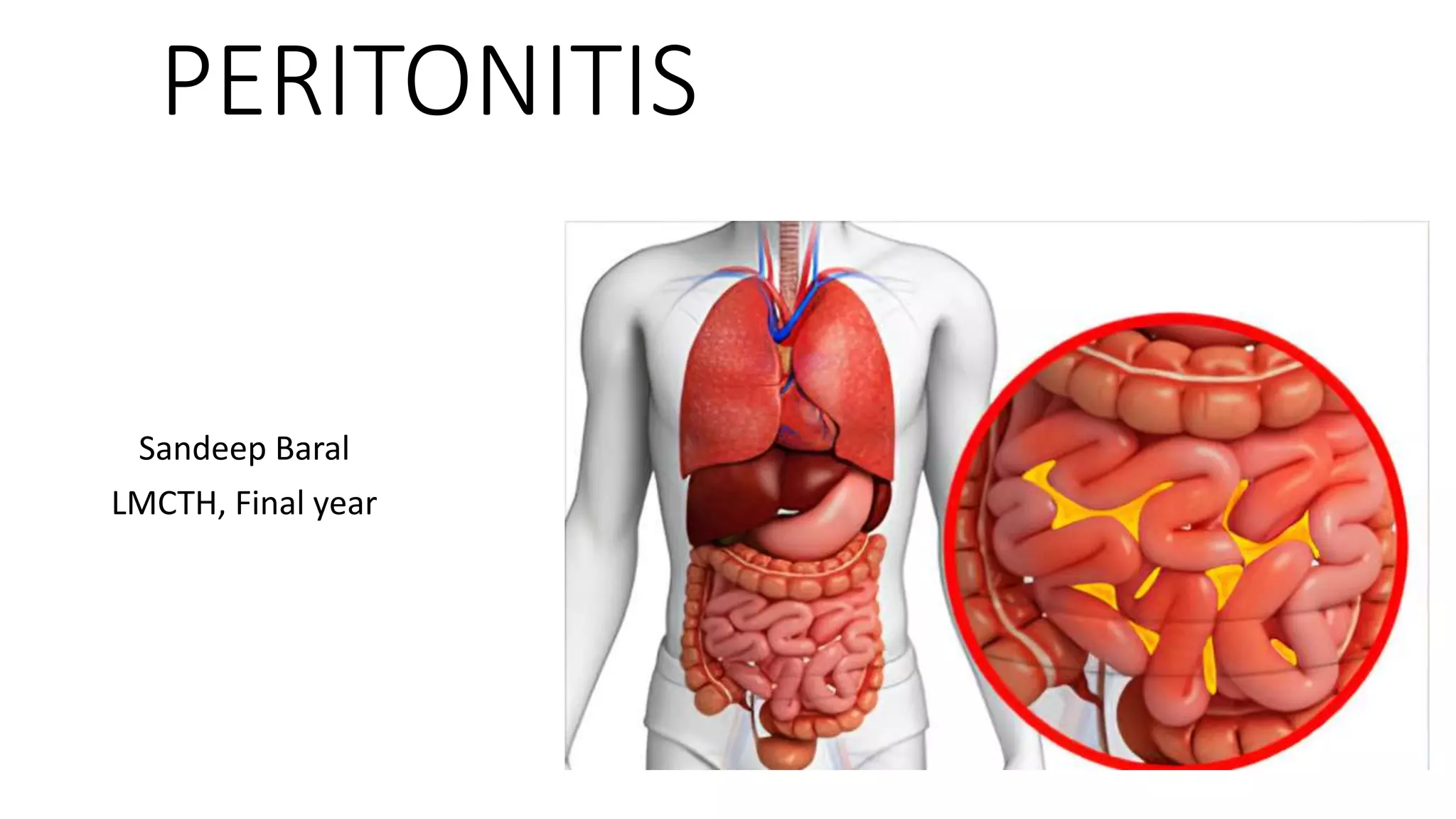
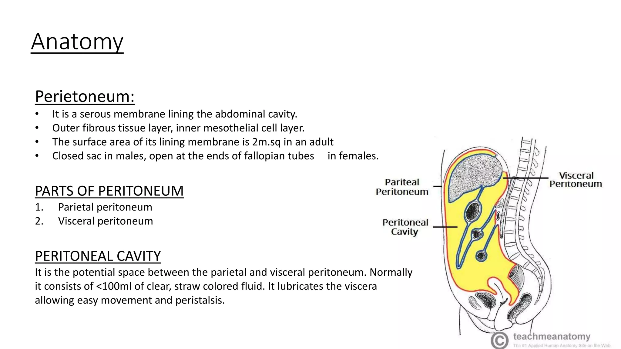
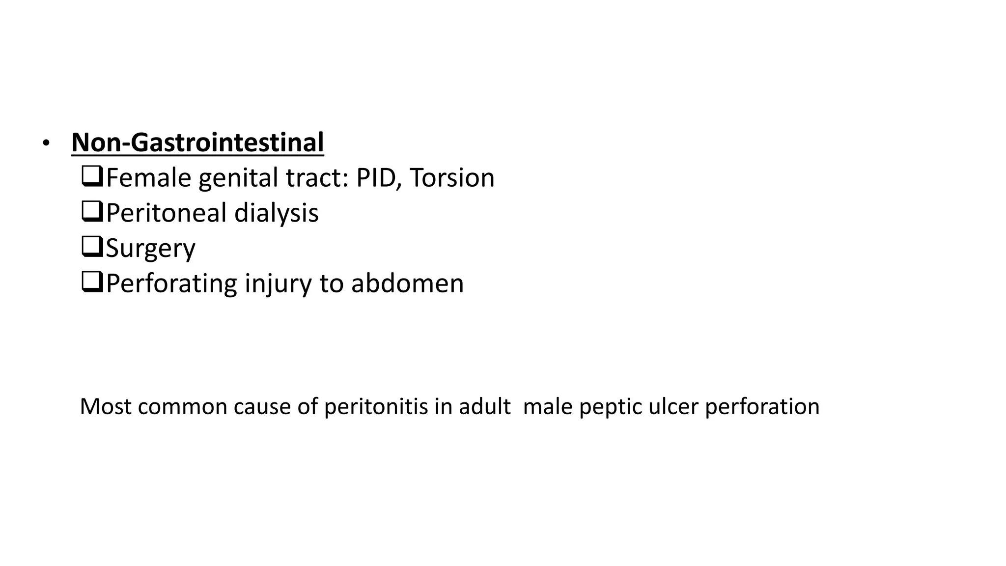
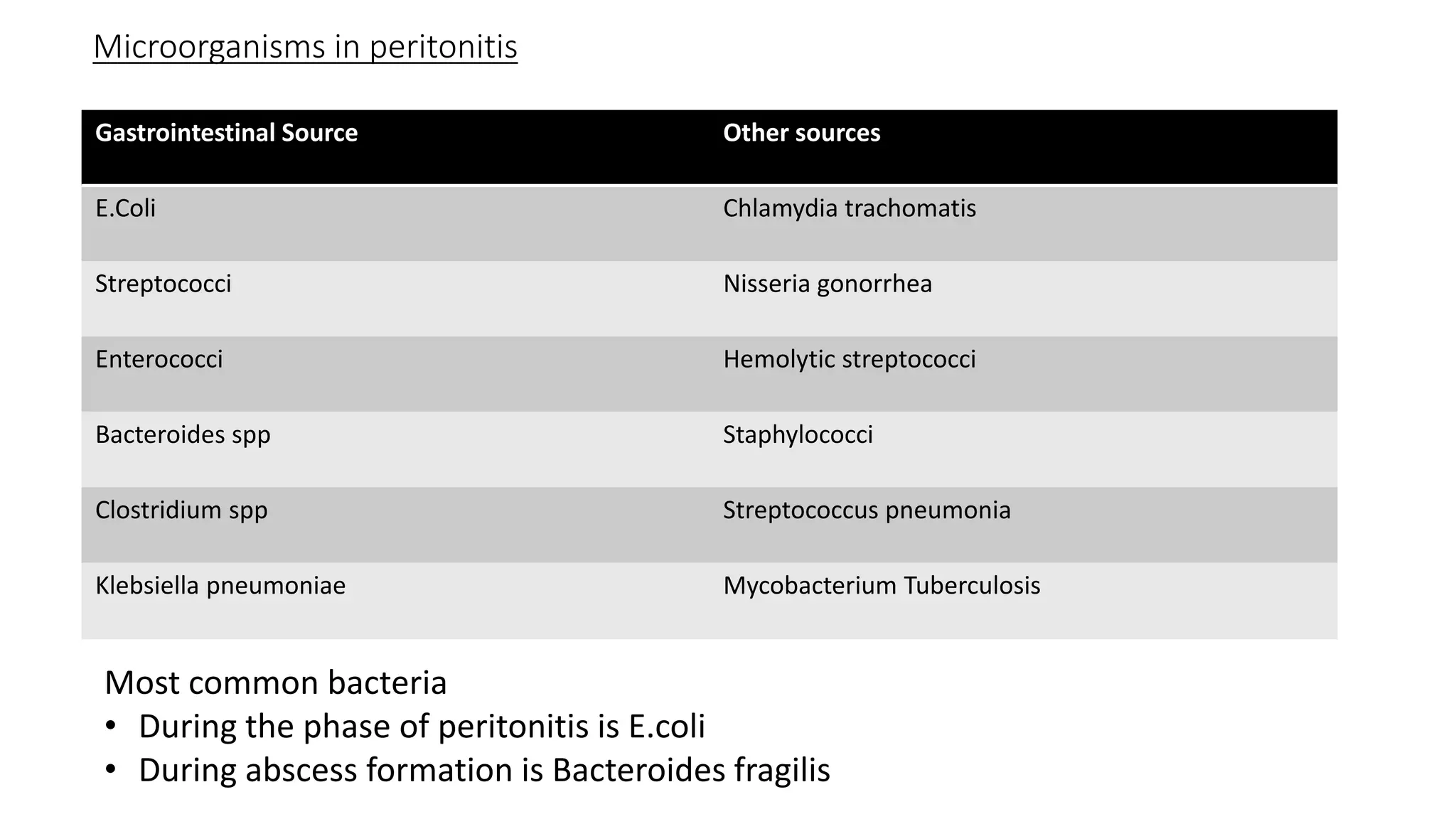
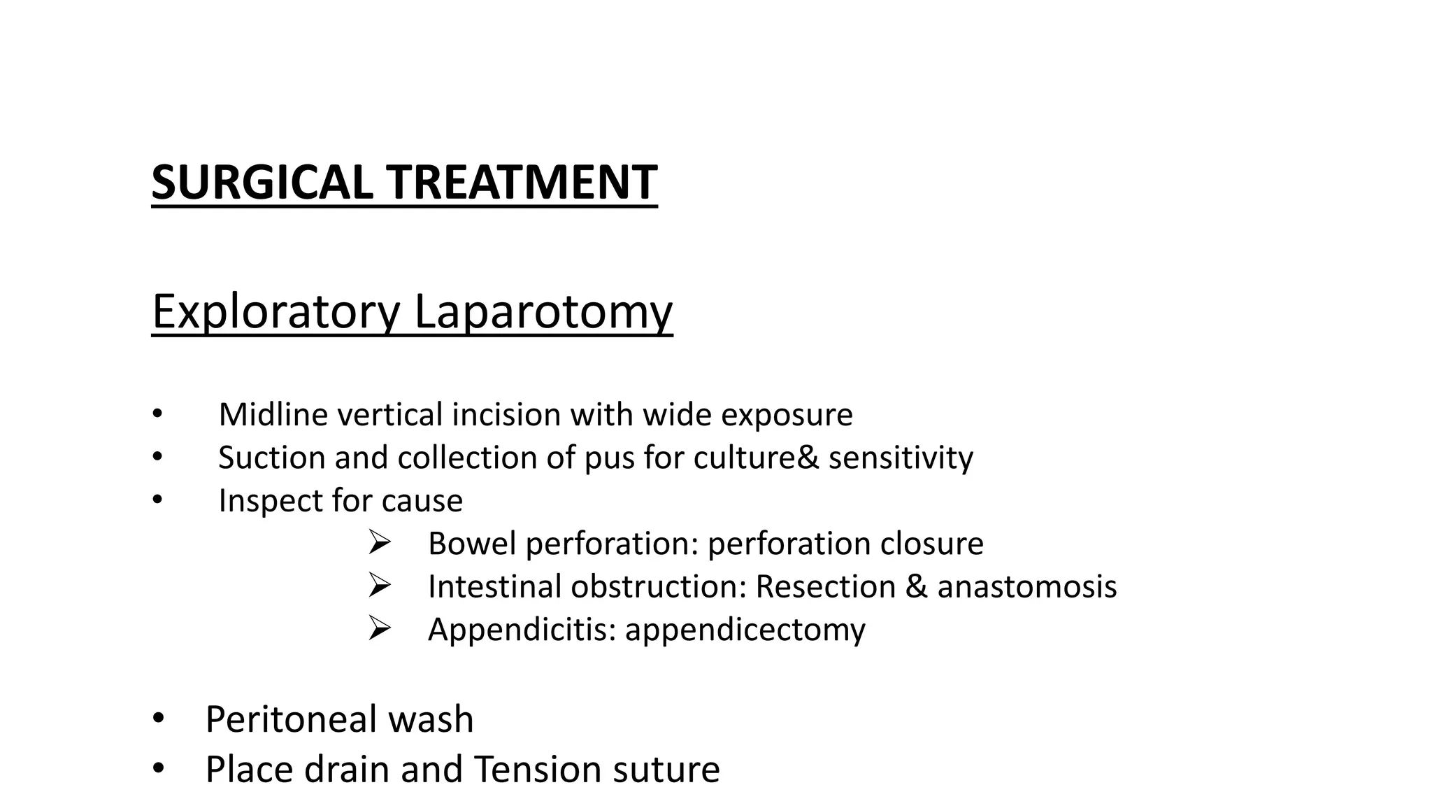
PERITONITIS
Sandeep Baral
LMCTH, Final year
Anatomy
Perietoneum:
• It is a serous membrane lining the abdominal cavity.
• Outer fibrous tissue layer, inner mesothelial cell layer.
• The surface area of its lining membrane is 2m.sq in an adult
• Closed sac in males, open at the ends of fallopian tubes in females.
PARTS OF PERITONEUM
1. Parietal peritoneum
2. Visceral peritoneum
PERITONEAL CAVITY
It is the potential space between the parietal and visceral peritoneum. Normally
it consists of <100ml of clear, straw colored fluid. It lubricates the viscera
allowing easy movement and peristalsis.
Function of peritoneum
IN HEALTH
• Visceral lubrication
• Fluid and particulate absorption
IN DISEASE
• Pain perception
• Inflammatory and immune responses
• Fibrinolytic activity
The peritoneum has the capacity to absorb large volumes of fluid, this ability is used during peritoneal
dialysis in the treatment of renal failure
Peritonitis
Peritonitis is defined as inflammation of the parietal
and serosal layer of peritoneum either due to chemicals
like gastric acids/bile or due to bacterial infection which
may be localized or generalized.
Causes of peritoneal inflammation:
Bacterial: gastrointestinal and non-gastrointestinal
Chemical: eg; bile, barium
Traumatic: operative handling
Ischemic: strangulated bowel, vascular occlusion
Types of acute peritonitis
Peritonitis can be chemical or bacterial or initially chemically induced later
bacterial.
TYPES
1. Primary
2. Secondary
3. Tertiary
Can also be classified as:
1. Localized
2. Generalized
1. Primary:
• Common in cirrhotic patient with ascites , as spontaneous bacterial
peritonitis(SBP)
• Common in young girls between 3 to 9 years.
• Results from bacterial, fungal or mycobacterial infection in absence of
GI perforation.
• 90% of SBP infection is monomicrobial: E.coli( 40%)
• Commonly due to Pneumococci, ocassionly due to streptococci and
hemophilus and othe gram negative( E.coli)
• Ascitis fluid WBC count if more than 250 cells/mm3 with more than
50% cells are polymorphonuclear cell suggestive of primary
peritonitis.
• Total count is very high > 30,000/mm3
. Secondary:
• It occurs in GI perforation
• Duodenal perforation and brust appendicitis are
commonest cause.
• E.coli is most common organism involved.
Tertiary:
• Defined as persistent/ recurrent intraabdominal infection
after an adequate treatment for primary or secondary
peritonitis usually after 48 hours.
• It occurs after any abdominal surgeries which is usually
severe and patient may go in for SIRS/ MODS early.
• Common in immunocompromised individuals
Paths to peritoneal infection
• Gastrointestinal Perforation: e.g. perforated ulcer, appendix, diverticulum
• Transmural Translocation: e.g. pancreatitis, ischemic bowel, primary bacterial
peritonitis
• Exogenous contamination: e.g. drains, open surgery, trauma, peritoneal dialysis
• Female genital tract infection: Pelvic inflammatory disease
• Hematogenous: septicemia
CAUSES OF PERITONEAL INFLAMMATION
• Gastrointestinal:
Perforation of bowel
Spontaneous/transmural translocation of bacteria
Pancreatitis
• Non-Gastrointestinal
Female genital tract: PID, Torsion
Peritoneal dialysis
Surgery
Perforating injury to abdomen
Most common cause of peritonitis in adult male peptic ulcer perforation
Microorganisms in peritonitis
Gastrointestinal Source Other sources
E.Coli Chlamydia trachomatis
Streptococci Nisseria gonorrhea
Enterococci Hemolytic streptococci
Bacteroides spp Staphylococci
Clostridium spp Streptococcus pneumonia
Klebsiella pneumoniae Mycobacterium Tuberculosis
Most common bacteria
• During the phase of peritonitis is E.coli
• During abscess formation is Bacteroides fragilis
Pathogenesis
Clinical Features
Abdominal pain, worse on movement, coughing and deep respiration
Constitutional upset: anorexia, malaise, fever, lassitude
GI upset: nausea and vomiting
Pyrexia( may be absent)
Raised pulse rate
Tenderness: guarding/rigidity/rebound of abdominal wall
Pain/tenderness on rectal/vaginal examination
Absent / reduced bowel sounds
Eventually leading to Hippocrates facies
Septic shock, SIRS, and MODS in later satges
Investigations
BLOOD
• Total Leucocyte count: increased
• Amylase(if 4 times normal value then
significant)
• Lipase
• Urea and Creatinine
• Electrolytes
Imaging
• Plain x-ray abdomen
Erect: gas under diaphragm
Supine: ground glass appearance
• CT scan abdomen
Invasive
• Diagnostic laparoscopy
• Diagnostic peritoneal lavage
Differential diagnsosis
Pancreatitis
Intestinal Obstruction
Ruptured Ectopic Pregnancy
Acute mesenteric ischemia
Principle of therapy in peritonitis
1.To control source of infection
2.To eliminate bacteria and sepsis
3.To maintain vital organ functions- Cardiac, Pulmonary
and Renal
4.Nutrition and metabolic support
MANAGEMENT
GENERAL CARE OF THE PAIENT
• Correction of fluid and electrolyte loss and circulating
volume
• Urinary catheterization and nasogastric drainage insertion
• Antibiotics therapy
Systemic antibiotic therapy
• Analgesia
• Vital system support
SURGICAL TREATMENT
Exploratory Laparotomy
• Midline vertical incision with wide exposure
• Suction and collection of pus for culture& sensitivity
• Inspect for cause
 Bowel perforation: perforation closure
 Intestinal obstruction: Resection & anastomosis
 Appendicitis: appendicectomy
• Peritoneal wash
• Place drain and Tension suture
Post operative management
1.Proper critical care (icu)
2.Ventilatory support; monitoring vitals with urine
output, TLC, DLC, blood urea, serum creatinine,
LFT
3.Proper fluid and electrolyte management
4.Prevention of DVT
Complications
SYSTEMIC COMPLICATIONS
1.Septic sock
2.Systemic Inflammatory Response
Syndrome
3.Multi-organ dysfunction syndrome
4.Death
ABDOMINAL COMPLICATIONS
1.Paralytic ileus
2.Residual or recurrent abscess
3.Portal pyaemia
4.Adhesional small bowel obstruction
REFRENCES
• Bailey and love’s short practice of surgery 27th edition
• SRB’s manual of Surgery 5th edition
Peritonitis

Peritonitis

  • 1.
  • 2.
    Anatomy Perietoneum: • It isa serous membrane lining the abdominal cavity. • Outer fibrous tissue layer, inner mesothelial cell layer. • The surface area of its lining membrane is 2m.sq in an adult • Closed sac in males, open at the ends of fallopian tubes in females. PARTS OF PERITONEUM 1. Parietal peritoneum 2. Visceral peritoneum PERITONEAL CAVITY It is the potential space between the parietal and visceral peritoneum. Normally it consists of <100ml of clear, straw colored fluid. It lubricates the viscera allowing easy movement and peristalsis.
  • 3.
    Function of peritoneum INHEALTH • Visceral lubrication • Fluid and particulate absorption IN DISEASE • Pain perception • Inflammatory and immune responses • Fibrinolytic activity The peritoneum has the capacity to absorb large volumes of fluid, this ability is used during peritoneal dialysis in the treatment of renal failure
  • 4.
    Peritonitis Peritonitis is definedas inflammation of the parietal and serosal layer of peritoneum either due to chemicals like gastric acids/bile or due to bacterial infection which may be localized or generalized.
  • 5.
    Causes of peritonealinflammation: Bacterial: gastrointestinal and non-gastrointestinal Chemical: eg; bile, barium Traumatic: operative handling Ischemic: strangulated bowel, vascular occlusion
  • 6.
    Types of acuteperitonitis Peritonitis can be chemical or bacterial or initially chemically induced later bacterial. TYPES 1. Primary 2. Secondary 3. Tertiary Can also be classified as: 1. Localized 2. Generalized
  • 7.
    1. Primary: • Commonin cirrhotic patient with ascites , as spontaneous bacterial peritonitis(SBP) • Common in young girls between 3 to 9 years. • Results from bacterial, fungal or mycobacterial infection in absence of GI perforation. • 90% of SBP infection is monomicrobial: E.coli( 40%) • Commonly due to Pneumococci, ocassionly due to streptococci and hemophilus and othe gram negative( E.coli) • Ascitis fluid WBC count if more than 250 cells/mm3 with more than 50% cells are polymorphonuclear cell suggestive of primary peritonitis. • Total count is very high > 30,000/mm3
  • 8.
    . Secondary: • Itoccurs in GI perforation • Duodenal perforation and brust appendicitis are commonest cause. • E.coli is most common organism involved.
  • 9.
    Tertiary: • Defined aspersistent/ recurrent intraabdominal infection after an adequate treatment for primary or secondary peritonitis usually after 48 hours. • It occurs after any abdominal surgeries which is usually severe and patient may go in for SIRS/ MODS early. • Common in immunocompromised individuals
  • 10.
    Paths to peritonealinfection • Gastrointestinal Perforation: e.g. perforated ulcer, appendix, diverticulum • Transmural Translocation: e.g. pancreatitis, ischemic bowel, primary bacterial peritonitis • Exogenous contamination: e.g. drains, open surgery, trauma, peritoneal dialysis • Female genital tract infection: Pelvic inflammatory disease • Hematogenous: septicemia
  • 11.
    CAUSES OF PERITONEALINFLAMMATION • Gastrointestinal: Perforation of bowel Spontaneous/transmural translocation of bacteria Pancreatitis
  • 12.
    • Non-Gastrointestinal Female genitaltract: PID, Torsion Peritoneal dialysis Surgery Perforating injury to abdomen Most common cause of peritonitis in adult male peptic ulcer perforation
  • 13.
    Microorganisms in peritonitis GastrointestinalSource Other sources E.Coli Chlamydia trachomatis Streptococci Nisseria gonorrhea Enterococci Hemolytic streptococci Bacteroides spp Staphylococci Clostridium spp Streptococcus pneumonia Klebsiella pneumoniae Mycobacterium Tuberculosis Most common bacteria • During the phase of peritonitis is E.coli • During abscess formation is Bacteroides fragilis
  • 14.
  • 15.
    Clinical Features Abdominal pain,worse on movement, coughing and deep respiration Constitutional upset: anorexia, malaise, fever, lassitude GI upset: nausea and vomiting Pyrexia( may be absent) Raised pulse rate Tenderness: guarding/rigidity/rebound of abdominal wall Pain/tenderness on rectal/vaginal examination Absent / reduced bowel sounds Eventually leading to Hippocrates facies Septic shock, SIRS, and MODS in later satges
  • 16.
    Investigations BLOOD • Total Leucocytecount: increased • Amylase(if 4 times normal value then significant) • Lipase • Urea and Creatinine • Electrolytes
  • 17.
    Imaging • Plain x-rayabdomen Erect: gas under diaphragm Supine: ground glass appearance • CT scan abdomen
  • 18.
    Invasive • Diagnostic laparoscopy •Diagnostic peritoneal lavage
  • 19.
  • 20.
    Principle of therapyin peritonitis 1.To control source of infection 2.To eliminate bacteria and sepsis 3.To maintain vital organ functions- Cardiac, Pulmonary and Renal 4.Nutrition and metabolic support
  • 21.
    MANAGEMENT GENERAL CARE OFTHE PAIENT • Correction of fluid and electrolyte loss and circulating volume • Urinary catheterization and nasogastric drainage insertion • Antibiotics therapy Systemic antibiotic therapy • Analgesia • Vital system support
  • 22.
    SURGICAL TREATMENT Exploratory Laparotomy •Midline vertical incision with wide exposure • Suction and collection of pus for culture& sensitivity • Inspect for cause  Bowel perforation: perforation closure  Intestinal obstruction: Resection & anastomosis  Appendicitis: appendicectomy • Peritoneal wash • Place drain and Tension suture
  • 23.
    Post operative management 1.Propercritical care (icu) 2.Ventilatory support; monitoring vitals with urine output, TLC, DLC, blood urea, serum creatinine, LFT 3.Proper fluid and electrolyte management 4.Prevention of DVT
  • 24.
    Complications SYSTEMIC COMPLICATIONS 1.Septic sock 2.SystemicInflammatory Response Syndrome 3.Multi-organ dysfunction syndrome 4.Death
  • 25.
    ABDOMINAL COMPLICATIONS 1.Paralytic ileus 2.Residualor recurrent abscess 3.Portal pyaemia 4.Adhesional small bowel obstruction
  • 26.
    REFRENCES • Bailey andlove’s short practice of surgery 27th edition • SRB’s manual of Surgery 5th edition