The document provides instructions for compiling a complete lab report from four biology labs on genomic databases, primer design, PCR, and molecular cloning. It outlines the necessary sections for the report, including an introduction describing the overall question and background, materials and methods, results with data and figures, and a discussion/conclusion section. It also provides additional details on designing a transgene reporter gene based on knowledge gained from the lab exercises, including defining a transgene, necessary gene elements, and ideas for using the transgene.

![Introduction ( for all 4 labs results) and ( see the instructions for all 4 labs)
methods (for all 4 labs results) and ( see the instructions for all 4 labs)
Results (Data) (for all 4 labs results) make sure to include table, graph and ( see the
instructions for all 4 labs)
Discussion/Conclusion ( for all 4 labs results) and ( see the instructions for all 4 labs)
Reference(for also 4 labs results)
Grammer
Requirements: depends | .doc file
Biology 366LLab Report Due First Meeting Week 7
Exercises Completed in Module I•Introduction to genomic databases and BLAST: Why do
we use BLAST? What does the output of BLAST searches tell us?•Primer Design and PCR:
What is the object of designing good primers? How will you use the
primers?•MolecularCloning:Why do we clone genes? What
doyouneedtocloneageneandamplifyit?•You have been compiling the “Results” of your
report. Let’s put it all together.
Group Research Report1)Introduction: Describe the major question, problem, or technical
issue addressed. Explain relevance and background information, including previous
work.2)Materials/Methods: Summarize the materials and methods used to test the
hypothesis for an assigned gene.3)Results: Explain the results and how they were obtained.
Refer specifically to data in figures and/or tables. You will be trained to prepare scientific
illustrations for images or quantitative data. This section will discuss the methods used
(refer to the Methods section of the report).4)Conclusions and Discussion: Briefly conclude
findings and raises future questions. Summarize and explain the significance of the work
presented in the paper. When appropriate, you are encouraged to discuss other relevant
approaches or articles from the literature.
Discussion•Discussion: Based on the knowledge you have built from the lab exercises,
include a discussion on building a reporter gene or a transgene: a) define a transgene; b)
discuss what gene elements are necessary for gene expression of artificial genes; c) discuss
ideas for how to use the transgene (see articles posted in Module II in Canvas).
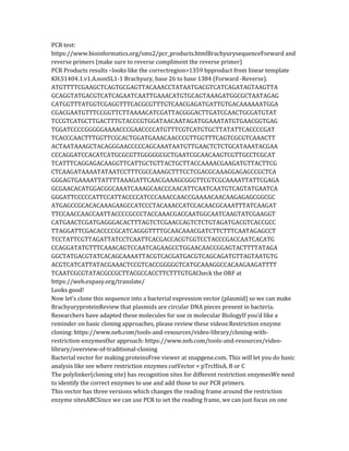
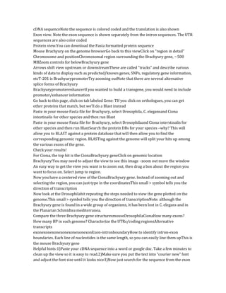
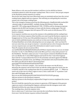
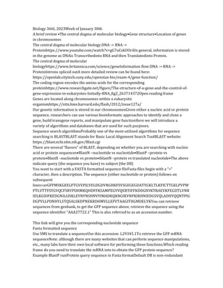
Gene expression in living cells is often difficult to detect because of limited access of
substrates to marker enzymes. Here gene expression in specific neurons of the nematode
Caenorhabditis elegans is monitored by the bright green fluorescence of the green
fluorescent protein (GFP) from the jellyfish Aequorea victoria. The GFP fills entire neurons,
including in one neuron an extended, fanned growth cone visible in the tail end (upper
portion) of the nematode. See page 802. [Photo: Martin
Chalfie]https://www.nobelprize.org/prizes/chemistry/2008/summary/
Group No.:Biology 366L: Spring 2023Grader:Laboratory Report Rubric Total Points
100ExcellentGoodFairNeeds ImprovementIntroduction (26 pts)1. Clearly communicates
the question that is trying to be answered. 2. States a clear a goal or
hypothesis that is predictable and testable. 3.](https://image.slidesharecdn.com/onecompletereportfromallthe4labs-230307014155-7d6d93d7/85/one-complete-report-from-all-the-4-labs-pdf-2-320.jpg)

![approaches or articles from the literature.
Discussion•Discussion: Based on the knowledge you have built from the lab exercises,
include a discussion on building a reporter gene or a transgene: a) define a transgene; b)
discuss what gene elements are necessary for gene expression of artificial genes; c) discuss
ideas for how to use the transgene (see articles posted in Module II in Canvas).
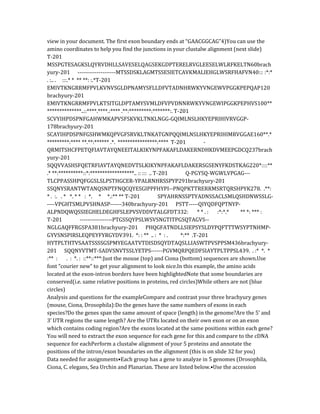
Gene expression in living cells is often difficult to detect because of limited access of
substrates to marker enzymes. Here gene expression in specific neurons of the nematode
Caenorhabditis elegans is monitored by the bright green fluorescence of the green
fluorescent protein (GFP) from the jellyfish Aequorea victoria. The GFP fills entire neurons,
including in one neuron an extended, fanned growth cone visible in the tail end (upper
portion) of the nematode. See page 802. [Photo: Martin
Chalfie]https://www.nobelprize.org/prizes/chemistry/2008/summary/
Group No.:Biology 366L: Spring 2023Grader:Laboratory Report Rubric Total Points
100ExcellentGoodFairNeeds ImprovementIntroduction (26 pts)1. Clearly communicates
the question that is trying to be answered. 2. States a clear a goal or
hypothesis that is predictable and testable. 3.
Provides evidence to support hypothesis from background/research. 4.
Incorporated edits to improve introduction from previous report.Some of the "excellent"
items are missing.Methods (8 pts)A description or step-by-step list of how the experiment
was performed. Description, unclear could not be repeated.Results (Data) (26 pts)1. Results
are clearly recorded and organized. 2. Figures are of publish
quality and clearly labeled and easy to follow with written results.
3. Easy for the reader to see trends in the results. 4. Clearly
transitioned and incorporated new data into results.One of the "excellent" items is
missing.Two of the "excellent" items are missing.Three of the "excellent" items are
missing.Discussion/Conclusion (28 pts)1. Summarizes essential data used to draw
conclusions. 2. Discusses application and
implications of results. ("real world connections"). 3. Puts findings
into context of previously published results. 4. Describes future
direction of project. 5. Concludes with an overall statement (e.g., In
summary, this work...). One of the "excellent" items is missing.Two of the "excellent"
items are missing.Three of the "excellent" items are missing.References (6 pts)Clearly
organized.Missing appropriate citations.Format and Grammar (6 pts) Neat organized with
clear headings, few grammatical and spelling errors.Somewhat lacking in organization,
multiple spelling and grammar errors, not neat.Comments:
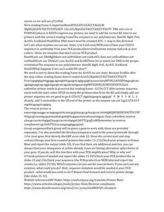
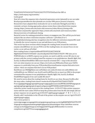
Transgenes –definition, design and testing
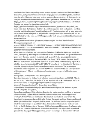
A Typical Eukaryotic GeneThe typical eukaryotic gene is composed of a cis-regulatory
domain as well as the sequences required to encode the protein Start of
transcriptionExons/intronsCis-regulatory region
The cis-regulatory domain contains binding sites for transcription factors –the binding of
these proteins regulates gene expression
In multicellular organisms, genes can be expressed in different cell types (like the
Cionaembryos to the left –green color), at different times and at different levelsThe cis-](https://image.slidesharecdn.com/onecompletereportfromallthe4labs-230307014155-7d6d93d7/85/one-complete-report-from-all-the-4-labs-pdf-14-320.jpg)


![Reporter gene[2]](https://cdn.slidesharecdn.com/ss_thumbnails/reportergene2-160424161339-thumbnail.jpg?width=640&height=640&fit=bounds)