
Recommended
More Related Content
What's hot
What's hot (20)
Viewers also liked
Viewers also liked (13)
Similar to Normal oral radiographic anatomy
Similar to Normal oral radiographic anatomy (20)
More from shyasaman
Recently uploaded
Recently uploaded (20)
Normal oral radiographic anatomy
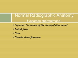
- 1. Superior Foramina of the Nasopalatine canal Latral fossa Nose Nasolacrimal foramen Normal Radiographic Anatomy (yasaman sherafatmand)
- 2. Radiographic features of Superior Foramina of the Nasopalatine canal The nasopalatine canal originates at two foramina in floor of the nasal cavity. Radiographically, it can be recognized as two radiolucent areas above the apices of the central incisors in floor of the nasal cavity near its anterior border and both the sides of the septum. Lateral wall of nasopalatine canalSuperior foramina
- 3. A : latral walls of the nasopalatine canal extended from the incisive foramen to the floor of the nasal fossa
- 8. RADIOGRAPHIC FEATURES OF THE LATERAL FOSSA Also called as INCISIVE FOSSA. Appears as depression in the maxilla near the apex of the lateral incisor . Appears diffusely radiolucent in the IOPA.
- 11. Latral fossa between canine and latral
- 12. RADIOGRAPHIC FEATURES OF NOSE The soft tissue of the nose is frequently seen in the projections of the maxillary central and lateral incisors ,superimposed over the roots of these teeth. Image appears uniformly opaque with a sharp border.
- 15. Green-Anterior nasal spine. Red- Zygomatic process of maxilla
- 16. Floor of nasal cavity
- 18. RADIOGRAPHIC FEATURES OF THE NASOLACRIMAL CANAL The nasal and maxillary bones form the nasolacrimal canal. It runs from the medial aspect of the antero inferior border of the orbit inferiorly, to drain under the inferior conchae into the nasal cavity.
- 22. Thank You